Modulating long-range energetics via helix stabilization: a case study using T4 lysozyme - bioRxiv
←
→
Page content transcription
If your browser does not render page correctly, please read the page content below
bioRxiv preprint first posted online Jun. 22, 2018; doi: http://dx.doi.org/10.1101/353649. The copyright holder for this preprint
 (which was not peer-reviewed) is the author/funder, who has granted bioRxiv a license to display the preprint in perpetuity.
                              It is made available under a CC-BY-NC-ND 4.0 International license.

        Modulating long-range energetics via helix stabilization: a case
                          study using T4 lysozyme
                     Sabriya N. Rosemond1,2, Kambiz M. Hamadani 1,3, Jamie H.D. Cate1,2,4,5
                                         and Susan Marqusee1,2,4,5

   1
       California Institute for Quantitative Biosciences, University of California, Berkeley CA 94720
   2
   Department of Molecular and Cell Biology, University of California, Berkeley, CA 94720-3220,
   USA.
    3
        California State University San Marcos, San Marcos, CA 92096, USA
   4
       Department of Chemistry, University of California, Berkeley CA 94720
   5
       Correspondence should be addressed to S.M. (510-642-7678, marqusee@berkeley.edu)

   Total number of manuscript pages: 21 manuscript pages, 1 page of tables, 4 pages of figures
bioRxiv preprint first posted online Jun. 22, 2018; doi: http://dx.doi.org/10.1101/353649. The copyright holder for this preprint
(which was not peer-reviewed) is the author/funder, who has granted bioRxiv a license to display the preprint in perpetuity.
It is made available under a CC-BY-NC-ND 4.0 International license.
Rosemond et al.
Cooperative protein folding requires distant regions of a protein to interact and provide
mutual stabilization. The mechanism of this long-distance coupling remains poorly
understood. Here, we use T4 lysozyme (T4L*) as a model to investigate long-range
communications across a globular protein. T4L* is composed of two structurally distinct
subdomains, although it behaves in a two-state manner at equilibrium. The subdomains
of T4L* are connected via two topological connections: the N-terminal helix that is
structurally part of the C-terminal subdomain (the A-helix) and a long helix that spans
both subdomains (the C-helix). To understand the role that the C-helix plays in
cooperative folding, we analyzed a circularly permuted version of T4L* (CP13*), whose
subdomains are connected only by the C-helix. We demonstrate that when isolated as
individual fragments, both subdomains of CP13* can fold autonomously into marginally
stable conformations. The energetics of the N-terminal subdomain depend on the
formation of a salt bridge known to be important for stability in the full-length protein. We
show that the energetic contribution of the salt bridge to the stability of the N-terminal
fragment increases when the C-helix is stabilized, such as occurs upon folding of the C-
terminal subdomain. These results suggest a model where long-range energetic coupling
is mediated by helix stabilization.
Keywords: protein folding, effective concentration, cooperativity, helix stabilization, T4 lysozyme,
In this work, we investigate how a helix spanning the two subdomains of T4 lysozyme* couples
distant regions of the protein. We find evidence for a model of long-distance coupling that relies
on the cooperative nature of helix formation to stabilize a long-range tertiary salt bridge
interaction at one end of the helix and thereby couple the folding of T4 lysozyme’s subdomains.
This mechanistic model may have implications for co-translational folding.
Introduction
Cooperativity is a hallmark of globular proteins. At equilibrium, many small (bioRxiv preprint first posted online Jun. 22, 2018; doi: http://dx.doi.org/10.1101/353649. The copyright holder for this preprint
(which was not peer-reviewed) is the author/funder, who has granted bioRxiv a license to display the preprint in perpetuity.
It is made available under a CC-BY-NC-ND 4.0 International license.
Rosemond et al.
communication between the subdomains must derive from the topology or backbone
connections between the two. There are two such connections: one at the end of the A-helix
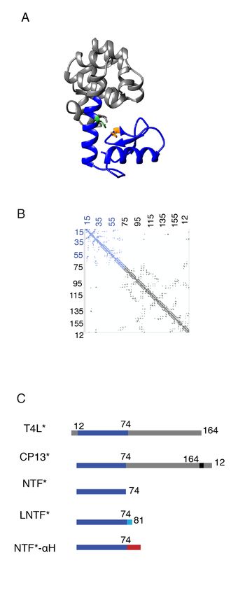
and one within the C-helix (Fig. 1). The A-helix (residues 1-12) is structurally part of the C-
terminal subdomain, making this subdomain discontinuous in sequence8 and connects the two
subdomains between residues 12 and 13. The C-helix is the central helix that spans both
subdomains (residues 59-81, Fig. 1A).
The role of this discontinuous subdomain architecture (the placement of helix A) was explored
using a circular permutant, CP13*8-11 (Fig. 1A). CP13* begins at residue 13 with the A-helix
(residues 1-12) appended to the C-terminus via a short flexible linker8. Thus, in CP13*, the C-
terminal subdomain is contiguous in sequence. In spite of this rearrangement, CP13* and T4L*
have similar structures (RMSDbioRxiv preprint first posted online Jun. 22, 2018; doi: http://dx.doi.org/10.1101/353649. The copyright holder for this preprint
(which was not peer-reviewed) is the author/funder, who has granted bioRxiv a license to display the preprint in perpetuity.
It is made available under a CC-BY-NC-ND 4.0 International license.
Rosemond et al.
denaturation curve can be fit to a two-state model14, yielding an extrapolated free energy
(ΔGunf(H2O) of 1.60 ±0.13 kcal/mol and an m-value =1.13 ±0.01 kcal/mol*M (Fig. 2B and Table
I). The m-value agrees with expectations based on the number of residues in the fragment (m-
values have been found to correlate with buried surface area and size of a given polypeptide15),
consistent with folding of the entire fragment.
The structure and energetics of NTF* are pH-dependent. CD spectra of NTF* at pH 5 and pH 7
show a difference in magnitude and shape (Fig. 2A). At pH 7, the fragment is no longer folded,
as judged by the loss of a cooperative unfolding transition with urea (Fig. 2B, inset). This pH
dependence may be explained by the partially buried salt bridge (H31-D70) within the N-
terminal subdomain7,8,13.
H31 and D70 are important for the folding and stability of NTF*
To probe the role of these residues in NTF*, we generated two single-site variants, NTF* H31N
and NTF* D70N. The CD spectra suggest that, unlike NTF*, both variants are predominately
unfolded under native-like conditions (Fig. 2A). Urea-induced denaturation of these variants (pH
5, 4°C) monitored by CD at 222 nm shows no notable folded baseline, demonstrating that, even
under conditions of no or low denaturant, the fragments are in the transition zone (Fig. 2B). To
estimate how these mutations affect NTF*’s stability, we carried out a two-state fit on these
data, fixing the folded baseline to the parameters from the wild-type NTF* fit. This analysis
yields an estimate of ΔGunf ~ 0.31 kcal/mol for NTF* H31N and ΔGunf ~ 0.09 kcal/mol for NTF*
D70N (Table I). The significant destabilization of NTF* that occurs upon mutating H31 and D70
suggests that these residues play a critical role in the stability of the isolated N-terminal
subdomain.
Extending the C-terminal helix enhances its stability
In full-length T4L*, the C-helix (residues 59-81) spans both subdomains: the majority of the helix
is part of the N-terminal subdomain, but the final seven residues are in the C-terminal
subdomain. To determine the effect of extending NTF* to include the complete C-helix, we
generated a longer fragment encoding the N-terminal subdomain with the entire C-helix
sequence, LNTF* (Long-NTF*, residues 13-81). At pH 5, LNTF* is folded as monitored by far-
UV CD and unfolds cooperatively as a function of urea (Fig. 2C and D). When fit with a two-
state linear extrapolation model14, this unfolding curve results in a ΔGunf = 2.16 ±0.04 kcal/mol
with an m-value = 1.20 ±0.02 kcal/mol*M. Therefore, under these conditions, an extended N-
terminal subdomain fragment is stabilized (0.56 ± .14 kcal/mol) compared to NTF*, which has
the shorter C-helix (Table I). Similar to NTF*, the folding of LNTF* is pH-dependent, with less
structure and a loss of cooperative unfolding at pH 7 compared to pH 5 (Fig. 2C and D, inset).
The same single-site variants investigated in the context of NTF* were also evaluated in the
background of LNTF* (LNTF* H31N and LNTF* D70N). CD spectra and denaturation curves of
these variants suggest that, like in the shorter fragment NTF*, they are predominately unfolded
under native-like conditions. Using a similar approach as above to estimate the energetics of
these variants, these curves were fit using a two-state model assuming the same baseline as
LNTF*. Based on this analysis, the variants are significantly destabilized compared to LNTF*
(LNTF* H31N: ΔGunf ~ -0.20 kcal/mol; LNTF* D70N ΔGunf ~ -0.86 kcal/mol) (Table I, Fig. 2C and
D).
To determine whether the increased stability of LNTF* as compared to NTF* is due to
sequence-specific interactions or simply the addition of a helical segment, we appended a
sequence known to form a stable helix16, (A(EAAAK)3A), to the C-terminus of NTF* , generating
NTF*-αH. When monitored by CD, NTF*-αH is folded and has a cooperative urea-induced
4bioRxiv preprint first posted online Jun. 22, 2018; doi: http://dx.doi.org/10.1101/353649. The copyright holder for this preprint
(which was not peer-reviewed) is the author/funder, who has granted bioRxiv a license to display the preprint in perpetuity.
It is made available under a CC-BY-NC-ND 4.0 International license.
Rosemond et al.
unfolding curve (Fig. 2D, Table I) (ΔGunf = 2.44 ±0.05 kcal/mol, m-value = 1.09 ±0.02 kcal/mol*M
(pH 5.0, 4°C)). The addition of this stable alpha helix increases NTF* stability by ~0.84 ±.14
kcal/mol at pH 5.
Full-length CP13* populates an equilibrium intermediate in the absence of the H31-D70
salt bridge
The above fragment studies suggest that the H31-D70 salt bridge is critical to the stability of the
isolated N-terminal subdomain (~1-2 kcal/mol). This same salt bridge is known to contribute 3-5
kcal/mol in full-length T4L*13. To probe the contribution of this interaction in full-length CP13*,
we investigated CP13*’s stability as a function of pH and mutation. Equilibrium urea-
denaturation studies analyzed with a two-state assumption showed a notable difference in the
free energy and calculated m-value at pH 5 and 7 (pH 5: ΔGunf = 12.94 ±0.17 kcal/mol, m-
value= 2.82 ±0.02 kcal/mol*M; pH 7: ΔGunf = 10.82 ±0.25 kcal/mol, m-value= 2.55± 0.06
kcal/mol*M, Fig. 3, Table I). The lower m-value at pH 7 suggests a breakdown in the two-state
assumption due to a measureable population of an equilibrium intermediate17-19, which could
indicate selective unfolding of the N-terminal subdomain at the higher pH.
To evaluate whether CP13* populates a partially folded intermediate at equilibrium, we
performed proteolysis experiments at both pH 5 and pH 7. Proteolysis of CP13* was monitored
as a function of time using the non-specific protease thermolysin20,21. Quenched reactions from
different time points were run on an SDS-PAGE gel to quantify the remaining full-length protein
(Fig 4A and Materials and Methods). There is an exponential decrease in the intensity of full-
length protein at both pH 5 and pH 7. Concomitant with this disappearance of the full-length
protein, a smaller band appears at approximately 17kDa (Ic) (Fig. 4A). The size of this
proteolysis-resistant fragment is consistent with the size of the C-terminal domain. Mass
spectrometry analysis indicates that the proteolysis fragment sequence maps onto the folded
region of the NSHX intermediate (Ic is the same as Ieq)10. Together, these results suggest that
the cleavable state consists of an unfolded N-terminal subdomain and a folded C-terminal
domain.
To gain more information about the difference in the energetics of CP13* under these two
conditions we carried out a quantitative analysis of the observed proteolysis rate (kp), utilizing
formalisms developed for hydrogen exchange21-23. This approach allows for the determination of
either the kinetics or thermodynamics of the opening reaction that leads to the formation of the
cleavable conformation, depending on the conditions of the experiment. To determine whether
proteolysis in our experiments was in the kinetic (EX1 exchange) regime or thermodynamic
(EX2 exchange) regime, we monitored kp as a function of the intrinsic rate of proteolysis, kint, by
modulating the concentration of thermolysin. If kp is constant as a function of kint, kp reports on
the kinetics of the partial unfolding reaction. If kp changes linearly as a function of kint, kp
provides thermodynamic information about the reaction, and the slope of this line is equal to the
equilibrium constant of the opening reaction (Kprot)23,24. As shown in Figure 4B, there is a linear
relationship between kp and kint for CP13* at both pH 5 and 7. Therefore, we are in the EX2
regime, and kp reports on the thermodynamics of the unfolding reaction. Using the slopes of
these lines (slopepH5= 0.0018, slopepH7=0.039) we find that ΔGprot, the free-energy difference
between the ground state (N) and the cleavable conformation (Ic), is 3.73 kcal/mol at pH 5 and
1.91 kcal/mol at pH 7. These data suggest that destabilizing the salt bridge, increases the
population of the intermediate which explains the difference in m-values under these two
conditions.
The pH dependence of the intermediate population implicates a role for the H31-D70 salt bridge
in coupling between the two subdomains of CP13*. To probe this directly, we performed
5bioRxiv preprint first posted online Jun. 22, 2018; doi: http://dx.doi.org/10.1101/353649. The copyright holder for this preprint
(which was not peer-reviewed) is the author/funder, who has granted bioRxiv a license to display the preprint in perpetuity.
It is made available under a CC-BY-NC-ND 4.0 International license.
Rosemond et al.
equilibrium denaturation experiments with the single-site variants of CP13* H31N and D70N.
Even at pH 5, two-state fits of the urea-induced denaturation curves for both CP13* H31N and
CP13* D70N result in m-values significantly lower than that for CP13* (CP13* H31N: 2.10 ±0.05
kcal/mol*M (Δm-value= 0.72 kcal/mol*M); CP13*D70N: 2.17 ±0.03 kcal/mol*M (Δm-value= 0.65
kcal/mol*M)) (Table I and Fig. 4). Interestingly, these m-values are quite similar to each other
and to the m-value of a protein fragment designed to mimic Ieq observed in previous NSHX
experiments9,10,12 LCTF, 2.06 ±0.02 kcal/mol*M (Table I). These lower m-values, together with
the decreased m-value of CP13* at pH 7 compared to pH 5, suggest that the salt bridge (H31-
D70) plays an important role in the folding cooperativity of the two subdomains in CP13*,
without which there is a significant population of an intermediate. Indeed, proteolysis studies on
these two variants show complete proteolysis to Ic within one minute (the first time point). This
rapid proteolysis suggests that, in these variants, the N-terminal subdomain is substantially
unfolded under native conditions.
Discussion
In this work, we investigate how a helix spanning the two subdomains of T4 lysozyme*, CP13*,
couples distant regions of the protein. CP13* is a small globular protein composed of two
distinct subdomains with almost no side-chain interactions between the two7,8, yet it exhibits
typical two-state equilibrium unfolding behavior6,8-10, suggesting that the coupled folding of the
two subdomains must arise from sources other than interactions at the subdomain interface. We
investigate the role of the single topological connection between the subdomains, the C-helix in
coupling these subdomains.
By comparing the stability of the N-terminal subdomain in different contexts - in isolation, with
additional residues to complete the C-helix, with additional residues encoding a stable helix, and
in the context of the full-length protein - we find that the energetics of this subdomain are
tunable and that this malleability relies on the addition of a stable helical sequence to the N-
terminal subdomain, thus suggesting a role for the C-helix in stabilizing this region. By
investigating the pH dependence of the subdomain’s stability and the effect of site-specific
mutations, we find evidence that these changes in stability directly correlate with a well-known
salt bridge within the N-terminal subdomain13. The stability of this interaction increases as the
stability of the helix is increased remotely (its C-terminus). In the context of the full-length
protein, where interactions between the C-terminal end of the C-helix and other residues of the
C-terminal subdomain maximally stabilize the helix, we find that removing the salt bridge
decouples the two subdomains such that a conformation with an unfolded N-terminal
subdomain is populated at equilibrium. Because D70 is in the C-helix, we posit that stabilizing
the helix orients D70 to strengthen the D70-H31 salt bridge. Thus, the stability of the C-helix
provides the conduit for long-range energetic coupling of the two subdomains.
Coupling of the subdomains via the C-helix
Our current work, together with previous studies of the C-terminal subdomain, demonstrates
that both subdomains can fold autonomously, as suggested by analyses of side-chain contacts7
(see figure 1B for a contact map of CP13*). Independently these regions are marginally stable,
but when fused in the context of CP13* the stability is much greater than the sum of the parts
(3.75 kcal/mol and 1.60 kcal/mol vs. 12 kcal/mol (Table I)).
In CP13*, the two subdomains are joined only by a shared 23 amino-acid helix, the C-helix. The
first sixteen residues are part of the N-terminal subdomain while the last seven are part of the C-
terminal subdomain8. While helical in the full-length protein, these residues in isolation do not
encode a stable helix: a peptide corresponding to the isolated C-helix is unstructured in
aqueous solutions when monitored by far-UV CD25, and AGADIR, an algorithm parameterized
6bioRxiv preprint first posted online Jun. 22, 2018; doi: http://dx.doi.org/10.1101/353649. The copyright holder for this preprint
(which was not peer-reviewed) is the author/funder, who has granted bioRxiv a license to display the preprint in perpetuity.
It is made available under a CC-BY-NC-ND 4.0 International license.
Rosemond et al.
to predict peptide helix propensity, suggests a very low helical propensity for the C-helix
sequence (4.3 %)26.
The C-helix is stabilized in the context of the C-terminal subdomain. Previously we determined
the high-resolution crystal structure of a fragment encoding the C-terminal subdomain with the
entire C-helix (an additional 15 residues: 60-74), LCTF10. LCTF has a native-like fold and
residues 65-81 form a helix, indicating that all but the first turn of the C-helix (residues 59-64)
folds in this context. NSHX data shows that C-helix residues 75-81, which make contacts with
other residues in the C-terminal subdomain, are significantly more stable than the remainder of
the C-helix10,12. Thus, the C-terminal subdomain and its contacts with the last seven residues of
the C-helix (75-81) stabilize the entire helix10. Such contact-assisted helix formation has been
observed in molecular dynamic simulations of barnase and Protein A27.
The presence of the complete C-helix sequence also appears to stabilize the C-terminal
subdomain. Interactions between the C-helix and the rest of the C-terminal subdomain stabilizes
that subdomain, as LCTF is 5.5 kcal/mol more stable than CTF (75-164, 1-12)9,10. In the present
work, we find that the N-terminal subdomain shows similar, albeit much less, stabilization and
linkage between the C-helix and the subdomain. The presence of the complete C-helix
sequence stabilizes both subdomains in isolation.
Long-distance coupling and communication via helix stability
In proteins, relatively weak bimolecular interactions are strengthened when they occur between
components of a single polypeptide chain due to an increase in effective concentration28. For
example, the interaction between imidazole and carboxylic acid is quite weak; however, in the
case of the H31-D70 salt bridge in T4L*, it is worth 3-5 kcal/mol13. The stabilization of this
interaction can be thought of as a result of the increased effective concentration of H31 and D70
in the context of the protein.
Here, we observe an increase in the stability of the H31-D70N interaction when the NTF*
sequence is extended either by completion of the C-helix (LNTF*), by appending a stable
engineered alpha helix (NTF-αH*), or within the context of the full-length protein (CP13*). These
data suggest that increasing the stability of the C-terminal end of the C-helix modulates the
effective concentration of H31 and D70, presumably by positioning them in an optimal
orientation for interaction.
Our model for the role of the C-helix in coupling the N- and C-terminal subdomains is that
energetic information is transmitted through helix stabilization. We suggest that side-chain
interactions between residues in the C-terminal subdomain stabilize the alpha helix via
interactions with the C-terminus of the C-helix. The helix-coil transition is known to be a
cooperative process – nucleation or stabilization at one end of the helix, will propagate to the
rest of the helix29,30. Thus, these side-chain interactions in the C-terminal subdomain stabilize
the entire C-helix, including the portion that extends into the N-terminal subdomain. The
resulting proper and stable orientation of D70, which resides on the C-helix, increases its
effective concentration and its interaction with H31, thereby stabilizing the N-terminal
subdomain.
It appears that an important aspect of our model is the low intrinsic stability of the C-helix25.
This property allows helix stability to be tuned via side-chain interactions in the individual
subdomains, which provides the needed coupling to ensure two-state folding. If the C-helix
sequence encoded an intrinsically stable helix, this would likely prohibit the communication and
coupling between the subdomains. Therefore, it seems likely that the C-helix’s lack of intrinsic
7bioRxiv preprint first posted online Jun. 22, 2018; doi: http://dx.doi.org/10.1101/353649. The copyright holder for this preprint
(which was not peer-reviewed) is the author/funder, who has granted bioRxiv a license to display the preprint in perpetuity.
It is made available under a CC-BY-NC-ND 4.0 International license.
Rosemond et al.
helicity in aqueous solution25,26 is what allows it to act as a conduit of structural and energetic
information in the T4L* sequence and prevent substantial build-up of a partially folded
intermediate that may lead to deleterious aggregation or amyloid formation31.
Several studies on the nature of coupling between regions of a cooperatively folded protein
have focused on side-chain interactions at the interfaces between each region. For example,
elegant studies on variants of ankryin-repeat proteins have highlighted the importance of the
interface interactions between neighboring repeat elements and their relationship to the intrinsic
stability of each unit in creating a cooperatively folded protein32. For titin, a multi-subunit protein
with many Ig-like β-sheet modules, the domains are coupled by mutually stabilizing interactions
between domains 33 .
Communication via a unit of secondary structure, such as an alpha helix, provides an alternative
and important mechanism for coupling systems without notable interfaces or side-chain contacts
between the modules. A helix-dependent mechanism of coupling has previously been identified
in the model protein spectrin. In nature, spectrin exists as a multidomain protein, with each
domain composed of three α-helices. In the multidomain context, a central helix spans two
domains. Similar to the subdomains of T4L*, isolated spectrin domains unfold in a two-state
manner at equilibrium, but they are more stable in the multidomain context, suggesting a role for
the central helix in coupling the energetics of these domains. Despite the similarities between
spectrin’s behavior and our results for T4L*, there is a notable distinction. A study of how
cooperativity is conferred in pairs of spectrin domains found that the sequence of the central
helix is important for proper coupling between spectrin domains18. This helix sequence-
dependent communication between these domains differs from our model, as it relies on the
specific sequence of the linking helix, whereas the necessary feature in our model is a lack of
encoded helicity.
Considerations for protein dissection
A common practice in biochemical studies of large mulitdomain proteins is to isolate a given
domain or domains using sequence homology and secondary structure predictions as guides.
Often regions with no predicted secondary structure are thought to serve as flexible linkers that
play an insignificant role in the folding of neighboring domains, and are most often used as
excision sites. The role of the C-helix in the cooperativity of T4L*, in spite of its lack of intrinsic
helicity, suggests a need for caution when evaluating seemingly inconsequential regions of
protein sequence during protein dissection studies as they might be critical to the stability and
folding of neighboring sequences.
Implications for co-translational folding
In addition to offering a mechanism to explain long-distance communication in fully translated
proteins, our helix stabilization model might be relevant to co-translational folding. The
ribosomal exit tunnel can accommodate34-37 and stabilize helices38, but the potential role of
these transient helices remains unclear. We posit that the stabilizing effect of the tunnel on
these transient helices might be transmitted to the exposed nascent polypeptide chain, thereby
stabilizing folding of the emerging N-terminus.
8bioRxiv preprint first posted online Jun. 22, 2018; doi: http://dx.doi.org/10.1101/353649. The copyright holder for this preprint
(which was not peer-reviewed) is the author/funder, who has granted bioRxiv a license to display the preprint in perpetuity.
It is made available under a CC-BY-NC-ND 4.0 International license.
Rosemond et al.
Materials and Methods
Plasmid Construction
The NTF* construct was subcloned from the CP13* sequence into a pET27a vector that
included a sequence that encodes an N-terminal TEV-cleavable hexahistidine tag. Site-directed
mutagenesis was used to convert the TEV-encoding site to a HRV-3C protease cleavable site.
The LNTF* construct was constructed from a CP13* construct containing encoded N-terminal
hexahisitidine tag and a HRV-3C site by site-directed mutagenesis to include a stop codon after
residue 81. NTF-αH* was created using the NTF* construct using site directed mutagenesis to
create the alanine-based helix sequence (AEAAAKEAAAKEAAAKA). All variants were made
using site-directed mutagenesis.
Protein Expression and Purification
Full-length proteins were expressed as described previously9,10. All fragments were
overexpressed in BL21 Codon plus cells. Cells were grown in Luria Broth containing kanamycin
(50μg/ml) at 37°C For NTF*, LNTF*, and NTF*-αH protein expression was induced with 1mM
IPTG at OD600~0.6. Cells were harvested after 3 hours post induction.
Pellets of cells that overexpressed NTF*, LNTF, NTF*-αH and their variants, were resuspended
in 20mM Tris pH 8.0 500mM NaCl 20mM imidazole 6M GdmCl. The resuspended pellets were
sonicated and whole cell lysates were spun and filtered. Filtered lysate was run on a column
packed with Ni-NTA agarose beads. The protein was eluted from resin using 20mM Tris pH 8.0
500mM NaCl 500mM imidazole and 6 M GdmCl. The eluent of NTF-αH was dialyzed into 20mM
KOAc 50mM KCl. The eluents of NTF, LNTF and their variants were concentrated and buffer
exchanged into 20mM KOAc 50mM KCl pH 5.0 and 4M urea and concentrated further. Aliquots
were dialyzed into denaturant-free buffer to ~1mg/ml prior to experiments.
Cell pellets containing overexpressed CP13*, were resuspended into 20mM Tris pH 8.0 10mM
NaCl. After sonication and centrifugation the lysate was run on an S column with a gradient
against 20mM Tris pH 8.0 300mM NaCl. The peak fractions were then diluted into 20mM
NaOAc 10mM NaCl pH 4.5 and run on an S column with a gradient against 20mM NaOAc 1M
sodium chloride pH 4.5. The peak fractions were collected, and dialyzed into 20mM KOAc
50mM KCl pH 5.0.
CP13* D70N and H31N were purified from inclusion bodies. The cells were lysed and
centrifuged. Pellets were washed by sonication in 20mM Tris pH 8.0 and 1% Triton X-100 and
centrifuged. The wash step was repeated without Triton and centrifuged. The pellet was
solubilized by sonication in 20mM Tris pH 8.0 6M GdmCl and centrifuged. The solubilized
protein was added dropwise into 20mM Tris 10mM NaCl pH 8.0 and purified over an S column
as described above.
Circular Dichroism Spectra
Far-UV experiments were performed on an Aviv 410 spectrophotometer. All data was taken in
either 20mM KOAc pH 5.0 50mM KCl or 20mM KPO4 pH 7.0 50mM KCl. CD spectra were
taken in an AVIV 410 CD spectrometer at 4°C in a 0.1 cm quartz cuvette at protein
concentrations ~500 μg/ml. Data were collected between 260-200 nm at 1 nm intervals with
each data point an average of 5 seconds of data. Data with dynode above 480v were not
included.
Equilibrium Denaturant Melts
All equilibrium experiments were carried out in aforementioned buffer conditions. Urea melts of
NTF*, LNTF* and NTF-αH* and variants were carried out at 4°C, those of CP13* and variants
were carried out at room temperature. CD signal was monitored at 222 nm and carried out in a
9bioRxiv preprint first posted online Jun. 22, 2018; doi: http://dx.doi.org/10.1101/353649. The copyright holder for this preprint
(which was not peer-reviewed) is the author/funder, who has granted bioRxiv a license to display the preprint in perpetuity.
It is made available under a CC-BY-NC-ND 4.0 International license.
Rosemond et al.
1cm quartz cuvette. Chemical denaturant melts were carried out using an automated titrator
with 5-minute equilibration times at each denaturant concentration. Protein concentrations
ranged from 20-50 μg/ml.
Proteolysis
Proteolysis rates were measured by incubating protein (400ug/ml) with various concentrations
(0.01mg/ml, 0.1mg/ml, 0.2mg/ml, and 0.4 mg/ml) of thermolysin at 25°C at pH 5 or 7 using the
buffers described above. Proteolysis reactions were quenched at various time points with 50mM
EDTA. The quenched reactions were run on SDS-PAGE gels. Gels were stained with SyproRed
(Lonza Rockland) and scanned using a Typhoon scanner (GE Healthcare). ImageJ (NIH) was
used to quantify the band corresponding to the full-length protein. The band intensities were
plotted as a function of time in Kaleidagraph (Synergy Software) and fit to a first-order rate
equation to calculate kp. The kp at various thermolysin concentrations were plotted against kint
which was calculated from the kcat/Km values calculated previously. The data were fit to a line
with a fixed resulting line was fit and the slope of that line (which is Kop) was used to calculate
ΔGprot (=-RTlnKprot)
Acknowledgements
This work used the Vincent J. Proteomics/Mass Spectrometry Laboratory at UC Berkeley,
supported in part by NIH S10 Instrumentation Grant S10RR025622. We thank the entire
Marqusee lab, Rachel Bernstein, Laura Rosen, and Katherine Tripp for helpful comments and
discussion. This work was supported by a grant from the NIH (R01-GM050945).
10bioRxiv preprint first posted online Jun. 22, 2018; doi: http://dx.doi.org/10.1101/353649. The copyright holder for this preprint
(which was not peer-reviewed) is the author/funder, who has granted bioRxiv a license to display the preprint in perpetuity.
It is made available under a CC-BY-NC-ND 4.0 International license.
Rosemond et al.
References
1. Barrick D (2009) What have we learned from the studies of two-state folders, and what are
the unanswered questions about two-state protein folding? Phys. Biol. 6:015001–10.
2. Lumry R, Biltonen R (1966) Validity of the “two-state” hypothesis for conformational
transitions of proteins. Biopolymers 4:917–944.
3. Kloss E, Courtemanche N, Barrick D (2008) Repeat-protein folding: New insights into origins
of cooperativity, stability, and topology. Archives of Biochemistry and Biophysics 469:83–99.
4. Aksel T, Majumdar A, Barrick D (2011) The Contribution of Entropy, Enthalpy, and
Hydrophobic Desolvation to Cooperativity in Repeat-Protein Folding. Structure 19:349–360.
5. Matsumura M, Becktel WJ, Levitt M, Matthews BW (1989) Stabilization of Phage-T4
Lysozyme by Engineered Disulfide Bonds. Proceedings of the National Academy of Sciences
86:6562–6566.
6. Baase WA, Liu L, Tronrud DE, Matthews BW (2010) Lessons from the lysozyme of phage T4.
Protein Sci. 19:631–641.
7. Fischer KF, Marqusee S (2000) A rapid test for identification of autonomous folding units in
proteins. Journal of Molecular Biology 302:701–712.
8. Llinas M, Marqusee S (1998) Subdomain interactions as a determinant in the folding and
stability of T4 lysozyme. Protein Sci. 7:96–104.
9. Cellitti J, Bernstein R, Marqusee S (2007) Exploring subdomain cooperativity in T4 lysozyme
II: Uncovering the C-terminal subdomain as a hidden intermediate in the kinetic folding pathway.
Protein Sci. 16:852–862.
10. Cellitti J, Llinas M, Echols N, Shank EA, Gillespie B, Kwon E, Crowder SM, Dahlquist FW,
Alber T, Marqusee S (2007) Exploring subdomain cooperativity in T4 lysozyme I: Structural and
energetic studies of a circular permutant and protein fragment. Protein Sci. 16:842–851.
11. Shank EA, Cecconi C, Dill JW, Marqusee S, Bustamante C (2010) The folding cooperativity
of a protein is controlled by its chain topology. Nature 465:637–640.
12. Llinas M, Gillespie B, Dahlquist FW, Marqusee S (1999) The energetics of T4 lysozyme
reveal a hierarchy of conformations. Nat. Struct. Biol. 6:1072–1078.
13. Anderson DE, Becktel WJ, Dahlquist FW (1990) pH-Induced denaturation of proteins: a
single salt bridge contributes 3-5 kcal/mol to the free energy of folding of T4 lysozyme.
Biochemistry 29:2403–2408.
14. Greene RF, Pace CN (1974) Urea and guanidine hydrochloride denaturation of ribonuclease,
lysozyme, alpha-chymotrypsin, and beta-lactoglobulin. J. Biol. Chem. 249:5388–5393.
15. Myers JK, Pace CN, Scholtz JM (1995) Denaturant m values and heat capacity changes:
relation to changes in accessible surface areas of protein unfolding. Protein Sci. 4:2138–2148.
11bioRxiv preprint first posted online Jun. 22, 2018; doi: http://dx.doi.org/10.1101/353649. The copyright holder for this preprint
(which was not peer-reviewed) is the author/funder, who has granted bioRxiv a license to display the preprint in perpetuity.
It is made available under a CC-BY-NC-ND 4.0 International license.
Rosemond et al.
16. Marqusee S, Baldwin RL (1987) Helix stabilization by Glu-...Lys+ salt bridges in short
peptides of de novo design. Proceedings of the National Academy of Sciences [Internet]
84:8898–8902.
17. Spudich G, Marqusee S (2000) A Change in the Apparent m Value Reveals a Populated
Intermediate under Equilibrium Conditions in Escherichia coli Ribonuclease HI †. Biochemistry
39:11677–11683.
18. Batey S, Randles LG, Steward A, Clarke J (2005) Cooperative Folding in a Multi-domain
Protein. Journal of Molecular Biology 349:1045–1059.
19. Connell KB, Horner GA, Marqusee S (2009) A Single Mutation at Residue 25 Populates the
Folding Intermediate of E. coli RNase H and Reveals a Highly Dynamic Partially Folded
Ensemble. Journal of Molecular Biology 391:461–470.
20. Chang Y, Park C (2009) Mapping Transient Partial Unfolding by Protein Engineering and
Native-State Proteolysis. Journal of Molecular Biology 393:543–556.
21. Park C, Marqusee S (2004) Probing the High Energy States in Proteins by Proteolysis.
Journal of Molecular Biology [Internet] 343:1467–1476.
22. Bai Y, Milne JS, Mayne L, Englander SW (1994) Protein stability parameters measured by
hydrogen exchange. Proteins 20:4–14.
23. Wildes D, Marqusee S (2004) Hydrogen-exchange strategies applied to energetics of
intermediate processes in protein folding. Meth. Enzymol. 380:328–349.
24. Hvidt A, Nielsen SO Hydrogen Exchange in Proteins. In: Advances in Protein Chemistry
Volume 21. Vol. 21. Advances in Protein Chemistry. Elsevier; 1966. pp. 287–386.
25. McLeish MJ, Nielsen KJ, Najbar LV, Wade JD, Lin F, Doughty MB, Craik DJ (1994)
Conformation of a peptide corresponding to T4 lysozyme residues 59-81 by NMR and CD
spectroscopy. Biochemistry 33:11174–11183.
26. Muñoz V, Serrano L (1994) Elucidating the folding problem of helical peptides using
empirical parameters. Nature Structural & Molecular Biology.
27. Scott KA, Alonso DOV, Pan Y, Daggett V (2006) Importance of Context in Protein
Folding: Secondary Structural Propensities versus Tertiary Contact-Assisted Secondary
Structure Formation †. Biochemistry 45:4153–4163.
28. Creighton TE Proteins: Structures and Molecular Properties. 1993.
29. Zimm BH, Bragg JK (1959) Theory of the Phase Transition between Helix and Random Coil
in Polypeptide Chains. The Journal of Chemical Physics 31:526–535.
30. Schellman JA (1958) The Factors Affecting the Stability of Hydrogen-bonded Polypeptide
Structures in Solution. J. Phys. Chem. 62:1485–1494.
31. Canet D, Last AM, Tito P, Sunde M, Spencer A, Archer DB, Redfield C, Robinson CV,
Dobson CM (2002) Local cooperativity in the unfolding of an amyloidogenic variant of human
12bioRxiv preprint first posted online Jun. 22, 2018; doi: http://dx.doi.org/10.1101/353649. The copyright holder for this preprint
(which was not peer-reviewed) is the author/funder, who has granted bioRxiv a license to display the preprint in perpetuity.
It is made available under a CC-BY-NC-ND 4.0 International license.
Rosemond et al.
lysozyme. Nat. Struct. Biol. 9:308–315.
32. Mello CC, Barrick D (2004) An experimentally determined protein folding energy landscape.
Proceedings of the National Academy of Sciences 101:14102–14107.
33. Steward A, Chen Q, Chapman RI, Borgia MB (2012) Two Immunoglobulin Tandem Proteins
with a Linking β-Strand Reveal Unexpected Differences in Cooperativity and Folding Pathways.
Journal of Molecular Biology 416:137–147.
34. Bhushan S, Gartmann M, Halic M, Armache J-P, Jarasch A, Mielke T, Berninghausen O,
Wilson DN, Beckmann R (2010) α-Helical nascent polypeptide chains visualized within distinct
regions of the ribosomal exit tunnel. Nature Publishing Group 17:313–317.
35. Tu L, Deutsch C (2017) Determinants of Helix Formation for a Kv1.3 Transmembrane
Segment inside the Ribosome Exit Tunnel. Journal of Molecular Biology 429:1722–1732.
36. Lu J, Deutsch C (2005) Folding zones inside the ribosomal exit tunnel. 12:1123–1129.
37. Javed A, Christodoulou J, Cabrita LD, Orlova EV (2017) The ribosome and its role in protein
folding: looking through a magnifying glass. Acta Cryst (2017). D73, 509-521
[doi:10.1107/S2059798317007446]:1–13.
38. Ziv G, Haran G, Thirumalai D (2005) Ribosome Exit Tunnel Can Entropically Stabilize α-
Helices. Proc. Natl. Acad. Sci. U.S.A. 102:18956–18961.
13bioRxiv preprint first posted online Jun. 22, 2018; doi: http://dx.doi.org/10.1101/353649. The copyright holder for this preprint
(which was not peer-reviewed) is the author/funder, who has granted bioRxiv a license to display the preprint in perpetuity.
It is made available under a CC-BY-NC-ND 4.0 International license.
Rosemond et al.
Table I
Energetics of fragment and full-length sequences derived from urea-denaturant melts
Cm
ΔGunf (kcal/mol) m-value (kcal/mol*M) ΔΔGunf (kcal/mol)
(M)
NTF 1.60 ± 0.01 1.13 ± 0.01 1.42 -
#
NTF* H31N ~ 0.38 - 0.32 -1.22
#
NTF* D70N ~ 0.09 - 0.11 -1.51
LNTF* 2.16 ± 0.04 1.20 ± 0.02 1.80 -
LNTF*
# ~ -0.20 - 0.69 -2.36
H31N
LNTF* -3.02
# ~ -0.86 - 0.96
D70N
NTF*-αH 2.44 ± 0.05 1.09 ± 0.02 2.23 -
4.58
CP13* pH5 12.94± 0.17 2.82 ± 0.02 -
CP13* pH7 10.80 ± 0.25 2.55± 0.06 4.23 -
CP13* H31N 8.43 ± 0.21 2.10 ± 0.05 4.01 -4.51
CP13* D70N 8.36 ±0.13 2.17 ± 0.03 3.85 -4.58
*-denotes cysteine-free sequence
#- Energetics calculated using two-state fit with a fixed folded baseline from wild-type sequence
14bioRxiv preprint first posted online Jun. 22, 2018; doi: http://dx.doi.org/10.1101/353649. The copyright holder for this preprint
(which was not peer-reviewed) is the author/funder, who has granted bioRxiv a license to display the preprint in perpetuity.
It is made available under a CC-BY-NC-ND 4.0 International license.
Rosemond et al.
Figure 1. CP13* structure, subdomain architecture and constructs. A. Ribbon diagram of CP13*. N-terminal
subdomain (dark blue) (PDB ID 2O4W) and C-terminal subdomain (gray) are connected by the long central C-helix.
Residues involved in salt bridge are colored His31(orange) Asp70 (green). B. Contact map of CP13*. Interresidue
contacts in the N-terminal subdomain in blue. Interresidue contacts in C-terminal subdomain in grey. C. Primary
sequence representation of T4L*, CP13*, NTF*, LNTF*, and NTF*-αH. N-terminal subdomain in dark blue, C-terminal
subdomain in grey. Extended N-terminal subdomain fragments LNTF (with residues 74-81 in light blue) and NTF-αH
with alanine helix residues in red.
15bioRxiv preprint first posted online Jun. 22, 2018; doi: http://dx.doi.org/10.1101/353649. The copyright holder for this preprint
(which was not peer-reviewed) is the author/funder, who has granted bioRxiv a license to display the preprint in perpetuity.
It is made available under a CC-BY-NC-ND 4.0 International license.
Rosemond et al.
Figure 2 NTF* folds into a marginally stable structure and extending its sequence alters its structure and
energetics. A. CD spectrum of NTF* at pH 5 (blue solid line) and pH 7 (blue dashed line) NTF* H31N (orange line)
NTF* D70N (green line). B. Representative equilibrium denaturation curves of NTF* and variants monitored by CD at
222nm at pH 5, of NTF* (blue circle), NTF* H31N (orange circles) and NTF* D70N (green circles). Inset of NTF* urea
denaturation melt at pH 7 (blue circles). C. CD spectra of LNTF* at pH 5 (light blue solid line) and pH 7 (light blue
dashed line), LNTF* H31N at pH5 (orange line), LNTF* D70N at pH 5 (green line) and NTF*-αH at pH 5 (red). D.
Representative equilibrium denaturation curves normalized to fraction folded. LNTF* (light blue circles) and NTF*-αH
(red circles), LNTF* H31N (orange circles) and LNTF* D70N (green circles) at pH5. Inset of LNTF* urea-denaturation
curve at pH 7 (light blue circles). All data taken at 4°C.
16bioRxiv preprint first posted online Jun. 22, 2018; doi: http://dx.doi.org/10.1101/353649. The copyright holder for this preprint
(which was not peer-reviewed) is the author/funder, who has granted bioRxiv a license to display the preprint in perpetuity.
It is made available under a CC-BY-NC-ND 4.0 International license.
Rosemond et al.
Figure 3 CP13* energetics are dependent on H31-D70 salt bridge. Representative denaturation melts of CP13* at
pH5 (black solid line), pH7 (black dashed line), and CP13* H31N (orange line) and CP13* D70N (green line) at pH5.
All data taken at 25°C.
17bioRxiv preprint first posted online Jun. 22, 2018; doi: http://dx.doi.org/10.1101/353649. The copyright holder for this preprint
(which was not peer-reviewed) is the author/funder, who has granted bioRxiv a license to display the preprint in perpetuity.
It is made available under a CC-BY-NC-ND 4.0 International license.
Rosemond et al.
Figure 4 Proteolysis of CP13* and variants. A. Representative gel of CP13* proteolysis. CP13* was incubated at
25°C with 0.20mg/ml of thermolysin at pH 5 or 7 (See Materials and Methods). Samples taken at designated time
points and run on a SDS-PAGE gel. Cleavage product denoted by (Ic). B. Regime test of proteolysis of CP13* at pH5
(black line) and pH7 (red line). kp was calculated from band intensities for full-length CP13* fit to a single exponential
equation. kint was calculated based on the concentration of thermolysin at a given pH. C. Boltzmann diagram of
CP13* at pH 5 and 7 determined from proteolysis experiments.
18You can also read