Effects of nano-sized titanium dioxide powder and ultraviolet light on superficial veins in a rabbit model
←
→
Page content transcription
If your browser does not render page correctly, please read the page content below
Original papers

Effects of nano-sized titanium dioxide powder
and ultraviolet light on superficial veins in a rabbit model
Fatih Ada1,A–D, Ferit Kasimzade2,C–E, Ali Sefa Mendil3,B,C, Hasan Gocmez4,E,F
1
  Department of Cardiovascular Surgery, School of Medicine, Sivas Cumhuriyet University,
2
  Department of Cardiovascular Surgery, Ministry of Health Ankara City Hospital, Turkey
3
  Faculty of Veterinary Medicine, Erciyes University, Kayseri, Turkey
4
  Faculty of Engineering, Kütahya Dumlupınar University, Turkey

A – research concept and design; B – collection and/or assembly of data; C – data analysis and interpretation;
D – writing the article; E – critical revision of the article; F – final approval of the article

    Advances in Clinical and Experimental Medicine, ISSN 1899–5276 (print), ISSN 2451–2680 (online)                                                      Adv Clin Exp Med. 2021

Address for correspondence
Fatih Ada
                                                                  Abstract
E-mail: drfatihada@gmail.com                                      Background. Titanium dioxide (TiO2) is widely used in many fields such as food, cosmetics, and paper
                                                                  industries. Studies on the photocatalytic properties of TiO2 on living tissue are limited.
Funding sources
None declared                                                     Objectives. To examine the histopathological effects of TiO2 solution on the marginal veins of rabbit ears
                                                                  under ultraviolet (UV) light.
Conflict of interest
None declared                                                     Materials and methods. In this study, 4 groups of rabbits (8 rabbits per group) were used: the 1st group
                                                                  was the control group, the 2nd group received 20% of nano-TiO2 only, the 3rd group received UV light only,
                                                                  and the 4th group received nano-TiO2 and UV light, simultaneously. The study lasted for 14 days and samples
Received on April 22, 2021                                        were taken from the marginal ear vein on the 15th day.
Reviewed on July 1, 2021
Accepted on August 21, 2021                                       Results. The ear tissues of rabbits in the control and TiO2 groups showed a normal histological appearance.
Published online on October 12, 2021
                                                                  In the UV group, the results showed severe chronic inflammation due to mononuclear cells around the hair
                                                                  follicles and perivascular areas. However, these findings decreased in the UV/nano-TiO2 group.
                                                                  Conclusions. The method applied in this study can be used in the treatment of telangiectasia in the future.
                                                                  However, this study investigating the effects of nano-TiO2 on vascular structures under UV light had a pre-
                                                                  dominantly histological and observational nature. Further studies involving genetic, cytogenetic, biochemical,
                                                                  histochemical, and immunohistochemical analyses need to be performed to test the theories we proposed.
                                                                  Key words: animal model, titanium dioxide, vein, nano-TiO2, ultraviolet light

Cite as
Ada F, Kasimzade F, Mendil AS, Gocmez H. Effects of nano-
sized titanium dioxide powder and ultraviolet light
on superficial veins in a rabbit model [published online
as ahead of print on October 12, 2021]. Adv Clin Exp Med. 2021.
doi:10.17219/acem/141501

DOI
10.17219/acem/141501

Copyright
© 2021 by Wroclaw Medical University
This is an article distributed under the terms of the
Creative Commons Attribution 3.0 Unported (CC BY 3.0)
(https://creativecommons.org/licenses/by/3.0/)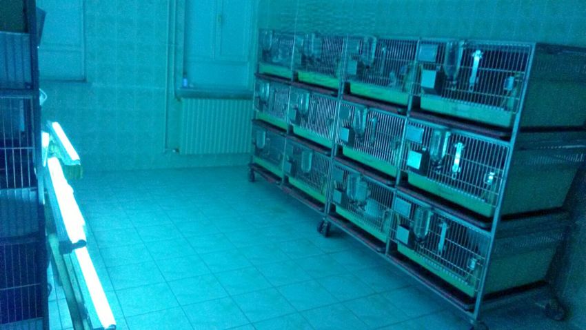
2 F. Ada et al. TiO2 powder effects on veins using UV light
Background in different fields. Watanabe et al. evaluated the photo-
catalytic activity of nano-sized TiO2 in an artificial skin
Titanium dioxide (TiO2), also known as titania, is widely model.4 They sequentially measured the CO2 levels on arti-
used in many fields such as paints, cosmetics, food prod- ficial skin with the addition of nano-sized TiO2, and found
ucts, and pharmaceuticals. With the discovery of the pho- an increase in ambient CO2 level with photocatalytic activ-
tocatalytic activity of TiO2, the use of this material has ity. Shen et al. encapsulated TiO2 with zeolite to increase
expanded.1 Titanium dioxide can be found in many crys- its photocatalytic activity in sunscreens, and found that
talline structures in nature, but it has 2 basic structures: the harmful effects of UV light were minimized.5
the rutile and anatase polymorphic phases. Titania also The present study aimed to investigate the effects
has many favorable properties such as semiconductivity, of nano-sized TiO2 on superficial veins under UV light
non-toxicity, white color, low cost, chemical stability, and with a 368 nm wavelength in a rabbit model.
photocatalytic activity.
The nano-TiO2 electron band gap is higher than 3 eV
and has a high absorption in the ultraviolet (UV) region. Materials and methods
However, due to its strong optical and biological properties,
it can be used in UV light protection applications. Many This study was approved by the Local Ethics Committee
studies have been conducted to examine the photocatalytic of Sivas Cumhuriyet University (Sivas, Turkey; approval
activity of TiO2. No. 65202830-050.04.04-62). All procedures were carried
Photocatalysts can be defined as semiconductors that be- out at Sivas Cumhuriyet University Laboratory of Experi-
come active when interacting with light, forming strongly mental Animals in accordance with the local rules of care
oxidized or reductive active surfaces. Sunlight promotes and use of experimental animals.
the purification of water systems in nature, such as rivers, The study was conducted as a controlled randomized
streams, lakes, and pools. The sunrays initiate the break- animal experiment. This study employed a total of 8 male
down of large organic molecules into smaller, simpler and female New Zealand white rabbits from 6 to 8 months
molecules. This breakdown reaction eventually results of age. The rabbits weighed between 3.2 kg and 3.5 kg for
in the formation of carbon dioxide, water and other molec- males and between 2.75 kg and 3 kg for females. All ani-
ular products. The results of laboratory studies in the early mals were fed a standard laboratory diet with free access
1980s indicated that semiconductors accelerated this natu- to water. The rabbits were housed with 1 animal in each
ral purification process induced by sunlight.2 cage. Rabbits that were able to perform their normal activi-
High-efficiency photocatalysts under UV light also have ties in the cages were kept in rooms with a temperature
high efficiency under visible light when various methods are of 22 ±2°C, a humidity level between 50% and 70%, and
applied. The S-doped TiO2 has high photocatalytic activity a 12 h day/12 h night cycle. All animals were kept un-
under visible light.3 For example, S-doped TiO2 has high ef- der observation for 1 day to determine whether they were
ficiency at a 500 nm wavelength, while Ru-doped TiO2 has healthy before the experiments. Healthy animals without
high photocatalytic activity at a 440 nm wavelength.3 any problems were included in the study.
Studies on the photocatalytic properties of TiO2 on liv- The rabbits were divided into the following 4 groups
ing tissue are limited. To address this research gap, this (8 rabbits per group; Table 1):
study investigated the histopathological effects of TiO2 – group 1 – control group: no treatment was performed;
on the superficial veins of rabbit ears under UV light by uti- – group 2 – 20% nano-TiO2 group: 0.2 mL of nano-sized
lizing its photocatalytic properties. 20% TiO2 solution was topically applied to the visible mar-
ginal vein of the right ears of the rabbits every day. No other
procedure was performed;
Objectives – group 3 – UV group: rabbits were exposed to ultra
violet A (UVA) light at a wavelength range of 368 nm for
Photocatalysts, which are commonly used for clean- 12 h a day, at a distance of approx. 150 cm. No other pro-
ing the environment, water and air, are now being used cedure was performed;
Table 1. Experimental groups and their characteristics
Groups Procedure Method of procedure Procedure modality and duration Procedure features
Group 1 control group – – –
Group 2 20% nano-TiO2 topical daily 0.2 mL solution
Group 3 UV 12 h 12 h a day, at a distance of 150 cm at a wavelength of 368 nm UV
0.2 mL of solution also
Group 4 20% nano-TiO2 + UV topical + UV 12 h a day, at a distance of 150 cm
at a wavelength of 368 nm UV
UV – ultraviolet light; TiO2 – titanium dioxide.
Adv Clin Exp Med. 2021 3
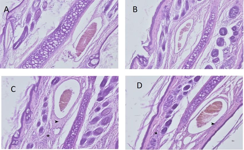
Fig. 1. Ultraviolet
light was applied at
a distance of 150 cm.
Each rabbit was kept
in a single cage
– group 4 – 20% nano-TiO2 and UV light group: 0.2 mL to assess histopathological changes. Sections were exam-
of nano-sized 20% TiO2 solution was topically applied ined under a light microscope and semi-quantitatively
to the visible marginal vein of the right ears of the rabbits scored as negative (−), mild (+), moderate (++), or severe
every day. Simultaneously, UVA at a wavelength of 368 nm (+++) for chronic inflammation. The increase in mac-
at a distance of 150 cm was applied for 12 h a day. rophage and lymphocyte density in the tissue was used
The procedures were performed for 14 days (Fig. 1). as a chronic inflammation marker. We evaluated the vas-
On the 15th day, punch biopsies were taken from the mar- cular samples and scored them according to the histo-
ginal vein under local anesthesia from all groups. The sam- pathological scale described by Kuwahara et al.6
ples were then examined histologically.
For the nano-TiO2, 20 nm TiO2 powder with the trade- Statistical analyses
name Degussa p25 (Nanoshel LLC, Wilmington, USA) was
used. The UVA light sources were Sylvania brand UV lamps Microsoft Excel 2010 (Microsoft Corp., Redmond, USA)
(Erlangen, Germany), measuring 40W/4FT/T12/BL368 with and SPSS v. 16.0 (SPSS Inc., Chicago, USA) were used to per-
a wavelength of 368 nm. No treatment or euthanasia was form the statistical analysis. Based on an α of 0.05, β of 0.20
performed on the animals at the end of the study. and 1-β of 0.80, we decided to include 8 rabbits in each group
(32 rabbits in total) and the power was calculated to be 0.8345.
Histopathological examination Since the distribution of the data was not normal, the Kruskal–
Wallis H test was conducted to assess significant differences
The ear tissue samples from the rabbits were placed between groups. For post hoc tests of differences between
in 10% buffered formalin solution. The samples were then groups, Dunn’s post hoc test was used, because it preserves
subjected to routine follow-up procedures and embedded the pooled variance for the tests implied by the Kruskal–Wallis
in paraffin blocks. Next, the 5-µm sections taken from null hypothesis (Table 2). When the data violated the normal-
the blocks were stained with hematoxylin and eosin (H&E) ity assumption (Table 3), we decided to use the nonparametric
Table 2. Statistical significance of differences between groups according to Dunn’s post hoc test
Sample 1 compared to sample 2 Test statistic SD error SD test statistic p-value
20% nano-TiO2 group compared to control group 1.063 4.464 0.238 0.812
20% nano-TiO2-group compared to 20% nano-TiO2 and UV group −12.687 4.464 −2.842 0.004
20% nano-TiO2 -group compared to UV group −20.000 4.464 −4.480 0.000
Control group compared to 20% nano-TiO2 and UV group −11.625 4.464 −2.604 0.009
Control group compared to UV group −18.937 4.464 −4.242 0.000
20% nano-TiO2 and UV group compared to UV group 7.313 4.464 1.638 0.101
UV – ultraviolet light; SD – standard deviation; TiO2 – titanium dioxide.4 F. Ada et al. TiO2 powder effects on veins using UV light
Table 3. Descriptive statistics of the treatment groups
Groups Mean (SD) Median Maximum Minimum
Control group 0.25 (0.463) 0.00 1 0
20% nano-TiO2 group 0.13 (0.354) 0.00 1 0
UV group 2.88 (0.354) 3.00 3 2
20% nano-TiO2 and UV group 1.88 (0.354) 2.00 2 1
UV – ultraviolet light; SD – standard deviation; TiO2 – titanium dioxide.
Kruskal–Wallis H test to compare the mean rank scores treatment methods (χ2(3) = 27.835, p < 0.001), with mean
of the groups. The effect size for the test was calculated as 4.92. rank scores of 9.12 for the control group, 8.06 for the 20%
nano-TiO2 group, 28.06 for the UV group, and 20.75 for
the group administered both 20% nano-TiO2 and UV.
Results Dunn’s pairwise post hoc test indicated significant dif-
ferences between the control group and the UV group
Four female and 4 male rabbits were included in each (p < 0.001), between the 20% nano-TiO2 group and
experimental group; thus, there were no statistically the UV group (p < 0.001), between the 20% nano-TiO2
significant differences between the groups in terms group and the group administered both 20% nano-TiO2
of gender (p > 0.05). The results showed that the ear tis- and UV light (p = 0.004), and between the control group
sues of the rabbits in the control and nano-TiO2 groups and the group administered both 20% nano-TiO2 and
had a normal histological appearance (Fig. 2). However, UV light (p = 0.009). There were no other significant
we observed the formation of severe chronic inflammation differences (Table 5). The histopathological changes de-
of mononuclear cells in the perivascular areas and around creased in the group that received both UV and nano-
the hair follicles in the UV group (Table 4). TiO2. Macroscopically, the results indicated that the vis-
The results of the Kruskal–Wallis H test indicated sig- ibility of rabbit ear veins decreased in the 20% nano-TiO2
nificant differences between groups subjected to different and UV light group (Fig. 3).
Fig. 2. A. Control group. Normal histological appearance. ×40 magnification, hematoxylin and eosin (H&E) staining; B. Titanium dioxide (TiO2) group.
Normal histological appearance. ×40 magnification, H&E staining; C. Ultraviolet (UV) group. Severe mononuclear cell infiltration is present around the vein
(arrowhead) and near the hair follicles (*). ×40 magnification, H&E staining; D. Ultraviolet light + TiO2 group. Moderate mononuclear cell infiltration
is present around the vessel (arrowhead) and near the hair follicles (*). ×40 magnification, H&E stainingAdv Clin Exp Med. 2021 5
Table 4. Inflammation degrees of the groups
Group 1 Group 2 Group 3 Group 4
Inflammation
(n = 8) (n = 8) (n = 8) (n = 8)
Negative [%] 75 87.5 0 0
Mild [%] 25 12.5 0 12.5
Moderate [%] 0 0 12.5 87.5
Severe [%] 0 0 87.5 0
Table 5. Kruskal–Wallis H test analysis table for all treatment groups
Groups Mean rank χ2 df p-value
Control group 9.12 27.835 3 0.000
20% nano-TiO2 group 8.06 – – –
UV light group 28.06 – – –
20% nano-TiO2 and UV light group 20.75 – – –
UV – ultraviolet light; TiO2 – titanium dioxide; df – degrees of freedom.
Discussion In a wide-ranging review, Iavicoli et al. investigated
the effects of nano-TiO2 on mammals organ by organ and
At present, TiO2 is widely used in many industries such system by system.10 Although they discussed the effects
as pharmaceuticals, cosmetics (especially in sunscreens), of nano-TiO2 on many systems, they emphasized that their
food, biomedical products, etc. Given its wide use, it is nec- effects on vascular structures were not clear. However,
essary to investigate the toxicological and histological effects they stated that the effects of TiO2 were directly related
of nano-TiO2, as well as its effects on the immune system. to the way it was administered, its dose, duration, and
Fabian et al. administered titanium dioxide nanoparticles particle size. Tang et al. examined the relationship between
intravenously to rats and found that they did not cause any administration routes and doses of TiO2.11 They found that
toxic effects at a 5 mg/kg dose and could be used safely TiO2 with a size of 5 nm could be passed to the blood intra
at this dose.7 Warheit et al. administered 300 nm of titanium tracheally; when it reached a dose of 0.8 mg/kg, it could
dioxide via inhalation in rabbits and showed that short-term reach the lungs and kidneys and it changed liver and kidney
exposure did not cause any lung problems.8 In the same function. However, they observed that these effects were
study, they reported that TiO2 of this size had no genotoxic reversible after 1 week without permanent damage.
effect and did not cause any irritation of the skin, but revers- There is a limited number of studies on the effects
ible conjunctival redness in the eyes of rabbits was observed. of TiO2 on vascular structures in the literature. In a study
In a review of the effects of nano-sized TiO2 on the lung, using porcine pulmonary artery endothelial culture, Han
size, exposure time, and the amount of nano-titanium were et al. reported that nano-TiO2 increased the inflammatory
identified as the most important risk factors.9 response in endothelial cells.12 They concluded that the in-
creased inflammation response occurred via the redox-
sensitive cell signaling pathway.
Montiel-Dávalos et al. conducted a study on human
umbilical vein endothelial cell culture (HUVEC) and
found that TiO2 inhibited the proliferation of endothe-
lial cells and accelerated apoptotic and necrotic death.13
Alinovi et al. also conducted a study using HUVEC and
found that TiO2 nanoparticles increased inflammation
in endothelial cells.14 Park et al. conducted a study us-
ing a mouse endothelial cell culture line and evaluated
whether the size of the nanotube affected the endothelial
adhesion and apoptosis rates of TiO2 nanotubes.15 They
observed more endothelial adhesion, less apoptosis, and
less inflammation in 15 nm nanotubes. Furthermore, their
results indicated an increase in inflammation and apopto-
Fig. 3. On the 14th day of the study, titanium dioxide (TiO2) and ultraviolet sis as the size of the nanotubes increased. These findings
(UV) light were applied to the rabbits. Titanium dioxide was applied
to the upper ear, but not lower ear. Note the difference between venous demonstrate the importance of size in terms of the ef-
structures fectiveness of nano-TiO2 to be used in vascular implants.6 F. Ada et al. TiO2 powder effects on veins using UV light Ge et al. conducted a study using HUVEC and their results high stability under lighting, nontoxic nature, and low indicated that thin film nano-TiO2 provided laminin im- price. Titanium dioxide performs its photocatalytic effect mobilization and an increase in endothelial adhesion.16 best under 340 nm wavelength light, but a wavelength range Ultraviolet light has been used in the treatment of skin of 320–380 nm is also good for photocatalyst activity.29 diseases since the beginning of the 20th century. Although In general, studies have demonstrated that organic cell the mechanism of UV light action is not clear, hypotheses degradation increases as the wavelength decreases. This that it increases cell proliferation, epidermal thickness and means that at a wavelength of 280 nm or less, TiO2 has its blood flow in cutaneous capillaries have been proposed.17 maximum photocatalytic effect, leading to the degrada- In a study that applied UV light to patients with venous tion of DNA and RNA.30,31 Therefore, the light wavelength ulcers, Dodd et al. concluded that UV light had no benefit selected in our study was in the range of 320–380 nm. in the treatment of venous ulcers.18 However, they observed The same ultraviolet light wavelength and application meth- that UV light increased skin oxygen permeability and in- ods used in previous animal experiments, as well as the dose, hibited normal vasoconstrictor response. concentration, and routes of TiO2 administration were ap- When light treatment is required in medicine, the pre- plied in the current study.32,33 What distinguishes this study ferred UV light wavelength is in the 300–320 nm range. from the other studies is that we investigated the effects In dermatology, psoralen ultraviolet A (PUVA) treatment of TiO2 and UV light on vascular structures simultaneously. for psoriasis, eczema, vitiligo, and cutaneous lymphoma Photodynamic therapy, especially in dermatology and is widely used.19 Although UV light has positive results when plastic surgery, is used effectively and widely for the treat- applied to appropriate patients for a certain period of time, ment of many dermatological diseases in the form of a com- dose and treatment, excessive exposure can lead to many bination of light and various creams applied to the skin. adverse conditions such as skin cancer, T cell damage, im- The main principle of creams with methyl aminolevulinate mune depression, sunburn, and premature skin aging.20 as the active ingredient is to be absorbed by the skin and Telangiectasias are enlarged venous vessels with a diam- subcutaneous tissue, and to provide local apoptosis with eter of 0.5–1 mm near the skin surface. Telangiectasias are the effect of light. In fact, Galvão found that telangiectasias enlarged postcapillary venules located in the papillary and on the face were not visible after photodynamic therapy superficial reticular dermis with histologically complete in a patient with facial actinic keratosis.34 meaning.21 Photodynamic therapy consists of 3 main elements: Engel et al. conducted a field study with a large popula- a photosensitive material, a light source and oxygen. When tion in the USA and found that the rate of telangiectasia these 3 factors are combined effectively, they can be ap- in the legs was 29–41% in women and 6–15% in men.22 Fa- plied for the successful treatment of many skin lesions. cial telangiectasia affects tens of millions of people around In photodynamic therapy, the therapeutic effect is achieved the world.23 Sclerotherapy is the standard approach for by a slight activation of a light-sensitive substance, and the treatment of telangiectasias. Sclerosis of the telangi- reactive oxygen intermediates are formed in the pres- ectatic vein is the main target in sclerotherapy. In order ence of oxygen. These intermediates irreversibly oxidize to achieve sclerosis, various chemical sclerosing agents are the essential cellular components that cause apoptosis administered directly into the enlarged telangiectatic vein, and necrosis, thereby providing the treatment of lesions resulting in the formation of a thrombosis and endothelial on the skin. 35 Essentially, our study has shown similar damage within the vein.24 results to photodynamic therapy. In this sense, the study Apart from sclerotherapy, many different laser treatments, demonstrated that substances with photocatalytic activa- such as argon ion lasers, diode lasers and intense pulsed tion may be an appropriate choice for this treatment. light sources, are applied.25,26 Laser treatment was first per- formed in the treatment of telangiectasias in 1960 using Limitations light at a 694 nm wavelength, followed by different systems and wavelengths.27 The underlying logic of laser treatment The method applied in the present study could be used in telangiectasia is to cause the loss of veins close to the skin in the treatment of telangiectasia in the future. However, surface by creating thermal damage to the vascular struc- our study has a predominantly histological and observa- tures through the heat generated by UV light. Hemoglobin tional nature. Further studies involving genetic, cytoge- in the vessel absorbs light best at 418 nm, 542 nm and 577 nm netic, biochemical, histochemical, and immunohistochemi- wavelengths.28 However, the wavelengths used in the treat- cal analyses are needed to test the theories described above. ment of vascular lesions are usually 488 nm and 600 nm.28 Photocatalysts can be described as semiconductors that create a strongly oxidized environment on the surface Conclusion through the effect of UV light. Therefore, photocatalysts can cause degradation in organic tissues close to the region The results of this study revealed that nano-TiO2 where they are applied. Among the known semiconductor protected the skin and the main vascular structures photocatalysts, TiO2 is widely used due to its high activity, close to the skin from the harmful effects of UV light.
Adv Clin Exp Med. 2021 7
Furthermore, this study determined that telangiectatic 13. Montiel-Dávalos A, Ventura-Gallegos JL, Alfaro-Moreno E, et al. TiO2
nanoparticles induce dysfunction and activation of human endothe-
structures close to the skin were not observed in the UV
lial cells. Chem Res Toxicol. 2012;25(4):920–930. doi:10.1021/tx200551u
and nano-TiO2 group. 14. Alinovi R, Goldoni M, Pinelli S, et al. Oxidative and pro-inflamma-
Several theories were put forward regarding the absence tory effects of cobalt and titanium oxide nanoparticles on aortic
of telangiectatic structures in the rabbit ear on macro- and venous endothelial cells. Toxicol In Vitro. 2015;29(3):426–437.
doi:10.1016/j.tiv.2014.12.007
scopic observation. The 1st is that nano-TiO2 and UV 15. Park J, Bauer S, Schmuki P, von der Mark K. Narrow window in nanos-
light induced apoptosis in telangiectatic structures, just cale dependent activation of endothelial cell growth and differen-
as in photodynamic therapy. The 2nd is that thermal dam- tiation on TiO2 nanotube surfaces. Nano Lett. 2009;9(9):3157–3164.
doi:10.1021/nl9013502
age was caused by heating during the photocatalytic acti- 16. Ge SN, Chen JY, Leng YX, Huang N. Laminin immobilized on titani-
vation of nano-TiO2. The 3rd theory is that collagen tissue um oxide films for enhanced human umbilical vein endothelial cell
deposited under the epidermis compressed the telangi- adhesion and growth. Key Eng Mater. 2007;342:305–308. doi:10.4028/
www.scientific.net/KEM.342-343.305
ectatic veins or precipitated them towards the dermis. 17. Thai TP, Houghton PE, Keast DH, Woodbury MG. Ultraviolet C in the tre-
The 4th is damage of hemoglobin in the telangiectatic vein. atment of chronic wounds with MRSA: A case study. Ostomy Wound
The 5th is selective photothermolysis that led to panendo- Manage. 2002;48(11):52–60. PMID:12426452
thelial obliteration. Any combination of these theories, 18. Dodd HJ, Sarkany I, Gaylarde PM. The short‐term benefit and long‐
term failure of ultraviolet light in the treatment of venous leg ulcers.
or some other mechanism of action, may have led to these Br J Dermatol. 1989;120(6):809–818. doi:10.1111/j.1365-2133.1989.
results. tb01379.x
19. Hönigsmann H, Szeimies RM, Knobler R, et al. Photochemotherapy
ORCID iDs and photodynamic therapy. In: Goldsmith LA, Katz SI, Gilchrest BA,
Paller AS, Leffell DJ, Wolff K. Dermatology in General Medicine. New
Fatih Ada https://orcid.org/0000-0002-6953-5906 York, USA: McGraw-Hill; 1999:2477–2493.
Ferit Kasimzade https://orcid.org/0000-0003-3646-3181 20. Elmets CA, Bergstresser PR, Tigelaar RE, Wood PJ, Streilein JW. Analy-
Ali Sefa Mendil https://orcid.org/0000-0003-2722-3290 sis of the mechanism of unresponsiveness produced by haptens
Hasan Gocmez https://orcid.org/0000-0003-3748-0311 painted on skin exposed to low dose ultraviolet radiation. J Exp Med.
1983;158:781–794. doi:10.1084/jem.158.3.781
References 21. Walker JG, Stirling J, Beroukas D, et al. Histopathological and ult-
1. Şam ED, Ürgen M, Tepehan FZ. TiO2 fotokatalistleri. İTÜ Dergisi/D rastructural features of dermal telangiectasias in systemic sclerosis.
Mühendislik. 2007;6(5–6):81–92. http://itudergi.itu.edu.tr/index.php/ Pathology. 2005;37(3):220–225. doi:10.1080/00313020500033262
itudergisi_d/article/viewFile/395/337. Accessed November 1, 2020. 22. Engel A, Johnson ML, Hynes SG. Health effects of sunlight exposure
2. Ochiai T, Fujishima A. Design and optimization of photocatalytic in the United States: Results from the first National Health and Nutri-
water purification reactors. In: Pichat P, ed. Photocatalysis Water Puri- tion Examination Survey 1971–1974. Arch Dermatol. 1988;124:72–79.
fication: From Fundamentals to Recent Applications. Weinheim, Ger- doi:10.1001/archderm.1988.01670010036018
many: Wiley-VCH; 2013:361. doi:10.1002/9783527645404.ch14 23. Cassuto DA, Ancona, DM, Emanuelli G. Treatment of facial telangiecta-
3. Ohno T, Akiyoshi M, Umebayashi T, et al. Preparation of S-doped sias with a diode-pumped Nd: YAG laser at 532 nm. J Cutan Laser Ther.
TiO2 photocatalysts and their photocatalytic activities under visib- 2000;2(3):141–146. doi:10.1080/14628830050516399
le light. Appl Catal A Gen. 2004;265(1):115–121. doi:10.1016/j.apca- 24. Rabe E, Breu FX, Cavezzi A, et al. European guidelines for sclerothe-
ta.2004.01.007 rapy in chronic venous disorders. Phlebology. 2014;29(6):338–354.
4. Watanabe E, Fukaya M, Nishizawa K, Miki T, Taoda H. Test method for skin doi:10.1177/0268355513483280
damage of titania photocatalyst nanoparticles in vitro. Mater Sci Forum. 25. Landthaler M, Hohenleutner U. Laser therapy of vascular lesions.
2008;569:9–12. doi:10.4028/www.scientific.net/MSF.569.9 Photodermatol Photoimmunol Photomed. 2006;22(6):324–332. doi:10.
5. Shen B, Scaiano JC, English AM. Zeolite encapsulation decreases TiO2‐ 1111/j.1600-0781.2006.00254
photosensitized ROS generation in cultured human skin fibroblasts. 26. Goldman MP, Martin DE, Fitzpatrick RE, Ruiz-Esparza J. Pulsed dye
Photochem Photobiol. 2006;82(1):5–12. doi:10.1562/2005-05-29-RA-551 laser treatment of telangiectases with and without subtherapeutic
6. Kuwahara T, Asanami S, Kubo S. Experimental infusion phlebitis: Tole- sclerotherapy: Clinical and histologic examination in the rabbit ear
rance osmolality of peripheral venous endothelial cells. Nutrition. vein model. J Am Acad Dermatol. 1990;23(1):23–30. doi:10.1016/0190-
1998;14(6):496–501. doi:10.1016/s0899-9007(98)00037-9 9622(90)70180-p
7. Fabian E, Landsiedel R, Ma-Hock L, Wiench K, Wohlleben W, van 27. Apfelberg DB, Maser MR, Lash H, White DN, Flores JT. Use of the argon
Ravenzwaay B. Tissue distribution and toxicity of intravenously admi- and carbon dioxide lasers for the treatment of superficial venous
nistered titanium dioxide nanoparticles in rats. Arch Toxicol. 2008; varicosities of the lower extremity. Lasers Surg Med. 1984;4:221–232.
82(3):151–157. doi:10.1007/s00204-007-0253-y doi:10.1002/lsm.1900040302
8. Warheit DB, Hoke RA, Finlay C, Donner EM, Reed KL, Sayes CM. Deve- 28. Spendel S, Prandl EC, Schintler MV, et al. Treatment of spider leg veins
lopment of a base set of toxicity tests using ultrafine TiO2 particles with the KTP (532 nm) laser: A prospective study. Lasers Surg Med.
as a component of nanoparticle risk management. Toxicol Lett. 2007; 2002;31(3):194–201. doi:10.1002/lsm.10088
171(3):99–110. doi:10.1016/j.toxlet.2007.04.008 29. Thiruvenkatachari R, Vigneswaran S, Moon IS. A review on UV/TiO2 pho-
9. Warheit DB. How to measure hazards/risks following exposures to tocatalytic oxidation process. Korean J Chem Eng. 2008;25(1):64–72.
nanoscale or pigment-grade titanium dioxide particles. Toxicol Lett. doi:10.1007/s11814-008-0011-8
2013;220(2):193–204. doi:10.1016/j.toxlet.2013.04.002 30. Matthews RW, McEvoy SR. Photocatalytic degradation of phenol in
10. Iavicoli I, Leso V, Fontana L, Bergamaschi A. Toxicological effects the presence of near-UV illuminated titanium dioxide. J Photochem
of titanium dioxide nanoparticles: A review of in vitro mammalian Photobiol. 1992;64(2):231–246. doi:10.1016/1010-6030(92)85110-G
studies. Eur Rev Med Pharmacol Sci. 2011;15(5):481–508. doi:10.1155/ 31. Li PG, Yue PL. Photocatalytic oxidation of chlorophenols in single
2012/964381 component and multicomponent systems. Ind Eng Chem Res. 1999;
11. Tang M, Zhang T, Xue Y, et al. Dose dependent in vivo metabolic cha- 38(9):3238–3245. doi:10.1021/ie9807598
racteristics of titanium dioxide nanoparticles. J Nanosci Nanotechnol. 32. Emerson JA, Whittington JK, Allender MC, Mitchell MA. Effects of ult-
2010;10(12):8575–8583. doi:10.1166/jnn.2010.2482 raviolet radiation produced from artificial lights on serum 25-hydro
12. Han SG, Newsome B, Hennig B. Titanium dioxide nanoparticles incre- xyvitamin D concentration in captive domestic rabbits (Oryctola-
ase inflammatory responses in vascular endothelial cells. Toxicology. guscuniculi). Am J Vet Res. 2014;75(4):380–384. doi:10.2460/ajvr.75.
2013;306:1–8. doi:10.1016/j.tox.2013.01.014 4.3808 F. Ada et al. TiO2 powder effects on veins using UV light
33. Lansdown ABG, Taylor A. Zinc and titanium oxides: Promising UV-absor- 35. Wan MT, Lin JY. Current evidence and applications of photodynamic
bers but what influence do they have on the intact skin? Int J Cosmet Sci. therapy in dermatology. Clin Cosmet Investig Dermatol. 2014;7:145–163.
1997;19(4):167–172. doi:10.1111/j.1467-2494.1997.tb00180.x doi:10.2147/CCID.S35334
34. Galvão LEG. Terapia fotodinâmica com luz do dia: benefício clíni-
co e estético com sessões repetidas para ceratoses actínicas faciais.
Surg Cosmet Dermatol. 2016;8(4):40–42. doi:10.5935/scd1984-8773.
201683103You can also read