Case Report: Paratesticular Rhabdomyosarcoma - Frontiers
←
→
Page content transcription
If your browser does not render page correctly, please read the page content below
CASE REPORT
                                                                                                                                           published: 17 March 2021
                                                                                                                                     doi: 10.3389/fonc.2021.629878

                                              Case Report: Paratesticular
                                              Rhabdomyosarcoma
                                              Yiyi Zhu 1, Ziwei Zhu 2, Yunyuan Xiao 2 and Zaisheng Zhu 2*
                                              1Department of Endocrinology, Peking Union Medical College Hospital, Peking Union Medical College, Chinese Academy of
                                              Medical Sciences, Beijing, China, 2 Department of Urology, Jinhua Hospital Affiliated to Zhejiang University School of
                                              Medicine, Jinhua, China

                                              Paratesticular rhabdomyosarcoma (RMS) accounts for only 7% of all the RMS cases. Due
                                              to the limited available data, there is no consensus on the diagnosis and management of
                                              the paratesticular tumors. Here, we interrogated two paratesticular RMS cases in 25 and
                                              27-year-old men presenting with painless and rapidly growing mass in the scrotum.
                                              Whereas the data showed no upregulation of tumor markers such as b-human chorionic
                                              gonadotropin (b-HCG), alpha-fetoprotein (AFP), and lactate dehydrogenase (LDH), scrotal
                                              ultrasonography and magnetic resonance imaging indicated the existence of
                                              paratesticular and inguinal lesions respectively. There was local recurrence in one
                                              patient who underwent radical orchiectomy for the sarcoma one year ago. In addition,
                        Edited by:
                                              the CT scans showed no occurrence of distant metastasis. The two patients underwent
           Mohamed Saad Zaghloul,             radical inguinal orchiectomy or resection of the recurrent tumors with nerve-sparing
             Cairo University, Egypt          retroperitoneal lymph node dissection. Histologic examination revealed embryonal RMS
                       Reviewed by:           (eRMS) without lymph node metastasis. We highlight the importance of multi-disciplinary
              Saum Ghodoussipour,
          Rutgers Cancer Institute of         participation for paratesticular RMS detection and preoperative ultrasound-guided needle
          New Jersey, United States           biopsy (UNB) for rapid confirmatory diagnosis. Complete surgical resection coupled with
                         Sanja Štifter,
          University of Rijeka, Croatia
                                              chemotherapy and radiotherapy is the main treatment option for the paratesticular RMS.
                  *Correspondence:
                                              In addition, sperm cryopreservation treatment and endocrine follow-up could increase the
                        Zaisheng Zhu          overall survival and quality of life of the patients.
               zaishengzhu@126.com
                                              Keywords: paratesticular rhabdomyosarcoma, nerve-sparing retroperitoneal lymph node dissection,
                                              chemotherapy and radiotherapy, sperm cryopreservation, endocrine follow-up
                   Specialty section:
         This article was submitted to
              Genitourinary Oncology,
               a section of the journal
                 Frontiers in Oncology
                                              CASE PRESENTATION
      Received: 16 November 2020              Case 1
       Accepted: 01 February 2021             In Jan 2018, a 25-year-old unmarried man with no past medical or family cancer history presented
        Published: 17 March 2021
                                              to the hospital with a progressively growing but painless swelling in his left hemi-scrotum. The
                            Citation:         swelling had lasted for one month. Physical examination confirmed the swelling measuring about
                       Zhu Y, Zhu Z,
                                              9.0 × 5.0 × 6.0 cm, and the light transmittance test was negative. It was a painless and rubbery mass
             Xiao Y and Zhu Z (2021)
          Case Report: Paratesticular
                                              with poorly defined edges. Interestingly, all the known and relevant tumor markers such as cancer-
                Rhabdomyosarcoma.             associated antigen 19-9 (CA19-9), carcinoembryonic antigen (CEA), alpha-fetoprotein (AFP), b-
            Front. Oncol. 11:629878.          human chorionic gonadotropin (b-HCG) and lactate dehydrogenase (LDH) were within normal
     doi: 10.3389/fonc.2021.629878            ranges. Scrotal ultrasonography demonstrated an irregular and isoechoic mass within the left

Frontiers in Oncology | www.frontiersin.org                                        1                                       March 2021 | Volume 11 | Article 629878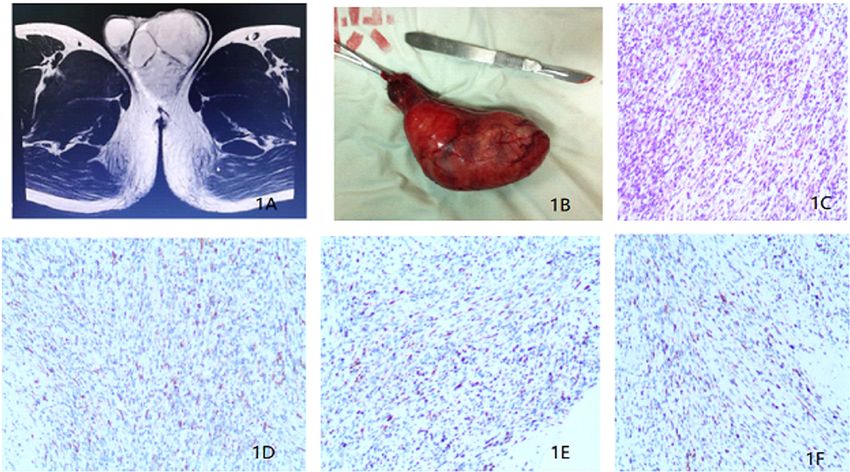
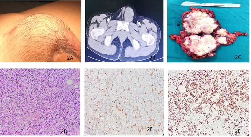
Zhu et al.                                                                                                            Case Report: Paratesticular Rhabdomyosarcoma

hemi-scrotum, while the testicular size was normal. In addition,                      operation, the patient recovered without any complication. The
Color Doppler ultrasound showed significant vascularity within                         postoperative pathological examination confirmed eRMS (Figure
the mass and a resistance index (RI) of 0.71, indicating a high                       1C) and a negative surgical margin. In addition, IHC analysis
probability of a tumor. On the other hand, magnetic resonance                         revealed the expression of Desmin, MyoD1 and Myogenin in the
imaging (MRI) showed that the soft tissue mass in the left scrotum                    excised tissue samples (Figures 1D–F).
was closely associated with the epididymis and had gradual                                Based on the staging evaluation by Intergroup
enhancement after contrast suggesting that the lesion might be                        Rhabdomyosarcoma Study Group (1), the case was classified as
arising from mesenchymal tissues (Figure 1A). A CT scan for the                       T1 clinical stage. Since there was no standard chemotherapy
chest, abdomen, and pelvis did not indicate evidence of metastasis.                   regimen for adult paratesticular RMS, the patient was
For a definitive diagnosis without post-operative bleeding and                         administered with a pediatric VAC regimen comprising of
tumor implantation along the needle track, preoperative                               vincristine, actinomycin D and cyclophosphamide (2, 3). Before
ultrasound-guided needle biopsy (UNB) using a coaxial needle                          chemoradiotherapy, he was referred to the Women’s hospital,
was adopted. The pathologic data from the UNB demonstrated                            Zhejiang University School of Medicine for sperm
presence of a myxoid stroma with a rich vascular weave in                             cryopreservation from 8 ml of semen. He then received adjuvant
paratesticular tissue samples. Spindle and irregular cells were                       radiotherapy of 30 Gy (retroperitoneal area) and 20 Gy (tumor
arranged in parallel or in a mesh-like pattern with abundant red-                     bed) and six cycles of adjuvant chemotherapy. Whereas the patient
staining cytosol. There was a significant presence of mitotic bodies.                  reported side effects such as pancytopenia, vomiting, loss of
Besides, immunohistochemical (IHC) experiments detected Ki-67                         appetite, and hair-loss during chemoradiotherapy, he completed
and Desmin, but not S-100, CEA, CD34, keratin and Caldesmon-H.                        the treatment plan. The patient was finally discharged from the
A multi-disciplinary team (MDT) of experts from urinary surgery,                      hospital in a tumor-free state. However, he was given traditional
medical oncology, endocrinology, reproductive health,                                 Chinese medicine named Guilingji combined with HCG
radiotherapy, medical imaging and pathology departments                               treatment scheme (1 500 IU intramuscular injection once every
concurred on the use of radical inguinal orchiectomy, coupled                         two weeks) as maintenance therapy. The patient underwent
with chemotherapy and radiotherapy to the retroperitoneal lymph                       follow-up schedules with scrotal ultrasound in the 6th month,
node region. During the operation, a pear-like lesion could be seen                   CT scan in the 12th month, and positron emission tomography-
on the cauda epididymis with a complete outer membrane (scrotal                       computed tomography (PET/CT) in the 24th month. Over the 24-
sheath) and a diameter of 9.5 × 5.0 × 4.5 cm (Figure 1B). After the                   month follow-up period, he remained free from local recurrence

 FIGURE 1 | (A) Magnetic resonance imaging revealing a soft tissue mass in the left scrotum. (B) Surgical piece of the left orchiectomy presenting a 9.5 × 5.0 × 4.5
 cm3 paratesticular tumor. (C) Histopathological section (hematoxylin and eosin staining). (D) Immunohistochemistry results showing the expression of Desmin.
 (E) Immunohistochemistry results showing the expression of MyoD1. (F) Immunohistochemistry results showing the expression of Myogenin.

Frontiers in Oncology | www.frontiersin.org                                       2                                         March 2021 | Volume 11 | Article 629878
Zhu et al.                                                                                                                Case Report: Paratesticular Rhabdomyosarcoma

and distant metastases. The last physical examination showed                             metastasis. There was, however, no evidence of metastasis to either
restoration of his erectile functions as well as serum endocrine                         the lymph nodes or other organs. After MDT discussion and the
hormones such as follicle-stimulating hormone (FSH), luteinizing                         patient’s permission, tumor resection was performed in
hormone (LH), estrogen (E), and testosterone (T). However, two                           combination with adjuvant chemotherapy. Before
semen examinations indicated oligospermia.                                               chemotherapy, the patient underwent sperm cryopreservation at
                                                                                         the reproductive center of Renji Hospital, Shanghai Jiaotong
Case 2                                                                                   University School of Medicine. Similarly, pediatric VAC
In Dec 2015, a 27-year-old man came to our department with a                             regimen was applied with vincristine 1.5 mg/m2, actinomycin D
history of orchiectomy to the left testicular mass. The surgery was                      1.5 mg/m2, and cyclophosphamide 500 mg/m2. Only three cycles
conducted a year ago. The patient reported to a urologist and                            of chemotherapy were administered due to the side effects, and
complained of presence of a mass in the left inguinal region that                        then the patient received nerve-sparing RPLND. Postoperative
persisted for 1 month. Previously, he was diagnosed with a                               pathologic examinations showed that the gross specimen had no
paratesticular tumor and underwent radical orchiectomy in a                              obvious capsule and had a pale profile with a fish-flesh appearance
different hospital. The postoperative pathological diagnosis                             (Figure 2C). In addition, the examination revealed that the
showed eRMS (5 cm in diameter) and no adjuvant therapy was                               recurrent tumor was eRMS and resection margins were negative
administered post-surgery. On physical examination, a surgical                           (Figure 2D). No metastases were found in the lymph nodes.
scar in the left inguinal region and a 4.0 × 5.0 cm mass localized                       Microscopically, the mass cells were poorly differentiated while the
above the inner ring of the inguinal canal were palpable. This mass                      rhabdomyoblasts had immense eosinophilic cytoplasm and
was hard and immobile, growing into the pelvic cavity (Figure                            necrotic tissues. The immunostaining results showed the
2A), but superficial lymph nodes were normal in size. His routine                         expression of Desmin and MyoD1 in the tumor cells (Figures
laboratory data such as complete blood cell count, renal and liver                       2E, F). Thereafter, adjuvant radiotherapy (30 Gy) was delivered to
function tests were normal. The serum tumor markers such as                              the tumor bed. To restore reproductive functions, the patient was
CEA, AFP, b-HCG and LDH showed no significant abnormalities.                              prescribed with traditional Chinese medicine named Guilingji,
Besides, a scrotum echography showed a heterogeneous mass                                HCG treatment scheme (1,500 IU intramuscular injection once
above the left groin and pelvis. In addition, chest, abdominal                           every two weeks) and sildenafil, 3 months following the surgery.
and pelvis CT and MRI scans showed an irregular lesion above the                         The patient’s erectile function gradually recovered and semen
left groin and pelvis (Figure 2B), indicating tumor recurrence and                       activity increased after nine months of surgery. The patient

 FIGURE 2 | (A) Recurrent tumors in the left inguinal region for a duration of 1 month. (B) Magnetic resonance imaging revealing presence of a soft tissue mass
 above the left groin and pelvis. (C) Surgical biopsy of the recurrent tumors from the left inguinal and pelvic region (D) Histopathological section (hematoxylin and
 eosin staining). (E) Immunohistochemistry results showing the expression of Desmin. (F) Immunohistochemistry results showing the expression of MyoD1.

Frontiers in Oncology | www.frontiersin.org                                          3                                          March 2021 | Volume 11 | Article 629878
Zhu et al. Case Report: Paratesticular Rhabdomyosarcoma
underwent regular follow-up (twice every year) by clinic visits and treatment regimen, but enables radical resection or RPLND at the
was monitored by scrotal ultrasound, abdominal CT scan and initial operation, avoiding multiple surgeries and reducing
serum endocrine hormones. The last follow-up was 39 months metastasis or recurrence. Besides, it protects the male
after the operation, and the PET-CT did not reveal any local reproductive function and improves the quality of life.
recurrence or distant metastases. His erectile function and semen
analysis returned to normal range. At present, his family moved to Histopathological Characteristics
another city (Guangzhou, China) with a newborn baby girl. According to the international RMS classification, the common
histological subtypes are alveolar, embryonal, botryoid embryonal,
spindle cell embryonal, and anaplastic (11). Embryonal RMS
(eRMS) is the most frequent type, which accounts for 60% of the
DISCUSSION cases. On the other hand, paratesticular RMS is rare in adults but
Rapid Confirmatory Diagnosis often occurs in children, representing 7 to 10% of the tumors in
Compared with other intra-scrotal tumors, the clinical male genitourinary system (12). Thus, since it’s uncommon in
manifestations of paratesticular RMS are non-specific. When adults, few studies have reported paratesticular eRMS (13, 14).
there is difficulty in physical examination of tumor localization, Pathologically, the eRMS presents the existence of poorly
ultrasonography (US) can be a useful alternative in the diagnosis differentiated cells and rhabdomyoblasts, with abundant
(4). The scrotal US results indicated that the lesions were eosinophilic cytoplasm indicating embryonal rhabdomyosarcoma.
heterogeneous echogenic mass and were extended to scrotum Cytogenetically, the eRMS is characterized by loss of heterozygosity
and inguinal region (5). In addition, the Color Doppler on the short arm of chromosome 11 (15). Whereas, electron
ultrasound image showed increased blood flow inside the mass microscopy and chromosome analysis are useful methods to
which help discriminate testicular from paratesticular improve the pathological diagnosis of the eRMS; they were not
localization (6). Previous studies reported that US of the examined in our cases.
scrotum and its contents can reliably distinguish testis and Histopathologically, the two cases showed a pale profile of
scrotal masses, with a sensitivity of >95% (7). Whereas both gross specimen with fish-like appearance. Microscopically, the
CT scan and MRI can accurately evaluate the location, size and tumor comprised of a significant number of rhabdomyoblasts
metastasis of the mass, they cannot be used as confirmatory with elongated eosinophilic cytoplasmic tales, commonly
diagnostic tools. Differential diagnosis of paratesticular RMS referred to as tadpole cells or strap cells. In addition, there
includes lesions such as leiomyosarcoma, liposarcoma and were cases of cross-striations within the cytoplasm. The lesion
fibrosarcoma. These tumors lack imaging features, thus presented a storiform growth pattern with abundant collagen
confirmatory diagnosis relies on postoperative pathology (8, 9). between the tumor cells. It showed an arrangement of tumor cells
The two cases lacked specific clinical manifestations but in bundles with a low to moderate amount of collagen,
presented to the hospital due to left scrotal or inguinal swelling resembling a leiomyosarcoma. Immunohistochemical findings
in a month. Thus, the lesions had rapid growth. In addition, both showed the expression of MyoD1, Myogenin and Desmin in the
cases lacked any serum tumor markers, such as CEA, AFP, b-HCG tumor cells, while keratin and CD34 were negative. On the basis
and LDH showing no significant abnormalities. Besides, the of these findings, a final diagnosis of spindle cell variant of the
scrotal US demonstrated irregular and isoechoic mass, while the rhabdomyosarcoma was conducted. The peripheral resection
Color Doppler US image showed significant vascular signals. Both margin and both seminal vesicles did not show presence of
the CT scan and MRI showed localization of the tumor in the tumor cells. Besides, there was no vessel or perineural invasion.
paratesticular region, with no definite distal metastasis. Thus, to
improve diagnosis precision, we suggest that adolescents should be Individualized Treatment Regimens
educated about self-examination. A thorough physical The RMS prognosis is related to age, histology, lesion site, and
examination is necessary at the discovery of the painless tumor diameter (16, 17). The 5-year survival rate is 22.2% in
scrotum or inguinal enlargement. Besides, US and MRI should eRMS patients with recurrence and metastasis and 94.6% in
be the main methods to examine localization of the lesion. They those without metastasis (18). The prognosis in adult patients is
are simple and do not emit any radiation. Further, we recommend worse than that in children, with a 5-year tumor-free survival
preoperative UNB for rapid confirmatory diagnosis. Some and 5-year overall survival rates of 28 and 40% respectively, in
previous case reports did not present preoperative diagnosis of adults (19).
the paratesticular RMS prior to the first surgery and underwent We, therefore, suggest that there should be a MDT discussion
simple resection of the lesion. The cases underwent remedial among specialties from the surgery department, endocrinology
radical operation after post-operative pathologic reports. In department, reproductive center and other departments after
recent years, testicular and epididymal puncture has been confirmation of the eRMS. A comprehensive and individualized,
accepted as a safe and reliable diagnostic method, which is not but optimal, treatment plan should include tumor resection and
associated with the risk of tumor needle implantation or tumor preservation of sexual and reproductive functions of the
metastasis (10). In our case report, preoperative UNB using coaxial male patients.
needle was adopted to confirm the pathological diagnosis. It not Surgical excision should be extensive and thorough.
only facilitates comprehensive preoperative MDT discussion and Paratesticular RMS often extends beyond the visual margin,
Frontiers in Oncology | www.frontiersin.org 4 March 2021 | Volume 11 | Article 629878Zhu et al. Case Report: Paratesticular Rhabdomyosarcoma
and therefore microscopic examination of the excision margin local recurrence or metastasis, with curative effect. In the second case,
might yield positive results. Therefore, surgery should be the patient had local relapse largely due to non-utilization of adjuvant
performed with extensive and thorough excision, and a precise chemoradiotherapy after first-time surgery. A complete surgery
circumstantial histopathological examination should be interference of the recurrent tumor was performed and medium
performed to determine the presence of microscopic tumor dose (30 Gy) radiotherapy in the tumor bed was administered after
residues. For the patients with a high risk of local recurrence or operation, which achieved a satisfactory curative effect.
positive margin, it is recommended to undergo reoperation of the
positive margin area or perform adjuvant radiotherapy (20, 21). Diagnosis, Treatment, and Follow-Up of
Although radical operation was performed in Case 2, local tumor the Reproductive Endocrine Function
recurrence occurred 1 year after operation. The recurrence was Paratesticular RMS often occurs in children and young adults
due to the large size of the primary tumor, insufficient scope of and usually has a better prognosis and higher survival rate after
resection, lack of postoperative pathological examination of the radical tumor resection, compared with other RMS (29).
resection margin, and lack of administration of adjuvant Previous reports have shown that the 5-year survival rates were
chemoradiotherapy in the abdominal and tumor bed area. 97 and 84% for patients diagnosed at the age of 10-
Nevertheless, the role of RPLND has always been controversial year-old respectively (30). In other words, it is an important issue
(22, 23) and any consensus tends to abandon lymphadenectomy for long-term survivors to restore and sustain their fertility and
(24, 25). RPLND is not only a therapeutic modality but also an other endocrine functions. In fact, surgery, chemotherapy and
established staging for patients. However, the postoperative radiotherapy are likely to have some negative impacts on the
complications depend on different surgeons. From our reproductive endocrine function of patients. Therefore, the
experience, as for eRMS, RPLND should be determined based treatment scheme should be evaluated in the first-time
on tumor staging and preoperative imaging (CT, MRI). RPLND is MDT discussion.
not preferred for clinical stage I–II patients with no retroperitoneal We suggest sperm cryopreservation before chemoradiotherapy
lymph node metastasis, and is thus substituted with to prepare for artificial insemination and assisted reproduction as
retroperitoneal radiotherapy. In Case 1, the primary tumor was in Case 2. Secondly, retroperitoneal lymph nodes should be treated
completely resected and further underwent chemoradiotherapy, with a higher dose (30 Gy) radiotherapy instead of RPLND to
which could better control the micro-metastasis and local avoid possible complications of surgery. In addition, long-term
recurrence. On the other hand, the patient in Case 2 follow-up of hormone function tests and personal mental health is
experienced local recurrence after 1 year following the required. For instance, in Case 2, spermatogenic Chinese medicine
operation, but the CT scan showed no lymph node enlargement named Guilingji, HCG treatment scheme and sildenafil were
and no metastasis in the retroperitoneal lymph node dissection. administered 3 months after the surgery. Indeed, the patient
For patients with stage III–IV clinical stage and suspected gradually restored his erectile function after 9 months following
metastasis of the enlarged retroperitoneal lymph node, unilateral the surgery. Besides, his semen returned to normal after 39
nerve-sparing RPLND is recommended to decrease the risk of months following the operation, aiding the early recovery of
relapse and preserve antegrade ejaculation. In Case 2, sexual male sexual function and quality of life.
function can be restored after the RPLND operation.
Moderate chemotherapy and radiation therapy are also
recommended. On the basis of imaging and histopathology, CONCLUSION
patients with stage I clinical stage were treated with the standard
VAC regimen (vincristine, actinomycin D and cyclophosphamide), Due to the rapid growth of adult paratesticular RMS, there is
which was effective in the treatment of pediatric rhabdomyosarcoma need for timely confirmatory diagnosis and comprehensive
(1). Retrospective analysis of the survival factors related to RWS treatment plan. Preoperative ultrasound-guided needle biopsy
found that tumor diameter of >5 cm and the diagnosis age of >10- is an available option for rapid diagnosis. Besides, extensive and
years old had a poor prognosis, and adjuvant chemotherapy was the thorough surgical excision is key to the treatment. Radiotherapy
main preventive measure to recurrence (26). On the other hand, or retroperitoneal lymph node dissection used based on the
Fagundes et al. observed that adjuvant radiotherapy could reduce tumor clinical stage. The adult chemotherapy regimen can refer
local recurrence (27). After local irradiation of spermatic sarcoma, to pediatric VAC regimen. Sperm cryopreservation should be
there was 100% inhibition of recurrence (28). Thus, we believe that performed before radiotherapy and chemotherapy. Treatment
recurrence and metastasis of the eRMS are similar across cancer types and follow-up of reproductive endocrine function after surgery
and the main goal for treatment is to prevention of recurrence and can significantly improve the quality of life.
metastasis. Adjuvant radiotherapy and sequential or cyclic
chemotherapy should aid extensive radical resection. As for the
Case 1, the patient was in clinical stage I because no signs of ETHICS STATEMENT
metastasis were found in the CT scan. After radical resection,
pediatric VAC adjuvant chemotherapy regimen with six cycles (6 The studies involving human participants were reviewed and
months) and adjuvant radiotherapy for retroperitoneal area (30 Gy) approved by Jinhua Hospital Affiliated to Zhejiang University
and tumor bed (20 Gy) were applied. The data demonstrated lack of School of Medicine. The patients/participants provided their
Frontiers in Oncology | www.frontiersin.org 5 March 2021 | Volume 11 | Article 629878Zhu et al. Case Report: Paratesticular Rhabdomyosarcoma
written informed consent to participate in this study. Written contributor in writing the manuscript. ZSZ revised the final
informed consent was obtained from the individuals for the manuscript. All authors contributed to the article and approved
publication of any potentially identifiable images or data the submitted version.
included in this article.
FUNDING
AUTHOR CONTRIBUTIONS
This work was supported by grants from Zhejiang Provincial
ZWZ and YX collected all the clinical and pathological data. YZ Technological Research Project for Public Welfare (grant
analyzed and interpreted the patient data and was a major number LGF18H050006).
REFERENCES 15. Visser M, Sijmons C, Bras J, Arceci RJ, Godfried M, Valentijn LJ, et al.
Allelotype of pediatric rhabdomyosarcoma. Oncogene (1997) 15:1309–14. doi:
1. Weiss AR, Lyden ER, Anderson JR, Hawkins DS, Spunt SL, Walterhouse DO, 10.1038/sj.onc.1201302
et al. Histologic and clinical characteristics can guide staging evaluations for 16. Newton WA Jr, Gehan EA, Webber BL, Marsden HB, van Unnik AJ,
children and adolescents with rhabdomyosarcoma: a report from the Hamoudi AB, et al. Classification of rhabdomyosarcomas and related
Children’s Oncology Group Soft Tissue Sarcoma Committee. J Clin Oncol sarcomas. Pathologic aspects and proposal for a new classification–an
(2013) 31(26):3226–32. doi: 10.1200/JCO.2012.44.6476 Intergroup Rhabdomyosarcoma Study. Cancer (1995) 76(6):1073–85. doi:
2. Ferrari A, Casanova M. Current chemotherapeutic strategies for 10.1002/1097-0142(19950915)76:6
rhabdomyosarcom. Expert Rev Anticancer Ther (2005) 5(2):283–94. doi: 3.0.CO;2-L
10.1586/14737140.5.2.283 17. Thompson LDR, Jo VY, Agaimy A, Llombart-Bosch A, Morales GN,
3. Walterhouse DO, Lyden ER, Breitfeld PP, Qualman SJ, Wharam MD, Meyer Machado I, et al. Sinonasal Tract Alveolar Rhabdomyosarcoma in Adults:
WH. Efficacy of topotecan and cyclophosphamide given in a phase II window A Clinicopathologic and Immunophenotypic Study of Fifty-Two Cases with
trial in children with newly diagnosed metastatic rhabdomyosarcoma: a Emphasis on Epithelial Immunoreactivity. Head Neck Pathol (2018) 12
Children’s Oncology Group study. J Clin Oncol (2004) 22(8):1398–403. doi: (2):181–92. doi: 10.1007/s12105-017-0851-9
10.1200/JCO.2004.05.184 18. Ferrari A, Bisogno G, Casanova M, Meazza C, Piva L, Cecchetto G,
4. Mak CW, Chou CK, Su CC, Huan SK, Chang JM. Ultrasound diagnosis of et al. Paratesticular rhabdomyosarcoma- report from the Italian and
paratesticular rhabdomyosarcoma. Br J Radiol (2004) 77(915):250–2. doi: German Cooperative Group. J Clin Oncol (2002) 20:449–55. doi: 10.1200/
10.1259/bjr/20564274 JCO.20.2.449
5. Akbar SA, Sayyed TA, Jafri SZH, Hasteh F, Neill JSA. Multimodality Imaging 19. Ferrari A, Dileo P, Casanova M, Bertulli A, Dileo P, Casanova M, et al.
of Paratesticular Neoplasms and Their Rare Mimics. RadioGraphics (2003) 23 Rhabdomyosarcoma in adults. A retrospective analysis of 171 patients
(6):1461–76. doi: 10.1148/rg.236025174 treated at a single institution. Cancer (2003) 98(3):571–80. doi: 10.1002/
6. Secil M, Bertolotto M, Rocher L, Pekindil G, Stocca T, Richenberg J, et al. cncr.11550
Imaging Features of Paratesticular Masses. J Ultrasound Med (2017) 36 20. Faure A, Diakite ML, Panait N, Chaumoitre K, Rome A, Merrot T.
(7):1487–509. doi: 10.7863/ultra.16.07015 Paratesticular rhabdomyosarcoma in children: a scrotal emergency. Arch
7. Frates MC, Benson CB, N.DiSalvo D, L.Brown D, C.Laing F, M.Doubilet P. Pediatr (2012) 19(12):1340–4. doi: 10.1016/j.arcped.2012.09.022
Solid extratesticular masses evaluated with sonography- pathologic 21. Terezakis SA, Wharam MD. Radiotherapy for rhabdomyosarcoma:
correlation. Radiology (1997) 204:43–6. doi: 10.1148/radiology.204.1.9205221 indications and outcome. Clin Oncol (R Coll Radiol) (2013) 25(1):27–35.
8. Burnette JO, Klaassen Z, Hatley RM, Neunert CE, Williams H, Donohoe JM. doi: 10.1016/j.clon.2012.07.009
Staging paratesticular rhabdomyosarcoma in the “as low as reasonably 22. Rodriguez D, Olumi AF. Management of spermatic cord tumors: a rare
achievable” age: the case for PET-CT. Urology (2013) 82(1):220–3. doi: urologic malignancy. Ther Adv Urol (2012) 4(6):325–34. doi: 10.1177/
10.1016/j.urology.2012.11.051 1756287212447839
9. Graiouid EM, Chakir Y, Gallouo M, Dakir M, Debbagh A, Aboutaieb R. 23. Alanee S, Holland B, Dynda D, Kamel O, Ganai S. Primary tumor size predicts
Paratesticular rhabdomyosarcoma: a case report. Pan Afr Med J (2019) 33:55. pathologic findings in the retroperitoneal lymph nodes in patients with
doi: 10.11604/pamj.2019.33.55.17269 paratesticular rhabdomyosarcoma. Virchows Arch (2014) 465(6):697–701.
10. Drudi FM, Maghella F, Martino G, Messineo D, Ciccariello M, Cantisani G, doi: 10.1007/s00428-014-1663-x
et al. Detection of small testicular masses in monorchid patients using US, 24. Dang ND, Dang PT, Samuelian J, Paulino AC. Lymph node management in
CPDUS, CEUS and US-guided biopsy. J Ultrasound (2016) 19(1):25–8. doi: patients with paratesticular rhabdomyosarcoma: a population-based analysis.
10.1007/s40477-015-0158-1 Cancer (2013) 119(17):3228–33. doi: 10.1002/cncr.28198
11. Qualman S, Lynch J, Bridge J, Parham D, Teot L, Meyer W, et al. Prevalence 25. Rogers T, Minard-Colin V, Cozic N, Jenney M, Merks JHM, Gallego S,
and clinical impact of anaplasia in childhood rhabdomyosarcoma : a report et al. Paratesticular rhabdomyosarcoma in children and adolescents-
from the Soft Tissue Sarcoma Committee of the Children’s Oncology Group. Outcome and patterns of relapse when utilizing a nonsurgical strategy
Cancer (2008) 113(11):3242–7. doi: 10.1002/cncr.23929 for lymph node staging: Report from the International Society of
12. Dangle PP, Correa A, Tennyson L, Gayed B, Reyes-Mugica M, Ost M. Current Paediatric Oncology (SIOP) Malignant Mesenchymal Tumour 89 and 95
management of paratesticular rhabdomyosarcoma. Urol Oncol (2016) 34 studies. Pediatr Blood Cancer (2017) 64(9). doi: 10.1002/pbc.26486
(2):84–92. doi: 10.1016/j.urolonc.2015.10.004 26. Stewart RJ, Martelli H, Oberlin O, Rey A, Bouvet N, Spicer RD,
13. Harel M, Ferrer FA, Shapiro LH, Makari JH. Future directions in risk et al. Treatment of children with nonmetastatic paratesticular
stratification and therapy for advanced pediatric genitourinary rhabdomyosarcoma: results of the Malignant Mesenchymal Tumors studies
rhabdomyosarcoma. Urol Oncol (2016) 34(2):103–15. doi: 10.1016/ (MMT 84 and MMT 89) of the International Society of Pediatric Oncology.
j.urolonc.2015.09.013 J Clin Oncol (2003) 21(5):793–8. doi: 10.1200/JCO.2003.06.040
14. Matias M, Carvalho M, Xavier L, Teixeira JA. Paratesticular sarcomas: two 27. Fagundes MA, Zietman AL, Althausen AF, Coen JJ, Shipley WU. The
cases with different evolutions. BMJ Case Rep (2014) 2014:1–5. doi: 10.1136/ Management of Spermatic Cord Sarcoma. Cancer (1996) 77(9):1873–6. doi:
bcr-2014-205808 10.1002/(SICI)1097-0142(19960501)77:93.0.CO;2-X
Frontiers in Oncology | www.frontiersin.org 6 March 2021 | Volume 11 | Article 629878Zhu et al. Case Report: Paratesticular Rhabdomyosarcoma
28. Cerda T, Martin E, Truc G, Crehange G, Maingon P. Safety and efficacy of Conflict of Interest: The authors declare that the research was conducted in the
intensity-modulated radiotherapy in the management of spermatic cord sarcoma. absence of any commercial or financial relationships that could be construed as a
Cancer Radiother (2017) 21(1):16–20. doi: 10.1016/j.canrad.2016.07.102 potential conflict of interest.
29. Hammond WJ, Farber BA, Price AP, Wolden SL, Heaton TE, Wexler
LH, et al. Paratesticular rhabdomyosarcoma: Importance of initial Copyright © 2021 Zhu, Zhu, Xiao and Zhu. This is an open-access article distributed
therapy. J Pediatr Surg (2017) 52(2):304–8. doi: 10.1016/j.jpedsurg. under the terms of the Creative Commons Attribution License (CC BY). The use,
2016.11.027 distribution or reproduction in other forums is permitted, provided the original author(s)
30. Kaefer M, Rink RC. Genitourinary rhabdomyosarcoma-Treatment options. and the copyright owner(s) are credited and that the original publication in this journal is
Urol Clin North Am (2000) 27(3):471–87. doi: 10.1016/S0094-0143(05) cited, in accordance with accepted academic practice. No use, distribution or
70095-1 reproduction is permitted which does not comply with these terms.
Frontiers in Oncology | www.frontiersin.org 7 March 2021 | Volume 11 | Article 629878You can also read