Original article / Article original Effect of sub-acute exposure to abamectin (insecticide) on liver rats (Rattusnorvegicus)
←
→
Page content transcription
If your browser does not render page correctly, please read the page content below
Ann Toxicol Anal. 2013; 25(2): 63-70                                      Available online at:

c Société Française de Toxicologie Analytique 2013                        www.ata-journal.org
DOI: 10.1051/ata/2013039

Original article / Article original
Effect of sub-acute exposure to abamectin (insecticide)
on liver rats (Rattus norvegicus )
Effet de la toxicité subaiguë de l’abamectine (insecticide) sur le foie des rats
(Rattus norvegicus)

Hassina Khaldoun-Oularbi1,2 , Camille Richeval3, Nadia Djenas4 , Michel Lhermitte3,5 , Luc Humbert3,
Ahcène Baz2
1
    Departement de Biologie, Faculté des Sciences Agrovétérinaires et biologiques, Université SAAD-Dahlab Blida, Algérie
2
    Laboratoire de recherche de Biologie et Physiologie Animale, E.N.S., Kouba, Alger, Algérie
3
    Laboratoire de Toxicologie, CHRU de Lille, France
4
    Laboratoire d’Anatomie Pathologie, CHRU Parnet, Alger, Algérie
5
    Université Lille Nord de France, Avenue du Professeur Leclercq, 59037 Lille Cedex, France

             Abstract – Objective: Extensive use of pesticides has harmful effects; they can damage human health as well as
             the environment. Abamectin (ABM) has been widely employed and is one of the most commonly used pesticides
             in Algeria. Methods: To evaluate the toxic effects of ABM, twenty-eight male and female rats (Rattus norvegicus)
             were randomly assigned to four groups. Groups 1 and 3 were the male and female control groups, respectively, which
             received distilled water. The experimental male and female groups, 2 and 4, received 2.13 mg/animal/day of abamectin,
             administered orally over 28 days. The animals were maintained in the same conditions without treatment for 14 days
             after this period. Plasma samples at 14, 28 and 42 days were used to determine biochemical parameters and avermectin
             B1a residues in rat plasma. Quantities of B1a in the liver were evaluated at the end of the experiment (day 42). In
             this study a UHPLC–MS/MS method was used to determine B1a residues in the plasma and liver. Results: Abamectin
             caused an increase (p < 0.05) in the glucose count and levels of ASAT, ALAT and γ-Gt in male and female rats at 14,
             28 and 42 days. All experimental animals showed the time-dependent presence of B1a residues in the plasma samples
             at 14 and 28 days but, after 42 days, there were no residues in the plasma of ABM-treated rats. B1a residues in the liver
             were detected in male and female experimental rats at the end of the experiment. Abamectin caused histopathological
             damage of the liver tissues in the form of dilated veins, leucocyte infiltration and degenerative hepatocytes. Conclusion:
             Our results show that ABM perturbs liver function in the rat.

             Key words: Abamectin, rat, liver, histology, biochemical parameters, UHPLC MS/MS

             Résumé – Objectif : L’utilisation intensive de pesticides a des effets néfastes sur la santé humaine et l’environ-
             nement. L’abamectine (ABM) a été largement utilisé partout dans le monde et est l’un des insecticides les plus
             utilisés en Algérie. Méthodes : Afin d’évaluer les effets toxiques de l’ABM sur la fonction hépatique, vingt-huit
             rats mâles et femelles « Rattus norvegicus » ont été répartis au hasard en quatre groupes. Deux groupes témoins
             mâle et femelle recevaient de l’eau distillée (1 mL/rat/jour). Deux groupes traités mâle et femelle recevaient
             2,13 mg/animal/jour d’abamectine, administré par voie orale pendant 28 jours. Les animaux ont été maintenus dans
             les mêmes conditions, sans traitement pendant 14 jours après cette période. Les échantillons de plasma aux 14e ,
             28e et 42e jours ont été utilisés pour déterminer les paramètres biochimiques et les résidus de l’abamectine B1a.
             Les quantités de B1a dans le foie ont été évaluées à la fin de l’expérience (J42). Dans cette étude, une méthode
             UHPLC-MS/MS a été utilisée pour déterminer les résidus de l’abamectine dans le plasma et le foie. Résultats :
             L’abamectine a provoqué une augmentation (p < 0,05) de la glycémie et des enzymes ASAT, ALAT et γ-GT chez
             les rats mâles et femelles. Les résidus de l’abamectine ont été détectés dans le plasma de tous les rats traités à
             J14 et J28 en fonction du temps alors qu’à la fin de l’expérimentation une absence totale de B1a a été constatée.
             Dans le foie, à J42, B1a a été détecté à différentes concentrations chez les rats traités (mâles : 83 ng/g et femelles :
             362 ng/g). Les résultats histopathologiques montrent que l’abamectine a provoqué des changements dans l’histologie


    Correspondence: H. Khaldoun-Oularbi, khaldounhassina@hotmail.fr

                                                    Article published by EDP Sciences                                                     63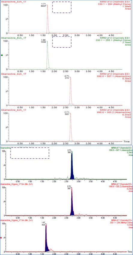
Annales de Toxicologie Analytique 2013; 25(2): 63-70                                                             H. Khaldoun-Oularbi et al.

           du foie, à savoir : une dilatation des veines, une infiltration des leucocytes et une dégénérescence des hépatocytes.
           Conclusion : Nos résultats montrent que l’ABM perturbe la fonction hépatique chez le rat.

           Mots clés : Abamectine, rat, foie, histologie, paramètres biochimiques, UHPLC MS/MS

           Received 26 February 2013, accepted after revision 4 June 2013
           Published online 17 September 2013

1 Introduction                                                         first detailed description of pathology in the liver during sub-
                                                                       acute ABM exposure, and to further investigate the effect of
     Pesticides are estimated to be responsible for approxi-           abamectin on some biochemical parameters in rats.
mately 4% of all deaths from accidental poisoning, mainly in
the developing world [1]. The use of substances toxic to man
and to a variety of forms of life has become an overall health         2 Materials and methods
problem.
     The macrocyclic lactons (MLs) include two chemical fam-           2.1 Chemicals
ilies: avermectins (abamectin, ivermectin, doramectin, epri-
nomectin and selamectin) and milbemycins (nemadectin,                      A commercial formulation of abamectin (Avermectin B1,
moxidectin, d-milbemycin, etc.) [2].                                   MK-936), named “Vertimec    r 1.8 EC” (Syngenta Agrochem-
     These chemical classes are naturally occurring products.          icals, Greensboro, USA) was used in the experiments. All
Avermectin is produced by the soil actinomycete bacterium              biochemical reagents used were obtained from commercial
“Streptomyces avermitilis” [3]. Avermectin derivatives are             sources (BIOLABO S.A., France).
among the most common active compounds applied as veteri-                  A pure reference standard of abamectin (>98.7%, purity,
nary drugs for food-producing animals, especially in aquacul-          Syngenta Agrochemicals, Greensboro, USA) was purchased
ture, and as plant protection agents in the agricultural sector,       from the Ministry of Agriculture and Rural Development (Di-
to control insects and mites on a wide range of agricultural           rection de la Protection des Végétaux et des Contrôles Tech-
products such as fruit, vegetable and ornamental crops [3–5].          niques DPVCT, Algeria). Acetonitrile and formic acid (HPLC
     Ivermectin (IVM) and abamectin (ABM) were the first               grade) were purchased from Biosolve Chimie (Valkenswaard,
macrocyclic lactones developed in the early 1980s. Other com-          the Netherlands). All other reagents were of analytical grade.
pounds have been marketed recently, such as moxidectin, do-
ramectin, eprinomectin and emamectin benzoate [6]. ABM is
known to be more toxic and moxidectin (MOX) less toxic                 2.2 Animals and abamectin exposure protocol
when compared with IVM [7].
     Abamectin (ABM) is a mixture of avermectin B1a (>90%)                 Twenty-eight male and female albino rats (Rattus norvegi-
and avermectin B1b (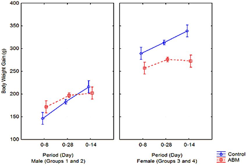
Annales de Toxicologie Analytique 2013; 25(2): 63-70                                                     H. Khaldoun-Oularbi et al.

Each rat received a single daily gavage dose of distilled wa-      0 L/h; desolvation gas flow rate and temperature: 200 L/h and
ter (controls) or abamectin at a dose of 2.13 mg/animal/day        250 ◦ C, respectively.
(abamectin-treated groups) for 28 consecutive days.                    All data were processed using the QuanLynx application
    The animals were weighed every day. The body weight            manager (Waters Corporation, MA, USA).
was determined throughout the acclimation, experimental and            In order to establish appropriate multiple reaction moni-
post-experimental periods. At the end of the experiments, the      toring (MRM) conditions, the cone voltage was adjusted to
rats were euthanized by cervical decapitation under light di-      maximize the intensity of the protonated molecular ion and
ethyl ether anesthesia and the liver was isolated and weighed.     collision-induced dissociation (CID) of both species was per-
                                                                   formed. Molecular ions (m/z 890.6 and 330.1 for abamectin
                                                                   and the IS, respectively), were selected in Ql and the corre-
2.2.1 Sample preparation
                                                                   sponding daughter ions (m/z 567.1 and 305.2 for abamectin
     An ultra-performance liquid chromatography tandem mass        and m/z 284.0 and 255.1 for the IS) were detected in Q3 after
spectrometry (UPLC–MS/MS) method employing electro-                collision with argon, used as the collision-activated dissocia-
spray ionization (ESI) was developed for the determination of      tion gas.
avermectin B1a in the plasma and liver of rats using methyl
clonazepam as the internal standard (EI).                          2.3 Biochemical levels
     The standard calibration curve samples of ABM at concen-
trations of 0, 1, 5, 10, 25, 50, 100, 250, 500 and 1 000 ng/mL         Blood samples (∼3 mL) were collected into EDTA tubes
were prepared by serial dilutions with plasma from stock solu-     via the supra-orbital sinus on days 14, 28 and 42. Plasma sam-
tion of 20 μg/mL.                                                  ples were obtained after centrifugation at 4 000 revolutions per
                                                                   minute for 15 min, and stored at –20 ◦ C until analysis. The
                                                                   following clinical chemistry parameters of plasma were mea-
2.2.2 Plasma and liver sample extractions                          sured using a Hitachi 912 Clinical Chemistry Analyzer: aspar-
     For the standard and plasma sample analyses, 100 μL EI        tate aminotransferase (AST), alanine aminotransferase (ALT)
(methyl clonazepam + hydroxyethyl theophiline) and 500 μL          and gamma-glutamyl transpeptidase (γ-Gt) enzyme activities,
of ACN were sequentially added into a 100-μ L plasma sample        and concentrations of glucose (GLUC).
then vortexed briefly. After centrifugation at 10800 rpm for           A commercial kit (BIOLABO S.A., France) was used ac-
8 min, the supernatant was evaporated.                             cording to the manufacturer’s instructions in these analyses.
     The rat liver samples were cut into small pieces and placed
in a glass tube; 1 000 μL of ACN were sequentially added to        2.4 Histopathogical analysis
2.5 g of liver sample, then vortexed. The mixing and extraction
steps were performed using a vortex for 2 min and ultrasoni-           The liver was excised from all rats and fixed in 10% neu-
cation for 15 min. Then, after centrifugation at 10 800 rpm for    tral formalin buffer. Tissue sections (5-μm thick) were cut
8 min, the clear liquid phase was collected and was evaporated.    and stained with hematoxylin and eosin for histopathological
     The plasma and liver residues were reconstituted in 100 μL    studies.
of the mobile phase [(A): (87%) ammonium formate solution
and (B): (13%) acetonitrile (ACN) and formic acid]. After cen-     2.5 Statistical analysis
trifugation at 4 000 rpm for 10 min, 15 μL of the supernatant
was then injected.                                                     Statistical analysis was performed using Statistica version
                                                                   10.0 (StatSoft Inc., Tulsa, USA). Data were calculated us-
                                                                   ing one-way analysis of variance (ANOVA) followed by the
2.2.3 UPLC-MS/MS conditions
                                                                   Newman-Keuls and Duncan’s post-hoc tests. Data were ex-
     The chromatographic separation was performed on an Ac-        pressed as the mean ±SE. P < 0.05 was considered as the
quity UPLC (Waters Corporation, MA, USA). Analytes were            level of significance.
separated using an Acquity UPLC HSS C18, 2.1 × 150 mm,
1.8-μm column (Waters Corporation, MA, USA) maintained             3 Results
at 50 ◦ C.
     The mobile phase was a binary mixture of formate buffer            The present study investigated the effect of repeat doses of
5 mM pH 3.0 (A) and acetonitrile containing 0.1% formic            2.13 mg/animal/day of abamectin on biochemical parameters,
acid (B) at a flow rate of 400 μL/min (0 min = 50% A;              residues of B1a in the plasma and liver, and the histopathology
1−3.25 min = 5% A; 4–5 min = 87% A). An injection vol-             of the liver on male and female rats.
ume of 15 μL was used.
     Detection was performed using a Waters TQ Detector
tandem quadrupole mass spectrometer (Waters Corporation,           3.1 Evaluation of body weight and liver weight
MA, USA) equipped with a Z-SprayTM source and ES probe.
The instrument was controlled using Waters MassLynxTMv4.1.             During the study period, there were no clinical signs of
Ionization was performed in ES+. Source conditions were as         toxicity in any treatment group. There were no differences be-
follows: source temperature was maintained at 100 ◦ C; cap-        tween females (control and ABM – female) and males (control
illary voltage: 3 kV; extractor cone: 3V; cone gas flow rate:      and ABM – male), while body weight was significantly lower

                                                                                                                                65
Annales de Toxicologie Analytique 2013; 25(2): 63-70 H. Khaldoun-Oularbi et al.
Fig. 1. Effect of abamectin (2.13 mg/animal/day) on the body weight of male (M) and female (F) rats throughout the acclimation (0–8 days),
experimental (0–28 days) and post-treatment periods (0–14 days).
Table I. Body weight, and absolute and relative liver weight (liver absolute weight/body weight at the end of the experiment ×100) (g ± SE) for
male (M) and female (F) rats in control and ABM-exposed (2.13 mg/animal/day) groups. [In the acclimation (1 week), experimental (28 days)
and post-treatment periods (14 days)].
Body weight Liver weight
Groups
Acclimation Experimentation Post-treatment At the end of experiment Absolute Relative
1: Control M 153.66 ± 4.91 183.49 ± 12.78 215.77 ± 6.62 221 ± 1.32 9.26 ± 0.65 4.19 ± 0.20
2: ABM M 165.10 ± 5.20 197.50 ± 11.54 202.11 ± 5.38* 199 ± 2.18* 9.70 ± 0.92* 4.87 ± 0.32*
3: Control F 283.55 ± 5.27 313.18 ± 11.49 276.41 ± 12.24* 342 ± 3.22 14.27 ± 1.87 4.17 ± 0.38
4: ABM F 262.99 ± 4.53 338.88 ± 3.89 272.55 ± 2.69* 271 ± 1.34* 11.71 ± 1.37* 4.32 ± 0.61*
∗
Statically significantly different (p < 0.05) from the control male and female groups (n = 7).
in the male than in the female groups during the acclimatiza- Table II also shows some biochemical variables that indi-
tion period. During the experimental period, the body weight cate liver injury in rats. Exposure of male and female rats to
of the animals in the ABM-female group (group 4) increased ABM produced a significant (p < 0.05) increase in plasma
significantly (p < 0.05) compared with the control group 3. ALAT, ASAT and γ-GT activities compared with those of the
At the end of the experimental study and after a post-treatment control groups.
period there was a significant decrease in body weight in male
and female ABM-treated groups. The relative organ weight 3.3 Plasma and liver abamectin concentrations
of the liver in ABM-treated groups 2 (male) and 4 (female)
showed a statistically significant increase (p < 0.05). The re- Abamectin was found in all plasma samples of male and
sults of the effects of 2.13 mg/animal/day of ABM on body female ABM-treated rats in a time-dependent manner at 14
and liver weight are summarized in figure 1 and table I. and at 28 days of treatment. However, at 42 days there were no
However, body weight gain was significantly lower in residues in the plasma of ABM-treated rats (figure 3).
ABM-treated groups 2 and 4 than in the control groups In male ABM-treated rats, the drug plasma concentration
(1 and 3) during the post-treatment period; lower mean food was 148 ng/mL at day 14 and 225 ng/mL at 28 days. How-
consumption was also noted in these groups. ever, in female ABM-treated rats, the drug plasma concen-
The results showed that treatment with abamectin caused tration was 193 ng/mL at day 14 and 279 ng/mL at day 28
an increase in the liver weight (1.77% in male and 7.51% in (figure 2A).
female ABM-treated rats compared with the controls). In the liver, abamectin concentrations at the end of the
experiment (day 42) were between 362 ng/g (female) and
83 ng/g (male). ABM concentrations were significantly lower
3.2 Biochemical results
in male compared with female rats at 14 days post-treatment
The effects of ABM on biochemical parameters in the (figure 2B).
rats are given in Table II. ABM exposure caused a significant The results showed that higher avermectin B1a concentra-
(p < 0.05) increase in glucose count levels in ABM-treated tions were recovered in the liver of female ABM-treated rats
rats but, after 14 days of non-treatment, there were no signifi- at 42 days than those measured in male rats. Mean ABM con-
cant differences between control and ABM-treated groups. centrations in the plasma and liver contents in male and female
66Annales de Toxicologie Analytique 2013; 25(2): 63-70 H. Khaldoun-Oularbi et al.
Fig. 2. (A) Plasma ABM [B1a (ng/mL)] concentrations at 14, 28 and 42 days obtained after administration of 2.13 mg/animal/day (daily for
28 days) to male and female rats. (B) Liver ABM [B1a (ng/mg)] concentrations at 42 days (post-treatment period) from male and female
ABM-treated rats. Mean (±SE) (n = 3) plasma abamectin (ABM) and mean (±SE) (n = 7) liver abamectin (ABM).
Table II. The overall means (± SE) (n = 7) of plasma glucose, alanine transaminase (ALT), aspartase aminotransferase (AST) and γ glutamyl
transpeptidase (γ-Gt); during the treatment (14 and 28 days) and post-treatment periods (42 days) of male (M) and female (F) rats with
2.13 mg/animal/day abamectin.
Time Group 1 Group 2 Group 3 Group 4
post-treatment Control M ABM M Control F ABM F
(days) (2.13 mg/animal/day) (2.13 mg/animal/day)
14 1.04 ± 0.01 1.08 ± 0.04 1.03 ± 0.02 1.03 ± 0.03
Glucose (g/dl) 28 1.03 ± 0.00 1.85 ± 0.14∗ 1.07 ± 0.00 2.47 ± 0.05*
42 1.04 ± 0.01 1.09 ± 0.09 1.00 ± 0.05 1.02 ± 0.06
14 84.00 ± 7.89 100.80 ± 5.30* 72.69 ± 5.31 105.33 ± 8.04∗
AST (IU/L) 28 75.25 ± 1.75 98.80 ± 6.17* 82.50 ± 6.63 95.66 ± 9.88∗
42 76.40 ± 3.29 73.50 ± 6.06 76.80 ± 3.73 74.33 ± 3.55
14 30.20 ± 1.65 38.50 ± 4.90* 46.33 ± 0.21 53.60 ± 0.97*
ALT (IU/L) 28 34.60 ± 2.71 43.75 ± 1.25∗ 44.33 ± 2.92 53.80 ± 0.28*
42 36.60 ± 3.28 41.50 ± 3.17∗ 34.33 ± 0.76 55.20 ± 1.10∗
14 7.23 ± 0.35 8.27 ± 0.73∗ 10.74 ± 0.87 20.00 ± 0.36∗
γ-Gt (UI/l) 28 7.34 ± 0.29 8.11 ± 0.68∗ 10.10 ± 0.20 12.89 ± 0.45∗
42 7.67 ± 0.34 8.99 ± 0.37∗ 11.12 ± 0.31 13.41 ± 0.95∗
∗
Statistically different from the control group of male and female rats (P < 0.05).
rats are compared in figure 2. Also, higher abamectin concen- nuclei and cell size, and cell granularity after chronic ABM
trations than those measured in male rats were recovered in the treatment, thus representing the active metabolizing state of
livers of female ABM-treated rats at 42 days. these cells.
3.4 Histological results
4 Discussion
The liver of control groups revealed normal hepatocytes
arranged in cords which are separated from each other by si- The present study provides additional information on
nusoids (figure 3A). Kupffer cells are also present along the ABM-induced toxicity in the rat, after subacute oral expo-
sinusoidal spaces. sure. The oral route seems to be the most relevant in long-term
Histological examination of the liver sections real exposure for the general population, due to residues in
(figures 4B−4D) of the ABM-treated groups revealed food [23] and also in tissues. Abamectin pesticide was chosen
massive congestion. The main findings were a narrowed in this study because it is used extensively all over the world,
appearance of Bowman’s space, degeneration of the tubular including Algeria where the study was performed. Based on
epithelial lining, a widened lumen, hemorrhage and cellular the results, the systemic effects of ABM may be described as
infiltration. The hepatocytes showed a significant increase in follows.
67Annales de Toxicologie Analytique 2013; 25(2): 63-70 H. Khaldoun-Oularbi et al.
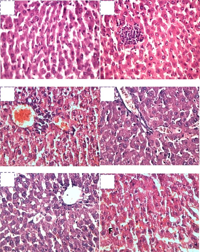
EI
A : Plasma
EI
B1a
B1a
B : Liver
B1a
B1a
EI
Fig. 3. Chromatograms of the two MRM transitions of ABM B1a in the plasma (A) and liver (B). The mass transitions used are as follows:
avermectin B1a: 890.6 > 567.1; 890.6 > 305.2 and EI: 330.1 > 284; 330.1255.1.
In the present study, cellular damage was evaluated by hepatocytes [24]. Our results indicated that the concentra-
measuring ALT and AST activity. In addition, the antioxidant tion of plasma ASAT, ALAT and γ-GT increased significantly
status of hepatocytes was determined by measuring the level (p < 0.05) in male and female rats treated with abamectin, a
of the GSH-related enzyme, γ-GT. Serum ALT and ALAT result in agreement with that of Hsu et al., [24] who showed
aminotransferases are usually employed in assessing the liver elevated levels of the cytosolic enzyme of the hepatocytes,
function; they are found in the cytoplasm and mitochondria aspartate aminotransferase (AST), in the blood serum of rats
of most cells. ABM poisoning can impair the function of exposed to 1−20 mg/kg body weight abamectin. However,
68Annales de Toxicologie Analytique 2013; 25(2): 63-70 H. Khaldoun-Oularbi et al.
A S B ABM exposure caused a significant (p < 0.05) increase in
glucose count in ABM-treated rats, but after 14 days of non-
treatment there were no significant differences between control
H CI and ABM-treated groups.
Different MLs have different toxicities in mammals. En-
zymes can metabolize MLs but the efflux of parental com-
pounds via active transport has been shown to be the main
route of ML elimination [7].
The presence of ATP-binding cassette (ABC) transporters,
including P-glycoprotein at the intestinal level, contributes ex-
C D tensively to the elimination of drugs in the feces and provides
VC
efficient barriers that protect the organism from the toxicity of
H most xenobiotics [28].
LI
CI All avermectins interfered with the transport activity of
P-glycoprotein, which acts as a trans-membrane protein, trans-
porting some drugs into and out of cells. Animals showing de-
creased P-gp activity show greater bioavailability of a drug af-
ter oral administration and accumulate greater levels of drugs
in their CNS tissue [6].
E P-gp is located on the apical surface of the enterocytes and
F hepatocytes, and thus expels drugs into the intestinal lumen
SD and the bile [29]. Nevertheless, previous studies support the
H extensive elimination of avermectins (IVM) by the fecal route
in rodents [30, 31]. Tests with laboratory animals show that
avermectin B1a is not readily absorbed into the bloodstream by
C mammals; it is rapidly eliminated from the body within 2 days
via the feces [30].
Accordingly, in the present investigation, the presence of
Fig. 4. Photomicrographs of the liver from a control rat (A) showing abamectin residues in all plasma samples of ABM-treated rats
normal architecture: hepatocytes (H) and sinusoids (S). From male at 14 and 28 days of treatment and their non-detection at
(B, C) and female (D, E, F) treated groups given ABM at the dose 42 days in the plasma of ABM- treated rats may be due to
of 2.13 mg/animal/day, showing vascular congestion (vc) and hem- their elimination by the fecal route.
orrhage (h), cellular infiltration (CI), sinusoidal dilatation (sd) and Pharmacokinetic parameters for IVM are strongly affected
foamy cytoplasm (FC) . (H&E × 400). by the presence or absence of P-gp [30]. The present data
showed that treatment with abamectin induced the accumu-
lation of residues in the liver even after a 14-day period of
in the present study, after 14 days of non-treatment, no non-treatment.
significant changes were observed in ALT activity of ABM- Dupuy et al. [32] showed an increased accumulation of
treated rats compared with controls. γ-glutamyl transpepti- moxidectin and avermectin in rat hepatocytes after inhibition
dase is the sensitive marker enzyme of renal cell injury [25]. of P-gp or other ABC transporters. Accordingly, the observed
El-Shenawy’s [26] study of the in vitro toxic action of some in- accumulation of residues in the liver suggests a corresponding
secticides, including ABA, on isolated rat hepatocytes showed inhibition of P-gp transporter, which leads to increased con-
a significant increase in ALT and AST activity when hepato- centration of this xenobiotic in the liver. Also, the lipophilic
cytes were incubated for 30 min with either concentration of nature of ABM may lead to its accumulation in the liver.
ABA. This activity persisted after 120 min, the longest time
for which data was collected.
These results reflect the increase in plasma membrane per- 5 Conclusion
meability resulting from the hepatic damage [27] and observed In summary, the UPLC MS/MS method was successfully
signs of abamectin liver toxicity, with increased activity of the applied to determine avermectin B1a in the plasma and liver
enzyme AST in rats treated with doses equivalent to 1/10 or after subacute abamectin exposure. Elevation of AST, ALAT
1/100 of the LD50 (18 mg/kg) in the diet of animals over and γ-GT activity, histopathological changes and persistence
30 consecutive days. of residues in the liver after 14 days without treatment confirm
The biochemical data (table II) showed that the increase liver injury in rats after ABM intoxication.
in plasma levels of ALAT and γ-glutamyl transpeptidase per-
sisted after 14 days with no further treatment.
References
The above findings were confirmed by histopathological
changes in the liver under the intoxication effect of abamectin. 1. Colosio C, Moretto A. Pesticides International Encyclopedia of
Abamectin caused marked damage of the liver tissues in the Public Health 2008, 59–66.
form of dilated veins, hemorrhagic spots and degenerative hep- 2. McKellar Q, Benchaoui H. Avermectins and milbemycins. J Vet
atocytes. Phamacol Ther. 1996; 19: 331–351.
69Annales de Toxicologie Analytique 2013; 25(2): 63-70 H. Khaldoun-Oularbi et al.
3. Burg RW, Miller BM, Baker EE, Birnbaum J, Currie SA, 18. Sheridan R, Desjardins L. Determination of abamectin, do-
Hartman R, Kong YL, Monaghan RL, Olson G, Putter I, Tunac ramectin, emamectin, eprinomectin, ivermectin, and mox-
JB, Wallick H, Stapley EO, Oiwa R, Omura S. Avermectins, idectin in milk by liquid chromatography–electrospray tandem
new family of potent anthelmintic agents: producing organism mass spectrometry. J AOAC Int. 2006; 89: 1088–1094.
and fermentation. Antimicrob Agents Chemother. 1979; 15: 19. Rübensam G, Barreto F, Barcellos Hoff R, Mara Pizzolato
361–367. T. Determination of avermectin and milbemycin residues in
4. Shoop W, Mrozik H, Fisher M. Structure and activity of aver- bovine muscle by liquid chromatography-tandem mass spec-
mectins and milbemycins in animal health. Vet Parasitol. 1995; trometry and fluorescence detection using solvent extraction
59: 139–156. and low temperature cleanup. Food Control. 2013; 29: 55–60.
5. Hernando MD, Suarez-Barcena JM, Bueno MJM, Garcia- 20. Ali MS, Sun T, McLeroy GE, Phillippo E. Confirmation of
Reyes JF, Fernandez-Alba AR. Fast separation liquid eprinomectin, moxidectin, abamectin, doramectin, and iver-
chromatography–tandem mass spectrometry for the confirma- mectin in beef liver by liquid chromatography/positive ion at-
tion and quantitative analysis of avermectin residues in food. J mospheric chemical ionization mass spectrometry. J AOAC
Chromatogr A. 2007; 1155: 62–73. Int. 2000; 83(1): 39–52.
6. Danaher M. Review of methodology for the determination 21. OECD. OECD Test Guideline for testing of chemicals, Section 4:
of macrocyclic lactone residues in biological matrices. Health Effects, OECD. Guideline 407, Repeated Dose 28-Day
J Chromatogr B. 2006; 844: 175–203. Oral Toxicity Study in Rodents 2003.
7. Castanha Zanoli JC, Maioli MA, Medeiros HCD, Mingatto FE.
22. Elbetieha A, Isa Daas S. Assessment of antifertility activities
Abamectin affects the bioenergetics of liver mitochondria: a
of ABM pesticide in male rats. Ecotoxicol Environm Safety.
potential mechanism of hepatotoxicity. Toxicol In Vitro. 2012;
2003; 55(3): 307–313.
26: 51–56.
8. Campbell WC, Fisher MH, Stapley EO, Albers-Schonberg G, 23. Cometa MF, Buratti FM, Fortuna S, Lorenzini P, Volpe MT,
Jacob TA. Ivermectin: a potent antiparasitic agent. Science. Parisi L, Testai E, Meneguz A. Cholinesterase inhibition and
1983; 221: 823–828. alterations of hepatic metabolism by oral acute and repeated
9. Kita K, Shiomi K, Omura S. Advances in drug discovery and bio- chlorpyrifos administration to mice. Toxicology. 2007; 234:
chemical studies. Trends in Parasitology. 2007; 23: 223−229. 90–102.
10. Horvat AJM, Petrovic M, Babic S, Pavlovic DM, Asaperger˛ D, 24. Hsu DZ, Hsu CH, Huang BM, Liu MY. Abamectin effects on
Pelko S, Mance AD, Kas telan-Macan M. Analysis, occur- aspartate aminotransferase and nitric oxide in rats. Toxicology.
rence and fate of anthelmintics and their transformation prod- 2001; 165: 189–193.
ucts in the environment. Trends Anal Chem. 2012; 31: 61–84. 25. Van der Harst JE, Fermont PCJ, Bilstra AE, Spruijt BM. Access
11. Yoon YJ, Kim ES, Hwang YS, Cho CY. Avermectin: biochemical to enriched housing is rewarding to rats as reflected by
and molecular basis of its biosynthesis and regulation. Appl their anticipatory bahaviour. Animal Behaviour. 2003; 66(3):
Microbiol Biotechnol. 2004; 63: 626–634. 493−504.
12. Valenzuela AI, Redondo MJ, Pico Y, Font G. Determination 26. El-Shenawy NS. Effects of insecticides fenitrothion, endosulfan
of abamectin in citrus fruits by liquid chromatography- and abamectin on antioxidant parameters of isolated rat hepa-
electrospray ionization mass spectrometry. J Chromatogr A. tocytes. Toxicol In Vitro. 2010; 24: 1148–1157.
2000; 871: 57−65. 27. Eissa FI, Zidan NA. Haematological, biochemical and
13. Turnipseed SB, Roybal JE, Andersen WC, Kuck LR. Analysis histopathological alterations induced by abamectin and
of avermectin and moxidectin residues in milk by liquid Bacillus thuringiensis in male albino rats. Acta Biol Hung.
chromatography–tandem mass spectrometry using an atmo- 2010; 61: 33–44.
spheric pressure chemical ionization/atmospheric pressure 28. Menez C, Mselli-Lakhal L, Foucaud-Vignault M, Balaguer P,
photoionization source. Anal Chim Acta. 2005; 529: 159−165. Alvinerie M, Lespine A. Ivermectin induces P-glycoprotein
14. Pozo OJ, Marin JM, Sancho JV, Hernandez F. Determination of expression and function through mRNA stabilization in
abamectin and azadirachtin residues in orange samples by liq- murine hepatocyte cell line. Biochem Pharmacol. 2012; 83:
uid chromatography–electrospray tandem mass spectrometry. 269–278.
J Chromatogr A. 2003; 992: 133–140. 29. Prichard R, Ménez C, Lespine A. Moxidectin and the aver-
15. Kolar L, Kuzner J, Erzen NK. Determination of abamectin and mectins: Consanguinity but not identity. Int J Parasitology:
doramectin in sheep faeces using HPLC with fluorescence de- Drugs Drug Resistance. 2012; 1–20.
tection. Biomed Chromatogr. 2004; 8: 507–511.
30. Laffont CM, Toutain PL, Alvinerie M, Bousquet-Melou A.
16. Chou HK, Lai CY, Chen T, Yen GC. A multiresidue method
Intestinal secretion is a major route for parent ivermectin elim-
for the determination of abamectin, doramectin, moxidectin,
ination in the rat. Drug Metab Dispos. 2002; 30: 626–630.
ivermectin, milbemectin A3, and milbemectin A4 residues
31. Kiki-Mvouaka S, Menez C, Borin C, Lyazrhi F, Foucaud-
in bovine muscle using HPLC with fluorescence detection. J
Vignault M, Dupuy J, Collet X, Alvinerie M, Lespine A. Role
Food Drug Anal. 2004; 12(2): 146–153.
of P-glycoprotein in the disposition of macrocyclic lactones:
17. Grimalt S, Pozo ÓJ, Marín JM, Sancho JV, Hernández F.
Evaluation of different quantitative approaches for the deter- a comparison between ivermectin, eprinomectin, and mox-
mination of noneasily ionizable molecules by different atmo- idectin in mice. Drug Metab Dispos. 2010; 38: 573–580.
spheric pressure interfaces used in liquid chromatography- 32. Dupuy J, Lespine A, Sutra JF, Alvinerie M. The interaction be-
tandem mass spectrometry: abamectin as case of study. J Am tween moxidectin and MDR transporters in primary cultures
Soc Mass Spectrom. 2005; 16: 1619–1630. of rat hepatocytes. J Vet Pharmacol Ther. 2006; 29: 107–111.
70You can also read