Hepatic triacylglycerol accumulation and insulin resistance
←
→
Page content transcription
If your browser does not render page correctly, please read the page content below
Hepatic triacylglycerol accumulation and insulin resistance

                         Cynthia A. Nagle, Eric L. Klett, and Rosalind A. Coleman1
                         Department of Nutrition, University of North Carolina at Chapel Hill, Chapel Hill, NC 27599

Abstract The association of hepatic steatosis with hepatic                  sequestering FA may lead to long-term morbidity with
insulin resistance and type 2 diabetes has prompted investi-                the development of further IR leading to type 2 diabetes
gators to elucidate the underlying mechanism. In this review                mellitus and the deterioration of hepatic function.
we focus on pathways of lipid metabolism, and we review                        Although fatty liver correlates with hepatic IR, it remains
recent data, primarily from mouse models, that link lipid in-
                                                                            unclear whether IR or excess TAG stores develop first and
termediates with insulin resistance. Most of the studies that
implicate acyl-CoA, lysophosphatidic acid, phosphatidic                     whether fatty liver invariably leads to IR. Peripheral IR may
acid, diacylglycerol, or ceramide rely on indirect associa-                 cause fatty liver by elevating plasma FA, glucose, and insu-
tions.     Convincing data to support the hypothesis that                   lin, which stimulate hepatic lipid synthesis and impair he-
specific lipid intermediates initiate pathways that alter insu-             patic b-oxidation. However, high-fat feeding may cause

                                                                                                                                                                       Downloaded from www.jlr.org by guest, on May 19, 2015
lin signaling will require studies in which the concentration               hepatic IR before systemic IR develops. We will focus on
of each purported signaling molecule can be manipulated                     the metabolic pathways involved in the development of
independently.—Nagle, C. A., E. L. Klett, and R. A.                         NAFLD, the role of altered hepatic TAG metabolism,
Coleman. Hepatic triacylglycerol accumulation and insulin                   and the role of lipid metabolites in the development of IR.
resistance. J. Lipid Res. 2009. 50: S74–S79.

Supplementary key words Hepatic steatosis • diacylglycerol • phospha-
tidic acid • lysophosphatidic acid • ceramide                                     METABOLIC PATHWAYS INVOLVED IN FATTY
                                                                                          LIVER DEVELOPMENT

   Nonalcoholic fatty liver disease (NAFLD) is character-                     Hepatic lipid accumulation can be caused by four differ-
ized by excessive lipid accumulation in the liver in the                    ent metabolic perturbations: increased FA delivery to he-
absence of ‘significant’ alcohol consumption and may                        patocytes from lipolyzed adipose TAG, dietary lipids, or
progress to nonalcoholic steatohepatitis, fibrosis, cirrhosis,              hepatic de novo lipogenesis (DNL); increased TAG syn-
and hepatocellular carcinoma. In the United States, the                     thesis; decreased hepatic FA oxidation; and inadequate
prevalence of NAFLD is estimated to be 20% (1); however,                    TAG secretion in VLDL (Fig. 1).
the true prevalence of NAFLD is unknown because many
individuals in earlier stages of the disease are asymp-                     Fatty acid delivery and synthesis
tomatic and remain undiagnosed. Although NAFLD was
                                                                              After consuming a meal containing fat, chylomicron-
previously thought to be a benign condition, its develop-
                                                                            TAG is delivered to the liver and is lipolyzed in lysosomes
ment parallels the development of the insulin resistance
                                                                            with the release of FA. FA can also arise from DNL in re-
(IR) syndrome and is associated with type 2 diabetes mel-
litus. Patients with the IR syndrome have a 4- to 11-fold in-
creased risk of developing NAFLD.
   With overnutrition and lack of exercise, liver and other                     Abbreviations: ACC, acetyl-CoA carboxylase; ACSL, acyl-CoA
                                                                            synthetase; AGPAT, 1-acylglycerol-3-phosphate acyltransferase; ChREBP,
tissues store excess energy as triacylglycerol (TAG). Shunt-                carbohydrate-responsive element-binding protein; CPT, carnitine-
ing carbon-energy into a storage form is likely protective                  palmitoyltransferase; DAG, diacylglycerol; DGAT, DAG acyltransferase;
against cytotoxic FA accumulation. Hepatic IR is asso-                      GPAT, glycerol-3-phosphate acyltransferase; HF, high fat; HF-SD, HF
ciated with the accumulation of TAG and FA metabolites                      safflower oil diet; IR, insulin resistance; IRS, insulin receptor substrate;
                                                                            KO, knock out; LCAD, long chain acyl-CoA dehydrogenase; LPA,
(fatty acyl-CoA, diacylglycerol (DAG), ceramide, and glyco-                 lysophosphatidic acid; LXR, liver-X-receptor; MCAD, medium chain
sphingolipid). The short-term protection achieved from                      acyl-CoA dehydrogenase; NAFLD, nonalcoholic fatty liver disease; DNL,
                                                                            de novo lipogenesis; PA, phosphatidic acid; PAP, phosphatidic acid
                                                                            phosphatase; PKC, protein kinase C; PPAR, peroxisome proliferator-
                                                                            activated receptor; TAG, triacylglycerol; SCAD, short-chain acyl-CoA
This work was supported by National Institutes of Health Grants DK56598,
DK59935, and DK56350.                                                       dehydrogenase; SCD, stearoyl-CoA desaturase; SREBP, sterol regulatory
                                                                            element binding protein; TCA, tricarboxylic acid; WT, wild type;
Manuscript received 16 October 2008 and in revised form 5 November 2008.    VLCAD, long chain acyl-CoA dehydrogenase.
                                                                                1
Published, JLR Papers in Press, November 6, 2008.                                 To whom correspondence should be addressed.
DOI 10.1194/jlr.R800053-JLR200                                                    email: rcoleman@unc.edu

                                                                           Copyright © 2009 by the American Society for Biochemistry and Molecular Biology, Inc.
S74       Journal of Lipid Research         April Supplement, 2009                                            This article is available online at http://www.jlr.org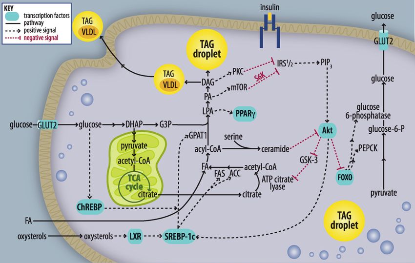
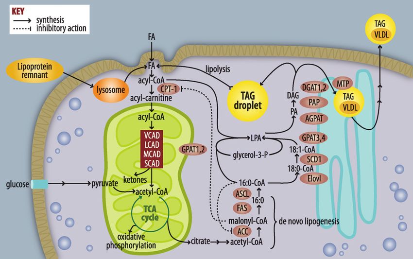
Fig. 1. Pathways of FA and triacylglycerol metabolism. FAs used for triacylglycerol (TAG) synthesis or b-
               oxidation are activated to form acyl-CoAs. If targeted toward b-oxidation, acyl-CoAs are converted to acyl-
               carnitines, enter the mitochondria, and are metabolized by a succession of dehydrogenases that are relatively

                                                                                                                                              Downloaded from www.jlr.org by guest, on May 19, 2015
               specific for very-long (VLAD), long (LCAD), medium (MCAD), and short (SCAD) chain acyl-CoAs. The end-
               product acetyl-CoA enters the tricarboxylic acid cycle (TCA), producing NADH and FADH, which contribute
               to ATP formation by oxidative phosphorylation. When excess calories from glucose are available, acetyl-CoA
               entering the TCA cycle is converted to citrate, which leaves the mitochondria and is used for FA synthesis via
               acetyl-CoA carboxylase (ACC) and FAS. FA elongases (Elovl) and desaturases (SCD) modify acyl-CoAs, which
               are used to esterify glycerol-3-phosphate by glycerol-3-P acyltransferases (GPAT). The lysophosphatidic acid
               (LPA) product is esterified to form phosphatidic acid (PA) by an acyl-glycerol-3-P acyltransferase, the PA is
               hydrolyzed by phosphatidic acid phosphohydrolase (PAP), and the final esterification step catalyzed by
               diacylglycerol acyltransferase (DGAT) produces TAG. Liver TAG can be stored in lipid droplets or trans-
               ferred to VLDL by the mitochondrial TAG transport protein.

sponse to a high carbohydrate meal, as excess glucose is                (AGPAT),1,2 three phosphatidic acid phosphatases (PAP),
metabolized to acetyl-CoA, the major substrate for FA                   and two DAG acyltransferases (DGAT) have been cloned3.
synthesis. Malonyl-CoA, the product of acetyl-CoA car-
boxylase (ACC), is not only a substrate for FAS , but also              Hepatic TAG lipolysis and b-oxidation
inhibits carnitine-palmitoyltransferase-1 (CPT-1), which                   During fasting, insulin levels fall, adipose TAG is hydro-
regulates long-chain FA entry into mitochondria for b-                  lyzed, and FAs are released and travel to the liver. It is not
oxidation. FAS sequentially adds two carbons from malonyl-              clear whether exogenously derived FAs must first be rees-
CoA to synthesize 16 and 18 carbon FA. These saturated                  terified and stored in lipid droplets before they can be oxi-
FAs must be activated by long-chain acyl-CoA synthetase                 dized, or whether acyl-CoAs can be directly converted to
(ACSL) before they can be elongated by an elongase                      acyl-carnitines and enter the oxidative pathway. Within
(ELOVL6), desaturated by stearoyl-CoA desaturase (SCD),                 the mitochondrial matrix, acyl-carnitines are converted
or used for the synthesis of glycerolipids or cholesterol esters.       to acyl-CoAs by CPT-2. Very long chain acyl-CoA dehydro-
                                                                        genase (VLCAD) acts on C12-C24 carbon acyl-CoAs, long
TAG synthesis                                                           chain acyl-CoA dehydrogenase (LCAD) acts on C8-C20
   Hepatic FAs are derived from entering chylomicron                    acyl-CoAs, medium chain acyl-CoA dehydrogenase (MCAD)
remnants, TAG lipolysis in lipid droplets, or DNL. After                acts on C4-C12 acyl-CoAs, and short-chain acyl-CoA de-
activation, the acyl-CoAs enter the glycerolipid synthetic              hydrogenase (SCAD) acts on C4-C6 acyl-CoAs. When the
pathway. In the liver, TAG may either be stored in cyto-                tricarboxylic acid (TCA) cycle cannot metabolize all the
plasmic lipid droplets or incorporated into VLDL particles
and secreted into the blood. The four-step synthesis of
TAG, first identified by Eugene Kennedy and his col-
                                                                           1
leagues (2), occurs on the cytosolic surface of the mitochon-                AGPAT8 and AGPAT6 are correctly termed GPAT3 and GPAT4,
drial outer membrane and the endoplasmic reticulum.                     respectively.
                                                                           2 CGI-58, and endophilin both have AGPAT activity.
Four glycerol-3-phosphate acyltransferases (GPAT), po-                     3
                                                                             Several additional enzymes exhibit some DGAT activity, includ-
tentially six 1-acylglycerol-3-phosphate acyltransferases               ing MGAT2.

                                                                                                   Fatty liver and insulin resistance   S75
acetyl-CoA generated from b-oxidation, excess acetyl-CoA of SREBP-1c (4). Transgenic hepatic over-expression of
is converted to ketone bodies. SREBP-1c produces a fatty liver and a 4-fold increase in
the rate of hepatic FA synthesis, with increases in lipogenic
VLDL secretion genes like FAS, ACC and SCD-1 (5), and GPAT1 (5). In-
The origin of VLDL-TAG remains poorly understood. A sulin increases LXRʼs ability to activate the SREBP-1c
precursor VLDL particle that contains apoB and a small promoter and increase hepatic lipogenesis (6). Thus, hy-
amount of TAG is formed in the endoplasmic reticulum perinsulinemia can activate LXR and SREBP-1c and result
(ER). This small VLDL precursor may fuse within the ER in increased lipogenesis, liver TAG accumulation, and wors-
with a larger droplet of TAG to form a TAG-rich particle. ening hepatic IR.
The TAG that fuses is thought to arise from cytosolic TAG ChREBP regulates hepatic lipid synthesis through tran-
derived primarily from plasma FA uptake and less from scriptional control of the lipogenic genes ACC and FAS in
DNL (7). Incorporation of cytosolic TAG into VLDL may response to glucose (7). Liver-specific ChREBP inhibition
require lipolysis and reesterification steps at the ER (3). results in decreased hepatic lipogenesis and ameliorated
hepatic steatosis in ob/ob mice (7).
TRANSCRIPTIONAL REGULATION OF DE NOVO
LIPOGENESIS AND FA OXIDATION DO INTERMEDIATES IN THE KENNEDY PATHWAY
OF GLYCEROLIPID BIOSYNTHESIS INITIATE
An excess of glucose, FA, and insulin ultimately lead to SIGNALING PATHWAYS?
hepatic steatosis and worsening hepatic IR via a network of
transcription factors (Fig. 2). Activation of hepatic liver-X- Although IR correlates highly with TAG content in skel-
receptor (LXR) by endogenous oxysterol ligands results in etal muscle and liver, TAG itself is believed to be a surro-
up-regulation of genes involved in cholesterol, lipid, and gate marker for the true disruptor of the insulin signal,
Downloaded from www.jlr.org by guest, on May 19, 2015
bile acid metabolism. In mice, oral LXR agonists result variously hypothesized to be a) a cytokine released from
in enhanced hepatic FA synthesis, hepatic steatosis, and hy- macrophages or adipocytes, b) activation of the NFkB path-
pertriglyceridemia, all mediated by increased expression way (8), or c) a signaling pathway initiated by a FA-derived
Fig. 2. Altered insulin signaling by lipid intermediates. Increased nutrient influx raises intracellular FA con-
centrations. With nutrient surfeit, elevated malonyl-CoA inhibits CPT-1 and b-oxidation and results in in-
creased availability of acyl-CoAs for TAG synthesis and storage or secretion. Intermediates in the TAG
biosynthetic pathway activate inhibitors of insulin signaling, including protein kinase C (PKC), mTOR,
and S6K, which suppress IRS-1 activation of PIP3. Ceramide inhibits Akt-mediated insulin signaling, dere-
pressing FOXO and leading to increases in transcription of phosphoenolpyruvate (PEPCK) and glucose-
6-phosphatase (G-6-Pase). These enhance gluconeogenesis and hepatic glucose output. Nutrients act on
transcription factors (LXR, ChREBP, SREBP-1c and PPAR g), which increase transcription of genes involved
in FA and TAG synthesis (ACC, FAS, GPAT1). These enhance steatosis and hepatic insulin resistance. DHAP,
dihydroxyacetone-P; G3P, glycerol-3-P; GSK, glycogen synthase kinase.
S76 Journal of Lipid Research April Supplement, 2009lipid. Lysophosphatidic acid (LPA), phosphatidic acid TAG and VLDL-TAG are 60%, 15%, and 24%, respectively
(PA), and DAG are well-established initiators of signaling (22). It is not known, however, whether the lipid inter-
pathways but have been studied primarily after their hydro- mediates derived from these pathways have similar effects
lysis from membrane phospholipids. In liver, however, the on the development of hepatic IR, or whether one path-
major production of LPA, PA, and DAG occurs via de novo way predominates. When over-expression of hepatic LPL
glycerolipid synthesis; lipid intermediates derived from this increases FA flux into the liver from lipoproteins, hepatic
pathway and related lipids [acyl-CoAs (9, 10), ceramide IR is associated with an increased hepatic content of TAG
(11) and acyl-carnitines] may also act as signaling mole- and acyl-CoA (23). Augmented DNL also appears to be im-
cules. Because incubating cells with FA or infusing FA into portant, because ob/ob mice deficient in either SREBP-1c
mice causes IR within a few hours, it appears that a direct or ChREBP are protected from hepatic steatosis and IR
FA metabolite can promote IR. (24, 25). Further, rodents with liver-specific decreases in
the synthesis of malonyl-CoA, ACC null mice, and rats that
over-express malonyl-CoA decarboxylase in liver have im-
BLOCK OF INSULIN SIGNALING BY proved hepatic insulin sensitivity due to lower DNL and
LIPID METABOLITES increased b-oxidation (26, 27). In contrast, mice with a
liver-specific knockout of FAS develop a fatty liver despite
With hepatic IR, insulin cannot effectively suppress he- suppressed DNL because PPARa is not activated (28). De-
patic glucose output in a hyperinsulinemic-euglycemic spite the fatty liver that results from decreased b-oxidation,
clamp study. IR can also be inferred by impaired tyrosine FAS null mice have impaired gluconeogenesis and are
phosphorylation of the insulin receptor and aberrant hypoglycemic and insulin sensitive.
phosphorylation of serine residues of insulin receptor sub- Increased SREBP-1c activity in ob/ob mouse liver in-
strate-1 (IRS-1) and downstream members of the insulin creases GPAT1 mRNA expression and hepatic de novo glyc-
signaling pathway. erolipid synthesis (29), whereas hepatic GPAT1 knockdown
Downloaded from www.jlr.org by guest, on May 19, 2015
Our studies implicate GPAT1-produced DAG as a cause in ob/ob mice lowers hepatic TAG and DAG content and
of IR (12, 13); when excess DAG is formed, protein kinase plasma glucose concentrations (30). Studies in GPAT1 knock
C (PKC)y is activated, and IRS-1 is phosphorylated on out (KO) mice and in rats with hepatic over-expression of
Ser307, which suppresses IRS-1 tyrosine phosphorylation GPAT1 suggest that the lipid accumulation from de novo
and diminishes phosphatidylinositol 3-kinase activation. glycerolipid synthesis contributes to the development of
These data suggest that the DAG formed during glycero- hepatic IR in the absence of obesity or a high fat diet
lipid synthesis can interact with and activate PKC at distant (12, 13). Unlike lipid accumulation from DNL, hepatic
membranes and show that GPAT1 modulates the cell con- steatosis caused by a block in VLDL-TAG secretion does
tent of lipid intermediates. This is not entirely surprising not cause hepatic IR, perhaps because lipid intermediates
because lipogenic diets normally increase SREBP-1c- do not accumulate, suggesting that lipids destined for
mediated GPAT1 mRNA expression (2). In addition to VLDL secretion are in a separate cellular pool that cannot
DAG, the glycerolipid intermediates LPA and PA can also affect inhibitors of insulin signaling (17).
initiate signaling pathways. LPA is a ligand for peroxisome
proliferator-activated receptor (PPAR) g (14) and might Impaired b-oxidation
be responsible for the up-regulation of PPARg target genes Obese mouse models largely demonstrate that activating
observed in NAFLD, whereas intracellular PA activates b-oxidation decreases hepatic lipid accumulation and im-
mammalian target of rapamycin (15), which down-regulates proves insulin sensitivity. For example, activating PPARa
the insulin signal by promoting serine phosphorylation of with Wy-14,643 increases b-oxidation, reduces hepatic TAG
IRS when nutrients are present in excess. accumulation, and improves insulin signaling in ob/ob mice
and in lipoatrophic A-ZIP/F-1 mice (31, 32). However, the
role of suppressed b-oxidation in the development of IR is
THE ROLE OF LIPID METABOLITES IN THE less clear. Both prolonged fasting and short-term pharma-
DEVELOPMENT OF HEPATIC INSULIN RESISTANCE cological inhibition of CPT-1 cause a fatty liver without IR
(18, 21). Unfortunately, DAG and other lipid intermedi-
Although fatty liver is associated with IR, several rodent ates were not measured in this study.
models with steatosis remain insulin sensitive (16–21). Fac-
tors that may explain the variable relationship between fatty Fasting-induced steatosis
liver and IR include the specific pathways that contribute to When hepatic TAG accumulates in lean fasted animals,
hepatic fat accumulation, the type of lipid metabolites it does not cause hepatic IR. Perhaps signaling lipids are
that accumulate, the cellular location and FA composition sequestered in pools that cannot interfere with insulin sig-
of those metabolites, the presence or absence of hepatic naling. Similar to pharmacological inhibition of b-oxidation,
or systemic inflammation, and the type of rodent diet used. genetic deficiency of PPARa, VLCAD, or MCAD causes a
fatty liver and mild to severe hypoglycemia under fasting
Altered de novo lipogenesis and glycerolipid synthesis conditions (33–35). PPARa KO mice are protected from he-
In humans with NAFLD, the estimated contribution of patic IR when fed a high fat (HF)-coconut oil diet (20), but
adipose lipolysis, dietary FA, and hepatic DNL to hepatic not when fed HF-palm oil or HF-lard diets (36). The insulin
Fatty liver and insulin resistance S77sensitivity of VLCAD and MCAD mice has not been studied, Elevated lipid intermediates without impaired
but LCAD KO mice develop hepatic steatosis and reduced insulin sensitivity
FA oxidation without hypoglycemia (37). Although fasting Mice can have elevated hepatic TAG, DAG, and ceramide
hepatic DAG and ceramide content do not differ between without hepatic IR (16, 17). This discrepancy might be
wild-type (WT) and LCAD KO mice, the hepatic DAG con- explained if different pools of DAG and ceramide exist
tent increases 3-fold in the LCAD KO livers after insulin within cells, with only certain pools able to regulate inhib-
stimulation during a hyperinsulinemic-euglycemic clamp, itors of insulin signaling. Thus, measuring hepatic DAG
and is associated with hepatic IR (43). Thus, inhibiting and ceramide total content instead of the content in spe-
b-oxidation in fasted lean mice causes hepatic steatosis with- cific membrane fractions may obscure the relevant mech-
out IR, whereas inhibiting b-oxidation in previously high- anism. Additionally, some DAG and sphingolipid species
fat-fed animals interferes with insulin signaling. Perhaps may be poor inhibitors of insulin signaling. The fact that
impaired b-oxidation is most detrimental to insulin sensitiv- diets high in different FA species have remarkably different
ity when DNL is activated in response to feeding, thereby effects on hepatic insulin sensitivity and gene expression
resulting in increased synthesis of FA and glycerolipid inter- (11, 43) suggests that DAG FA composition could be rele-
mediates. It is not clear what makes one kind of lipid accu- vant. Further, ceramide-induced IR may be caused by gan-
mulation differ from another, but differences in cellular glioside metabolites of ceramide (44). Future researchers
location or in lipid species may determine whether insulin should measure not only gross lipid content, but also the
signaling is impaired. Additionally, none of these models de- intracellular locations and FA composition of lipids, as well
scribes a liver-specific inhibition of b-oxidation. Inhibiting as inflammatory markers that are associated with IR.
b-oxidation in adipose tissue or muscle may alter plasma cy-
tokine levels that secondarily improve or inhibit hepatic in-
sulin signaling. SUMMARY AND FUTURE DIRECTIONS
Downloaded from www.jlr.org by guest, on May 19, 2015
Diet effects A major difficulty in assessing the importance or the sig-
Both DAG and ceramide can diminish hepatic insulin nificance of any lipid intermediate to impaired insulin sig-
sensitivity, and feeding rats a HF safflower oil diet (HF-SD) naling is the lack of direct experiments. Most of the studies
for three days increases hepatic DAG and PKCy activity that implicate acyl-CoA, LPA, PA, DAG, or ceramide rely
and hepatic IR (10). Conversely, a liver-specific knock-down on indirect associations. The effects of different diets are
of PKCy prevents HF-SD-induced hepatic IR, strength- not well documented or explained mechanistically, and
ening the hypothesis that PKCy and DAG mediate hepatic the inter-relationships between different putative signaling
IR in this model (38). Other mouse models that support pathways have been inadequately delineated. Convincing
this hypothesis include hepatic over-expression of GPAT1 data to support the hypothesis that specific lipid intermedi-
(13), LCAD KO mice (37), Elovl6 KO mice fed a high ates initiate signaling pathways that alter insulin signaling
fat-high sucrose diet (19), and mouse models fed with may require additional studies in cultured hepatocytes in
the HF-SD including: ACC2 KO (19), ACC1 and 2 knock- which the concentration of each purported signaling mole-
down (26), GPAT1 KO (12), and DGAT2 knockdown mice cule can be manipulated independently.
(16). Although these studies provide strong evidence that
DAG and PKCy are important mediators of hepatic IR in
HF-SD fed animals, several studies question the impor- REFERENCES
tance of DAG. For example, GPAT1 KO mice are protected
1. Ruhl, C. E., and J. E. Everhart. 2004. Epidemiology of nonalcoholic
from hepatic IR when fed the HF-SD, but not when fed a fatty liver. Clin. Liver Dis. 8: 501–519.
diet high in saturated fat (39, 40). Similarly, mice treated 2. Coleman, R. A., and D. P. Lee. 2004. Enzymes of triacylglycerol
with DGAT2 ASO are protected from HF-SD IR, but not synthesis and their regulation. Prog. Lipid Res. 43: 134–176.
from IR induced by a HF saturated diet (26, 41). These 3. Gibbons, G. F., D. Wiggins, A. M. Brown, and A. M. Hebbachi.
2004. Synthesis and function of hepatic very-low-density lipopro-
discrepancies suggest that different fatty acid species affect tein. Biochem. Soc. Trans. 32: 59–64.
mechanisms of hepatic IR differently. For example, a HF- 4. Grefhorst, A., B. M. Elzinga, P. J. Voshol, T. Plösch, T. Kok, V. W.
lard diet causes a ceramide-associated hepatic IR that Bloks, F. H. van der Sluijs, L. M. Havekes, J. A. Romijn, H. J.
Verkade, et al. 2002. Stimulation of lipogenesis by pharmacologi-
is reversed by inhibiting sphingolipid synthesis, whereas cal activation of the liver X receptor leads to production of large,
a HF-soy oil diet causes a DAG-associated IR that is not triglyceride-rich very low density lipoprotein particles. J. Biol. Chem.
affected by sphingolipid inhibition (11). Perhaps high- 277: 34182–34190.
5. Shimano, H., J. D. Horton, I. Shimomura, R. E. Hammer, M. S.
saturated, but not high-unsaturated fat diets induce a Brown, and J. L. Goldstein. 1997. Isoform 1c of sterol regulatory
low-grade systemic inflammation. Increased macrophage element binding protein is less active than isoform 1a in livers of
production of TNFa activates NFkB in hepatocytes and in- transgenic mice and in cultured cells. J. Clin. Invest. 99: 846–854.
6. Chen, G., G. Liang, J. Ou, J. L. Goldstein, and M. S. Brown. 2004.
creases ceramide and hepatic IR (8, 42). However, none of Central role for liver X receptor in insulin-mediated activation
the studies using a HF-SD to induce IR reported increases in of Srebp-1c transcription and stimulation of fatty acid synthesis in
inflammation, so this diet may not be as proinflammatory as liver. Proc. Natl. Acad. Sci. USA. 101: 11245–11250.
diets high in saturated fat. Human diets consist of both sat- 7. Uyeda, K., and J. J. Repa. 2006. Carbohydrate response element
binding protein, ChREBP, a transcription factor coupling hepatic
urated and unsaturated fats, and DAG and ceramide may glucose utilization and lipid synthesis. Cell Metab. 4: 107–110.
both be relevant lipid mediators of IR in human NAFLD. 8. Cai, D., M. Yuan, D. F. Frantz, P. A. Melendez, L. Hansen, J. Lee, and
S78 Journal of Lipid Research April Supplement, 2009S. E. Shoelson. 2005. Local and systemic insulin resistance resulting sense oligonucleotide inhibitors of acetyl-CoA carboxylases 1 and 2.
from hepatic activation of IKK-and NF-B. Nat. Med. 11: 183–190. J. Clin. Invest. 116: 817–824.
9. Chen, M. T., L. N. Kaufman, T. Spennetta, and E. Shrago. 1992. 27. An, J., D. M. Muoio, M. Shiota, Y. Fujimoto, G. W. Cline, G. I. Shulman,
Effects of high fat-feeding to rats on the interrelationship of body T. R. Koves, R. Stevens, D. S. Millington, and C. B. Newgard. 2004.
weight, plasma insulin, and fatty acyl-coenzyme A esters in liver and Hepatic expression of malonyl-CoA decarboxylase reverses muscle,
skeletal muscle. Metabolism. 41: 564–569. liver and whole-animal insulin resistance. Nat. Med. 10: 268–274.
10. Samuel, V. T., Z-X. Liu, X. Qu, B. D. Elder, S. Bliz, D. Befroy, A. J. 28. Chakravarthy, M. V., Z. Pan, Y. Zhu, K. Tordjman, J. G. Schneider, T.
Romanelli, and G. I. Shulman. 2004. Mechanism of hepatic insulin Coleman, J. Turk, and C. F. Semenkovich. 2005. “New” hepatic fat
resistance in non-alcoholic fatty liver disease. J. Biol. Chem. 279: activates PPARalpha to maintain glucose, lipid, and cholesterol
32345–32353. homeostasis. Cell Metab. 1: 309–322.
11. Holland, W. L., J. T. Brozinick, L. P. Wang, E. D. Hawkins, K. M. 29. Lindén, D., L. William-Olsson, M. Rhedin, A-K. Asztély, J. C. Clapham,
Sargent, Y. Liu, K. Narra, K. L. Hoehn, T. A. Knotts, A. Siesky, et al. and S. Schreyer. 2004. Overexpression of mitochondrial glycerol-3-
2007. Inhibition of ceramide synthesis ameliorates glucocorticoid-, phosphate acyltransferase in rat hepatocytes leads to decreased
saturated-fat-, and obesity-induced insulin resistance. Cell Metab. 5: fatty acid oxidation and increased glycerolipid biosynthesis. J. Lipid
167–179. Res. 45: 1279–1288.
12. Neschen, S., K. Morino, L. E. Hammond, D. Zhang, Z. X. Liu, A. J. 30. Xu, H., D. Wilcox, P. Nguyen, M. Voorbach, T. Suhar, S. J. Morgan,
Romanelli, G. W. Cline, R. L. Pongratz, X. M. Zhang, C. S. Choi, W. F. An, L. Ge, J. Green, Z. Wu, et al. 2006. Hepatic knockdown of
et al. 2005. Prevention of hepatic steatosis and hepatic insulin resis- mitochondrial GPAT1 in ob/ob mice improves metabolic profile.
tance in mitochondrial acyl-CoA:glycerol-sn-3-phosphate acyltrans- Biochem. Biophys. Res. Commun. 349: 439–448.
ferase 1 knock out mice. Cell Metab. 2: 55–65. 31. Ide, T., M. Tsunoda, T. Mochizuki, and K. Murakami. 2004. En-
13. Nagle, C. A., J. An, M. Shiota, T. P. Torres, G. W. Cline, Z-X. Liu, S. hancement of insulin signaling through inhibition of tissue lipid accu-
Wang, R. L. Catlin, G. I. Shulman, C. B. Newgard, et al. 2007. He- mulation by activation of peroxisome proliferators-activated receptor
patic overexpression of glycerol-sn-3-phosphate acyltransferase 1 in (PPAR) alpha in obese mice. Med. Sci. Monit. 10: BR388–BR395.
rats causes insulin resistance. J. Biol. Chem. 282: 14807–14815. 32. Chou, C. J., M. Haluzik, C. Gregory, K. R. Dietz, C. Vinson, O.
14. McIntyre, T. M., A. V. Pontsler, A. R. Silva, A. St. Hilaire, Y. Xu, J. C. Garvrilova, and M. L. Reitman. 2002. WY14,643, a peroxisome
Hinshaw, G. A. Zimmerman, K. Hama, J. Aoki, H. Arai, et al. 2003. proliferator-activated receptor alpha (PPARalpha) agonist, im-
Identification of an intracellular receptor for lysophosphatidic acid proves hepatic and muscle steatosis and reverses insulin resistance
(LPA): LPA is a transcellular PPAR agonist. Proc. Natl. Acad. Sci. USA. in lipoatrophic A-ZIP/F-1 mice. J. Biol. Chem. 277: 24484–24489.
100: 131–136. 33. Leone, T. C., C. J. Winheimer, and D. P. Kelly. 1999. A critical role
Downloaded from www.jlr.org by guest, on May 19, 2015
15. Foster, D. A. 2007. Regulation of mTOR by phosphatidic acid? for the peroxisome proliferator-activated receptor alpha in the cel-
Cancer Res. 67: 1–4. lular fasting response: the PPARalpha-null mouse as a model of fatty
16. Monetti, M., M. C. Levin, M. J. Watt, M. P. Sajan, S. Marmor, B. K. acid oxidation disorders. Proc. Natl. Acad. Sci. USA. 96: 7473–7478.
Hubbard, R. D. Stevens, J. R. Bain, C. B. Newgard, R. V. Farese, Sr., 34. Cox, K. B., D. A. Hamm, D. S. Millington, D. Matern, J. Vockley, P.
et al. 2007. Dissociation of hepatic steatosis and insulin resistance Rinaldo, C. A. Pinkert, W. J. Rhead, J. R. Lindsey, and P. A. Wood.
in mice overexpressing DGAT in the liver. Cell Metab. 6: 69–78. 2001. Gestational, pathologic and biochemical differences between
17. Minehira, K., S. G. Young, C. J. Villanueva, L. Yetukuri, M. Oresic, very long-chain acyl-CoA dehydrogenase deficiency and long-chain
M. K. Hellerstein, R. V. Farese, Jr., J. D. Horton, F. Preitner, B. Thorens, acyl-CoA dehydrogenase deficiency in the mouse. Hum. Mol. Genet.
et al. 2008. Blocking VLDL secretion causes hepatic steatosis but 10: 2069–2077.
does not affect peripheral lipid stores or insulin sensitivity in mice. 35. Tolwani, R., D. Hamm, L. Tian, J. Sharer, J. Vockley, P. Rinaldo, D.
J. Lipid Res. 49: 2038–2044. Matern, T. Schoeb, and P. Wood. 2005. Medium-chain acyl-CoA de-
18. Grefhorst, A., J. Hoekstra, T. G. Derks, D. M. Ouwens, J. F. Baller, R. hydrogenase deficiency in gene-targeted mice. PLOS Genet. 1: e23.
Havinga, L. M. Havekes, J. A. Romijn, and F. Kuipers. 2005. Acute 36. Patsouris, D., J. K. Reddy, M. Müller, and S. Kersten. 2006. Peroxi-
hepatic steatosis in mice by blocking beta-oxidation does not re- some proliferator-activated receptor alpha mediates the effects of high-
duce insulin sensitivity of very-low-density lipoprotein production. fat diet on hepatic gene expression. Endocrinology. 147: 1508–1516.
Am. J. Physiol. Gastrointest. Liver Physiol. 289: G592–G598. 37. Zhang, D., Z. X. Liu, C. S. Choi, L. Tian, R. Kibbey, J. Dong, G. W.
19. Matsuzaka, T., H. Shimano, N. Yahagi, T. Kato, A. Atsumi, T. Yamamoto, Cline, P. A. Wood, and G. I. Shulman. 2007. Mitochondrial dysfunc-
N. Inoue, M. Ishikawa, S. Okada, N. Ishigaki, et al. 2007. Crucial role tion due to long-chain Acyl-CoA dehydrogenase deficiency causes
of a long-chain fatty acid elongase, Elovl6, in obesity-induced insulin hepatic steatosis and hepatic insulin resistance. Proc. Natl. Acad.
resistance. Nat. Med. 13: 1193–1202. Sci. USA. 104: 17075–17080.
20. Guerre-Millo, M., C. Rouault, P. Poulain, J. André, V. Poitout, J. M. 38. Samuel, V. T., Z. X. Liu, A. Wang, S. A. Beddow, J. G. Geisler, M. Kahn,
Peters, F. J. Gonzalez, J. C. Fruchart, G. Reach, and B. Staels. 2001. X. M. Zhang, B. P. Monia, S. Bhanot, and G. I. Shulman. 2007. In-
PPAR-alpha-null mice are protected from high-fat diet-induced in- hibition of protein kinase Cepsilon prevents hepatic insulin resis-
sulin resistance. Diabetes. 50: 2809–2814. tance in nonalcoholic fatty liver disease. J. Clin. Invest. 117: 739–745.
21. Heijboer, A. C., E. Donga, P. J. Voshol, Z. C. Dang, L. M. Havekes, 39. Hammond, L. E., S. Neschen, A. J. Romanelli, G. W. Cline, O. R.
J. A. Romijn, and E. P. Corssmit. 2005. Sixteen hours of fasting Ilkayeva, G. I. Shulman, D. M. Muoio, and R. A. Coleman. 2005. Mito-
differentially affects hepatic and muscle insulin sensitivity in mice. chondrial glycerol-3-phosphate acyltransferase-1 is essential in liver for
J. Lipid Res. 46: 582–588. the metabolism of excess acyl-CoAs. J. Biol. Chem. 280: 25629–25636.
22. Donnelly, K. L., C. I. Smith, S. J. Schwarzenberg, J. Jessurun, M. D. 40. Yazdi, M., A. Ahnmark, L. William-Olsson, M. Snaith, N. Turner, F.
Boldt, and E. J. Parks. 2005. Sources of fatty acids stored in liver and Osla, M. Wedin, A. K. Asztély, A. Elmgren, M. Bohlooly-Y., S. Schreyer,
secreted via lipoproteins in patients with nonalcoholic fatty liver and D. Lindén. 2008. The role of mitochondrial glycerol-3-phosphate
disease. J. Clin. Invest. 115: 1343–1351. acyltransferase-1 in regulating lipid and glucose homeostasis in high-
23. Kim, J. K., J. J. Fillmore, Y. Chen, C. Yu, I. K. Moore, M. Pypaert, E. P. fat diet fed mice. Biochem. Biophys. Res. Commun. 369: 1065–1070.
Lutz, Y. Kako, W. Velez-Carrasco, I. J. Goldberg, et al. 2001. Tissue- 41. Yu, X. X., S. F. Murray, S. K. Pandey, S. L. Booten, D. Bao, X. Z.
specific overexpression of lipoprotein lipase causes tissue-specific Song, S. Kelly, S. Chen, R. McKay, B. P. Monia, et al. 2005. Antisense
insulin resistance. Proc. Natl. Acad. Sci. USA. 98: 7522–7527. oligonucleotide reduction of DGAT2 expression improves hepatic
24. Dentin, R., F. Benhamed, I. Hainault, V. Fauveau, F. Foufelle, J. R. steatosis and hyperlipidemia in obese mice. Hepatology. 42: 362–371.
Dyck, J. Girard, and C. Postic. 2006. Liver-specific inhibition of 42. Nieto-Vazquez, I., S. Fernández-Veledo, D. K. Krämer, R. Vila-Bedmar,
ChREBP improves hepatic steatosis and insulin resistance in ob/ob L. Garcia-Guerra, and M. Lorenzo. 2008. Insulin resistance associated
mice. Diabetes. 55: 2159–2170. to obesity: the link TNF-alpha. Arch. Physiol. Biochem. 114: 183–194.
25. Yahagi, N., H. Shimano, A. H. Hasty, T. Matsuzaka, T. Ide, T. Yoshikawa, 43. Buettner, R., K. G. Parhofer, M. Woenckhaus, C. E. Wrede, L. A.
M. Amemiya-Kudo, S. Tomita, H. Okazaki, Y. Tamura, et al. 2002. Kunz-Schughart, J. Schölmerich, and L. C. Bollheimer. 2006. De-
Absence of sterol regulatory element-binding protein-1 (SREBP-1) fining high-fat-diet rat models: metabolic and molecular effects of
ameliorates fatty livers but not obesity or insulin resistance in Lep different fat types. J. Mol. Endocrinol. 36: 485–501.
(ob)/Lep(ob) mice. J. Biol. Chem. 277: 19353–19357. 44. Aerts, J. M., R. Ottenhoff, A. S. Powlson, A. Grefhorst, M. van Eijk,
26. Savage, D. B., C. S. Choi, V. T. Samuel, Z. X. Liu, D. Zhang, A. Wang, P. F. Dubbelhuis, J. Aten, F. Kuipers, M. J. Serlie, T. Wennekes, et al.
X. M. Zhang, G. W. Cline, X. X. Yu, J. G. Geisler, et al. 2006. Rever- 2007. Pharmacological inhibition of glucosylceramide synthase en-
sal of diet-induced hepatic steatosis and insulin resistance by anti- hances insulin sensitivity. Diabetes. 56: 1341–1349.
Fatty liver and insulin resistance S79You can also read