Analysis of microbial lipids deposited on Mars Global Simulant (MGS-1) by geomatrix-assisted laser desorption/ionization-mass spectrometry
←
→
Page content transcription
If your browser does not render page correctly, please read the page content below
International Journal of                              Analysis of microbial lipids deposited on Mars
          Astrobiology
                                                                Global Simulant (MGS-1) by geomatrix-assisted
          cambridge.org/ija                                     laser desorption/ionization-mass spectrometry
                                                                Alef dos Santos1             , Edson Rodrigues-Filho1 and
          Research Article                                      Manoel Gustavo Petrucelli Homem2
                                                                1
          Cite this article: dos Santos A, Rodrigues-             Laboratório de Bioquímica Micromolecular de Micro-organismos (LaBioMMi), Departamento de Química,
          Filho E, Homem MGP (2021). Analysis of                Universidade Federal de São Carlos, São Carlos, 13565-905, SP, Brazil and 2Laboratório de Colisões Eletrônicas e
          microbial lipids deposited on Mars Global             Fotônicas (LCEF), Departamento de Química, Universidade Federal de São Carlos, 13565-905 São Carlos, SP, Brazil
          Simulant (MGS-1) by geomatrix-assisted laser
          desorption/ionization-mass spectrometry.
          International Journal of Astrobiology 20,               Abstract
          234–240. https://doi.org/10.1017/                       Lipids are among the organic substances that can work as biosignatures, indicating life in an
          S1473550421000100
                                                                  environment. We present an experimental investigation concerning analysis of lipids from a
          Received: 15 April 2020                                 microbial source deposited on the Mars Global Simulant (MGS-1) regolith by geomatrix-
          Revised: 1 March 2021                                   assisted laser desorption/ionization-mass spectrometry (GALDI-MS). Our results indicate
          Accepted: 10 March 2021                                 that lipids from intact microbial cells of a black yeast strain can be detected in these mimetic
          First published online: 7 April 2021                    samples of Martian soil. These lipid molecules are predominantly associated with the occur-
          Key words:                                              rence of adducts in the GALDI-MS spectra. The results can be helpful in the planning of
          Biosignatures; black yeast; GALDI; geomatrix;           future planetary missions.
          lipids; Mars; molecular biomarkers

          Author for correspondence:
          Alef dos Santos,
          E-mail: alef@estudante.ufscar.br
                                                                Introduction
                                                                The existence of life beyond Earth is a fascinating question that remains unanswered. The
                                                                answer to this question strongly depends on our ability to detect biosignatures indicative of
                                                                life, particularly under the extreme conditions of pressure, temperature and radiation to
                                                                which the solar system bodies are exposed, see, e.g. Röling et al. (2015). Specifically, the search
                                                                for extraterrestrial life on Mars occurs mainly due to its early geological history shared with
                                                                our planet (Vago et al., 2017). In this context, the search for evidence of past or present life
                                                                on this planet has been the subject of intense research in recent decades, including several
                                                                orbital and landed missions, as well as numerous laboratory-simulated experiments. In this
                                                                sense, experiments addressed to detect biosignatures in Mars-like conditions are valid for
                                                                shedding light on such investigations and guiding future planetary missions, e.g. ExoMars
                                                                (Westall et al., 2015).
                                                                    Extremophiles organisms, like some black fungi, lichens, and cyanobacteria species, are
                                                                extremely resistant to very unusual physical and chemical environmental conditions. Several
                                                                studies have recognized the capability of these organisms to survive Mars and space conditions
                                                                (Onofri et al., 2008, 2012, 2015; Scalzi et al., 2012; Zakharova et al., 2014). In agreement with
                                                                these studies, Tan et al. (2018) suggest that lipids from microbial sources can be preserved in
                                                                the Martian soil, and their occurrence, therefore, could be associated with fossil life. Although
                                                                it is known that fatty acids with a relatively short chain of around 2–12 carbons may be attrib-
                                                                uted to abiotic sources (Lai et al., 2019), lipids that have relatively long unsaturated carbon
                                                                chains are intrinsically related to biochemical processes. For instance, cell membranes gener-
                                                                ally have chains 12–20 carbons long (Deamer and Pashley, 1989). They are exciting biomarker
                                                                molecules since they have structural patterns and physical characteristics that can only result
                                                                from biological processes, discarding their origin from abiotic sources (Georgiou and Deamer,
                                                                2014). Therefore, studies that addressed the detection of these molecules on Mars are of cur-
                                                                rent interest.
                                                                    In the current research, we report an experimental study towards detecting lipid biosigna-
                                                                tures from an extremophile black yeast strain dispersed on a matrix composed of a mimetic of
                                                                Martian soil. For these studies, we have used the analogue to global basaltic regolith on Mars,
                                                                as represented by the Rocknest windblown deposit at Gale crater, namely Mars Global
                                                                Simulant (MGS-1), proposed by Cannon et al. (2019). Samples containing the black yeast
                                                                strain intact cells and a lipid extract derived from these cells were studied. Only the apolar por-
          © The Author(s), 2021. Published by                   tion of the lipid extract was studied to improve the identification of biosignatures from fatty
          Cambridge University Press                            acids of relatively long unsaturated carbon chains. Pure oleic and linoleic fatty acids dispersed
                                                                in MGS-1 and oleic acid dispersed in a matrix of Fe2O3 were also studied. All samples were
                                                                analysed using geomatrix-assisted laser desorption/ionization-mass spectrometry (GALDI-MS).
                                                                Additionally, an analysis of the lipid extract by gas chromatography (GC)-MS was carried out
                                                                to support our investigation.

Downloaded from https://www.cambridge.org/core. IP address: 46.4.80.155, on 12 Dec 2021 at 21:45:09, subject to the Cambridge Core terms of use, available at https://www.cambridge.org/core/terms.
https://doi.org/10.1017/S1473550421000100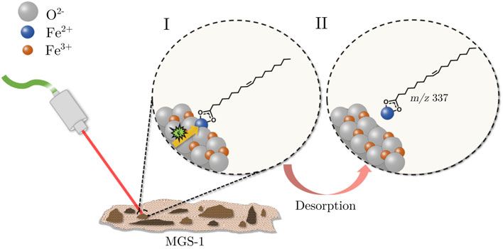
International Journal of Astrobiology                                                                                                                                       235

               Due to the samples’ mineral nature, GALDI-MS is a useful                               then shaken it in sterile water. The suspension was inoculated
           technique in identifying low molecular weight organic com-                                 in 70 ml autoclaved Czapek broth liquid in Erlenmeyer flasks
           pounds. To the best of our knowledge, this technique was first                             and allowed to grow for 7 days on a rotary shaker at 120 rpm
           used to investigate microbes’ interactions with heterogeneous                              and 25°C.
           minerals by Scott et al. (2006) and was proposed to identify
           mineral-associated by Yan et al. (2007). Later, the same research
                                                                                                      Microbial lipid extraction process
           group reported a detailed investigation about glycine identifica-
           tion in natural jarosites and its implications for the search for                          The total lipids of E. oligosperma were obtained by washing and
           life on Mars (Kotler et al., 2008) and how mineral thenardite                              filtering the biomass produced following the methodology
           improves detection and identification of glycine (Richardson                               described by Modenez et al. (2018). An amount of 20 g of freeze-
           et al., 2008) and aromatic amino acids (Richardson et al., 2009)                           dried yeast biomass was transferred to an Erlenmeyer flask, and
           by GALDI-MS. More recently, the same approach has been                                     the extraction process was carried out through the addition of
           applied by Li et al. (2015) to investigate the detection of organics                       AcOEt and shaking under an ultrasonic bath for 1 h. The extrac-
           trace in Mars-simulated samples containing perchlorate. Wörmer                             tion protocol was repeated three times. The mixture was filtered in
           et al. (2014) reported the only previous study focused on lipid                            a Buchner funnel and dried over anhydrous magnesium sulphate
           analysis using a GALDI-MS-based approach. This study shows                                 (MgSO4) and under vacuum to remove residual solvents. The
           the feasibility of detecting such molecules directly from marine                           extract was eluted with a mixture of hexane and AcOEt, 8 : 2
           sediment samples without any preparation. GALDI-MS is inter-                               (v : v), in a chromatography column packed with silica to remove
           esting because it does not require sample preparation. Unlike                              polar substances. The apolar compounds interact with the silica
           other techniques for desorption/ionization, GALDI makes use                                weakly than the polar ones, and then they come out of the column
           of the ability of minerals to perform desorption/ionization assist-                        first. Only the portion containing apolar or much less polar mole-
           ance. Thus, it is expected that the presence of Na-sulphate and                            cules, such as fatty acids, was collected, having obtained 0.115 g of
           other alkaline minerals on Mars, here represented by the                                   yellowish oil. Polar compounds, such as phospholipids, sugars
           MGS-1, and in other bodies of the Solar System, leads to the for-                          from the culture medium, secondary polar metabolites and
           mation of cation-attached organic ions like [M + Na]+, where M is                          amino acids were removed with this procedure.
           the molecular mass of analytes (Yan et al., 2007; Kotler et al.,
           2008; Richardson et al., 2009). The occurrence of alkali metal
                                                                                                      Derivatization and GC-MS analysis of lipid extract
           adducts in mass spectra obtained using condensed phases ioniza-
           tion techniques such as matrix-assisted laser desorption/ioniza-                           The derivatization reaction was performed in an ultrasonic water
           tion (MALDI) and GALDI is well documented in the literature                                bath (60–65°C) for 25 min by adding 5.0 ml of HMDS in 10 mg
           (Fuji, 2000; Ahn et al., 2016). Therefore, it may be useful in the                         of the lipid extract. The reaction content was then diluted to
           identification of organic matter in planetary geological                                   0.1 mg ml−1 in AcOEt. The GC-MS analyses were performed
           investigations.                                                                            using a QP-2010-plus Shimadzu system. The GC oven was fitted
                                                                                                      with a Restek Rxi-5ms fused silica column (J&W Scientific,
                                                                                                      10 m × 0.10 mm × 0.10 μm), and helium was used as a carrier
           Materials and methods                                                                      gas at a linear velocity of 45 cm s−1. The injector temperature
           Chemicals                                                                                  was set at 250°C. The column was kept initially at 60°C for
                                                                                                      1 min and then raised at a rate of 20°C min−1 to 300°C and main-
           Oleic acid (C18:1) and linoleic acid (C18:2), both of analytical                           tained at this temperature for 2 min. The ion source temperature
           grade, hexamethyldisilazane (HMDS/GC-MS grade), iron oxide                                 was set at 250°C and the interface at 270°C. The ions of m/z
           (Fe2O3) and silica gel (63–200 μm) were purchased from                                     between 40 and 500 were monitored from 1 to 15 min with 1 μl
           Sigma-Aldrich (Saint Louis, MO). Acetonitrile (ACN), hexane                                of injection volume in splitless mode. Identification of fatty acid
           and ethyl acetate (AcOEt), all of HPLC grade, were purchased                               trimethylsilyl ester was achieved by comparing the mass spectra
           from Panreac (Castellar del Vallès, Barcelona). The Mars Global                            with the NIST database library and mechanism fragmentation.
           Simulant (MGS-1) regolith was obtained from Exolith
           Laboratory at the University of Central Florida (Cannon et al.,
           2019).                                                                                     GALDI-time-of-flight (TOF) sample preparation
                                                                                                      Solutions of oleic and linoleic acids, and lipid extract isolated from
                                                                                                      E. oligosperma, were prepared in H2O : ACN 1 : 4 (v : v). The oleic
           Fungal strain and culture conditions
                                                                                                      acid solutions were prepared at concentrations of 0.002, 0.2, 200,
           The black yeast strain was surprisingly found and isolated from an                         and the linoleic acid and the lipid extract at a concentration of
           old HCl aqueous solution with pH 1.5 and was later identified as                           0.2 ppm. The volume of 1.0 ml of these solutions was transferred
           Exophiala oligosperma (Santos and Rodrigues-Filho, 2019). This                             to microtubes containing 0.04 g of MGS-1 (or Fe2O3) and left in
           strain is deposited in the culture collection of Micromolecular                            an ultrasonic bath for 5 min. The biological sample was prepared
           Biochemistry of Microorganisms Laboratory (LaBioMMi) of the                                by adding 0.008 g of lyophilized yeast in 0.08 g of MGS-1. This
           Department of Chemistry at the Federal University of São                                   mixture was suspended in 1.0 ml of H2O and homogenized in a
           Carlos (Brazil) under registration code LaBioMMi-1324. The                                 vortex at the frequency of 70 Hz. The methodology was adopted
           working Petri dishes were prepared using Czapek Agar medium,                               to promote a homogeneous mixture of the biomolecules with
           composed of glucose (26.7 g l−1), NaNO3 (3.0 g l−1), K2HPO4                                the matrix. A volume of 1 μl of each prepared suspension was
           (1.0 g l−1), MgSO4⋅7H2O (0.5 g l−1), KCl (0.5 g l−1), FeSO4 (0.01                          transferred to different sample holder spots using a micropipette
           g l−1) and Agar (30.0 g l−1). A cell suspension of E. oligosperma                          and dried for 15 min at room temperature before being intro-
           was prepared by collecting mycelium from the Petri dishes and                              duced and pumped into the spectrometer vacuum chamber.

Downloaded from https://www.cambridge.org/core. IP address: 46.4.80.155, on 12 Dec 2021 at 21:45:09, subject to the Cambridge Core terms of use, available at https://www.cambridge.org/core/terms.
https://doi.org/10.1017/S1473550421000100
236 Alef dos Santos et al.
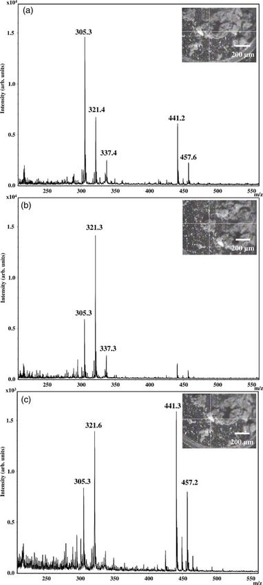
Fig. 1. GALDI-MS spectrum of the MGS-1 samples containing (a) intact cells of the black fungi E. oligosperma and (b) its corresponding lipid extract.
diameter spot was used to induce the ionization/desorption pro-
cesses. The positive ion mass spectra were acquired after TOF sep-
aration in the reflectron mode with an acceleration voltage of 19
kV. The ions were detected by microchannel plates. All spectra
were collected from an average of 100 laser shots. The mass cali-
bration was carried out using a standard mixture for low masses.
An MTP 384 polished steel target and matrices of mineral origin
(MGS-1 and Fe2O3) were used in the current study. The working
pressure of the spectrometer was around 10−7 mbar.
Results and discussion
GALDI-MS measurements in the 200–750 m/z range of samples
containing E. oligosperma intact cells and their lipid extract dis-
persed in MGS-1 are shown in Fig. 1(a) and (b), respectively.
Despite the MGS-matrix’s roughness, it was possible to obtain
spectra with sufficient mass resolution to identify the target com-
pounds. The GALDI-MS spectrum of the lipid extract was
obtained to support the assignments of the ions observed in
Fig. 1(a) and to provide information about the lipid pattern itself.
The lipid extract’s GC-MS analysis, whose details are given in the
Supplementary material, supports the present investigation.
Besides, the spectra were interpreted considering the possibility
of cation-attached organic ion formation due to the matrix min-
eral nature (Yan et al., 2007; Richardson et al., 2009). No attempt
was made to quantify relationships between the various ions
observed in the current study since they are dependent on the ion-
ization efficiencies of the elements and compounds and their
interactions with the matrix minerals (Yan et al., 2007; Kotler
et al., 2008). For instance, the difference in ion abundances
observed in Fig. 1(a) and (b) can be partially attributed to the
presence of several polar molecules in the former spectrum.
Fig. 2. (a) Expanded mass spectrum around ion m/z 617 region and (b) simulation of Since the GALDI-MS ionization processes occur by the acid–
the isotopic pattern of the DAG (C18:1/C16:0) sodium adduct by Compass
IsotopePattern® software.
base reaction in the gas phase induced by the laser, the polar
molecules will be more promptly ionized due to the presence of
heteroatoms, suppressing the ionization of apolar molecules,
such as fatty acids. In this sense, the remotion of the lipid extract’s
GALDI-TOF instrumentation and parameters
polar substances facilitates identifying the free fatty acids
The measurements were performed using an Autoflex Speed™ adsorbed on the matrix (Leopold et al., 2018). This procedure
Mass Spectrometer (Bruker Daltonics, Bremen, Germany). enables us to identify the fatty acids’ biosignatures that can be pre-
A 355 nm Nd:YAG laser source operating at a frequency of sent in the spectrum of Fig. 1(a). The lipid pattern study is also
10 Hz and 70% of the nominal power focused on a ≈5 μm essential, for instance, if we assume that free fatty acids are present
Downloaded from https://www.cambridge.org/core. IP address: 46.4.80.155, on 12 Dec 2021 at 21:45:09, subject to the Cambridge Core terms of use, available at https://www.cambridge.org/core/terms.
https://doi.org/10.1017/S1473550421000100International Journal of Astrobiology 237
Fig. 3. GALDI-MS spectrum of (a) oleic acid (C18:1) and (b) linoleic acid (C18:2), both at a concentration of 0.2 ppm, dispersed in the MGS-1 matrix.
desorption processes’ assistance is strongly related to the mineral
species, the observed pattern is also associated with the interaction
site’s predominant mineral. In the current study, no further study
about the ionization pattern’s dependence with the heterogeneity
of the matrix for the samples containing the intact cells and the
lipid extract was carried out. However, results showing this
dependence for a fatty acid sample are discussed below.
In Fig. 1(a), we attribute the occurrence of the base peak at m/z
617.6 to the presence of diacylglycerol (DAG). This molecule was
already reported in previous studies of yeast by using MALDI-MS
(Stübiger et al., 2016). From these studies, it is known that most
yeast DAGs contains oleic acid (C18:1) and palmitoleic acid
(C16:0) (Ganesan et al., 2016). Since the matrix is composed of
several minerals largely associated with Na+ and K+ ions, e.g. pyr-
oxene (Cannon et al., 2019), it is reasonable to assume that the
ion at m/z 617.6 probably correspond to the sodium adduct of
the C18:1/C16:0 DAG (theoretical molecular formula:
Fig. 4. GALDI-MS spectrum of oleic acid (0.2 ppm) in the Fe2O3 matrix. C37H70O5, calc. [M + Na]+ at m/z 617.5). This hypothesis is sup-
ported by the occurrence of such fatty acids in the sample ana-
lysed by GC-MS and also by the presence of ions at m/z 618.5,
619.5 and 620.6 corresponding to the carbon isotopic pattern of
on Mars rather than intact phospholipids. Finally, the laser beam the DAG (C18:1/C16:0) sodium adduct, as shown in Fig. 2(a).
spot size is smaller compared with the MGS-1 nominal mean This experimental data are also in excellent agreement with the
grain size of 122 μm (Cannon et al., 2019). Since the ionization/ simulated isotopic pattern shown in Fig. 2(b).
Fig. 5. Possible pathway to the formation of ion at m/z
337.4. Secondary electrons from the highly ionized
laser plume promotes the reduction of Fe3+ in Fe2+
and the production of [M − H + Fe2+]+ ion.
Downloaded from https://www.cambridge.org/core. IP address: 46.4.80.155, on 12 Dec 2021 at 21:45:09, subject to the Cambridge Core terms of use, available at https://www.cambridge.org/core/terms.
https://doi.org/10.1017/S1473550421000100238 Alef dos Santos et al.
200 ppm, along with the mass spectrum from pure MGS-1 matrix,
are shown in Fig. S4 available in the Supplementary material. The
observed variation in ion abundance between oleic acid concen-
trations could probably be related to the nature of the interaction
between the analyte and the matrix, as already mentioned. Even at
concentrations of 0.002 ppm, the peaks at m/z 305.2 and 321.2 are
still observed above the matrix background. These ions corres-
pond to the C18:1 sodium ([M + Na]+) and potassium ([M +
K]+) adducts, respectively. The designation of these ions as alka-
line adducts of C18:1 became evident when replacing it with
C18:2, where the decreasing displacement of two mass units in
its mass spectrum is observed, as shown in Fig. 3(b). Besides,
the mass separation of 16 u between the labelled peaks is a typical
signature of alkaline adducts in the mass spectrum corresponding
to the difference between the potassium (39 u) and sodium (23 u)
molar masses. Peaks with such mass differences were also
observed by Kotler et al. (2008). However, those authors argue
that this is most likely related to the replacement of O by S in
the cluster ions observed by them. In addition to the fact that
our matrix is less rich in S, our spectra’ isotopic pattern is not
consistent with such substitution. It is also chemically improbable
for the samples under study. Therefore, our results indicate that
the observed pattern is associated with sodium and potassium
cationization reactions. In this sense, the ion at m/z 279.3 can pre-
sumably be associated with the sodium adduct of C16:0.
An interesting situation is related to the ions at m/z 337.4 and
335.2 in Fig. 3(a) and (b), respectively. These ions are most likely
associated with iron adduct formation, corresponding to the add-
ition of 56 u from iron-cation to the mass of C18:1 (MW = 282 u)
and C18:2 (MW = 280 u), respectively. A high concentration
of iron oxides present in the minerals hematite (Fe3+), jarosite
(Fe2+) and magnetite (Fe3+ and Fe2+) contained in MGS-1 may
be responsible by the presence of these adducts in our mass spec-
tra. However, the complexation of Fe3+ with the carboxyl group of
fatty acids would result in a double charged state species, leading
to an ion at m/z 168.5, which was not observed in the current
study. To test the hypothesis to the formation of iron complexes,
we have used a matrix with only Fe3+. This oxidation state is the
most abundant on the Martian surface (Price et al., 2018) and,
therefore, of great interest. The mass spectrum of oleic acid dis-
persed on the Fe2O3 matrix is shown in Fig. 4 and contains the
ion m/z 337.4 as base peak. The presence of ions at m/z 335.3,
337.4 and 338.2 is in some agreement with the expected Fe iso-
topic pattern with the relative abundances of the ions 335.3 and
338.2 higher compared with the simulated pattern shown in
Fig. S3 (Supplementary material). Mathematically, the number
337.4 corresponds to the combination of the ionized carboxylic
fatty acid (281 u, [M − H]) with the most stable isotope of iron
(56Fe), resulting in a positively monocharged ion, detected in
our spectra as [M − H + Fe2+]+. The formation of this iron-adduct
Fig. 6. GALDI-MS spectra of pure oleic acid at concentrations of 0.2 ppm dispersed in
is only possible if we consider a reduction reaction Fe3+ to Fe2+
the MGS-1 matrix obtained from different positions on the same spot target.
probably induced by secondary electrons in the plume generated
by the laser pulse (Knochenmuss and Zenobi, 2003). In Fig. 5,
The ions in the range below m/z 500 shown in Fig. 1(a) are we present the possible pathway that leads to the formation of
probably associated with the fatty acid adducts since they are the [M − H + Fe2+]+ ion. A similar situation can hypothetically
also present in the mass spectrum of the lipid extract. To accur- occur on Martian soil due to secondary electrons’ presence
ately identify these ion adducts, we performed measurements from ionizing radiation to which the planet’s surface is exposed.
using solutions containing only oleic acid (C18:1) and linoleic Therefore, our results indicate the possibility of detecting iron
acid (C18:2) dispersed in the MGS-1 matrix. Both of them were adduct ions in lipid samples studied by GALDI-MS. This is in dis-
identified in the GC-MS. The results for the concentration of agreement with the results reported by Yan et al. (2007) that stud-
0.2 ppm are shown in Fig. 3(a) and (b), respectively. The spectra ied samples of amino acids in matrices of Fe2O3 by GALDI-MS
obtained for C18:1 fatty acid at concentrations of 0.002 and observed no iron-attached ions. They suggested that the lack of
Downloaded from https://www.cambridge.org/core. IP address: 46.4.80.155, on 12 Dec 2021 at 21:45:09, subject to the Cambridge Core terms of use, available at https://www.cambridge.org/core/terms.
https://doi.org/10.1017/S1473550421000100International Journal of Astrobiology 239
such ions is due to the fragmentation of the amino acids that most Cannon KM, Britt DT, Smith TM, Fritsche RF and Batcheldor D (2019)
likely occur due to the abundance of excited state Fe ions in the Mars Global Simulant MGS-1: a Rocknest-based open standard for basaltic
gas phase. However, the reason for the discrepancy observed Martian regolith simulants. Icarus 317, 470–478.
Deamer DW and Pashley RM (1989) Amphiphilic components of the
between our results and those of Yan et al. (2007) is not clear
Murchison carbonaceous chondrite: surface properties and membrane for-
to us. We believe that the detection of iron-attached ions may
mation. Origins of Life and Evolution of Biospheres 19, 21–38.
be linked to the fact that in the current study, the ratio between Fuji T (2000) Alkali metal ion/molecule association reactions and their
the amount of analyte and matrix is ∼1000 times less than that applications to mass spectrometry. Mass Spectrometry Reviews 18, 111–138.
used by those authors. Further studies are intended to be per- Ganesan S, Shabits BN and Zaremberg V (2016) Tracking diacylglycerol and
formed by our research group to clarify this issue. phosphatidic acid pools in budding yeast. Lipid Insights 8, 75–85.
Finally, further concern about the complexity of the observed Georgiou CD and Deamer DW (2014) Lipids as universal biomarkers of
spectra must be addressed. Figure 6 shows the mass spectra of extraterrestrial life. Astrobiology 14, 541–549.
pure oleic acid dispersed in MGS-1 obtained from different posi- Knochenmuss R and Zenobi R (2003) MALDI ionization: the role of
tions on the same spot target. The ions at m/z 441.3 and 457.2 in-plume processes. Chemical Review 103, 441–452.
Kotler JM, Hinman NW, Yan B, Stoner DL and Scott JR (2008) Glycine
could not be unambiguously identified. It is observed that the
identification in natural jarosites using laser desorption Fourier transform
relative abundance of the ions in the mass spectrum is strongly
mass spectrometry: implications for the search for life on Mars.
dependent on the heterogeneity of the matrix. The concentrations Astrobiology 8, 253–266.
of different minerals or the mineral phases throughout the matrix Lai JC-Y, Pearce BKD, Pudritz RE and Lee D (2019) Meteoritic abundances
significantly interfere with the observed signal’s intensity. As of fatty acids and potential reaction pathways in planetesimals. Icarus 319,
pointed out by Yan et al. (2007), this results from how different 685–700.
minerals assist the ionization/desorption process. In this context, Leopold J, Popkova Y, Engel KM and Schiller J (2018) Recent developments
the results of the current study differ from those obtained by trad- of useful MALDI matrices for the mass spectrometric characterization of
itional GALDI/MALDI-MS mainly due to the matrix’s complex- lipids. Biomolecules 8, 173.
ity. Despite this, our study shows that quite useful results in the Li X, Danell RM, Brinckerhoff WB, Pinnick VT, Van Amerom F, Arevalo
RD, Getty SA, Mahaffy PR, Steininger H and Goesmann F (2015)
identification of biomolecules can be obtained.
Detection of trace organics in Mars analog samples containing
perchlorate by laser desorption/ionization mass spectrometry. Astrobiology
Conclusion 15, 104–110.
Modenez IA, Sastre DE, Moares FC and Marques Netto CGC (2018)
We performed an analysis of microbial lipids deposited on MGS-1 Influence of glutaraldehyde cross-linking modes on the recyclability of
regolith using the GALDI-MS technique. The results obtained are immobilized lipase b from Candida Antarctica for transesterification of
encouraging and indicate that lipids from intact microbial cells of soy bean oil. Molecules 23, 2230.
a black yeast strain can be detected in Martian soil samples. Onofri S, Barreca D, Selbmann L, Isola D, Rabbow E, Horneck G, de Vera
These lipid molecules are predominantly associated with the occur- JPP, Hatton J and Zucconi L (2008) Resistance of Antarctic black fungi
rence of adducts in the GALDI-MS spectra. Sodium adducts of and cryptoendolithic communities to simulated space and Martian condi-
tions. Studies in Mycology 61, 99–109.
DAG and fatty acid adducts of sodium, potassium and iron are
Onofri S, D La Torre R, Vera J-P, Ott S, Zucconi L, Selbmann L, Scalzi G,
observed in the mass spectra. Different adducts arise due to the
Venkateswaran KJ, Rabbow E, Inigo FJS and Horneck G (2012) Survival
geological complexity of the soil, here represented by the MGS-1 of rock-colonizing organisms after 1.5 years in outer space. Astrobiology 12,
matrix. The detection of adducts instead of isolated molecules in 508–516.
the mass spectra can better identify lipid compounds or other Onofri S, De Vera JP, Zucconi L, Selbmann L, Scalzi G, Venkateswaran KJ,
organic molecules in Martian soil. Therefore, the regolith’s ability Rabbow E, De La Torre R and Horneck G (2015) Survival of Antarctic
to assist the ionization/desorption process makes the GALDI tech- cryptoendolithic fungi in simulated Martian conditions on board the inter-
nique very promising in the search for mineral-associated lipid, an national space station. Astrobiology 15, 1052–1059.
important group of organic molecules intrinsically linked to bio- Price A, Pearson VK, Schwenzer SP, Miot J and Olsson-Francis K (2018)
chemical processes. We hope that our investigation can contribute Nitrate-dependent iron oxidation: a potential Mars metabolism. Frontiers
in Microbiology 9, 513.
to efforts in the search for extraterrestrial life.
Richardson DC, Hinman NW, McJunkin TR, Kotler MJ and Scott JR
(2008) Exploring biosignatures associated with thenardite by geomatrix-
Supplementary material. The supplementary material for this article can assisted laser desorption/ionization Fourier transform ion cyclotron reson-
be found at https://doi.org/10.1017/S1473550421000100. ance mass spectrometry (GALDI-FTICR-MS). Geomicrobiology Journal 25,
432–440.
Acknowledgements. This research was supported by the Brazilian agencies: Richardson C, Hinman NW and Scott JR (2009) Effect of thenardite on the
Conselho Nacional de Desenvolvimento Científico e Tecnológico (CNPq) – direct detection of aromatic amino acids: implications for the search for life
Grant no. 311152/2016-3 and 304867/2017-9, Coordenação de in the Solar System. International Journal of Astrobiology 8, 291–300.
Aperfeiçoamento de Pessoal de Nível Superior (CAPES) – Finance Code 001 Röling WFM, Aerts JW, Patty CHL, Ten Kate IL, Ehrenfreund P and
and Fundação de Amparo à Pesquisa do Estado de São Paulo (FAPESP) – Direito SOL (2015) The significance of microbe-mineral-biomarker
Grant no. 2010/52312-8. A.S. is grateful to CAPES for his Ph.D. scholarship interactions in the detection of life on Mars and beyond. Astrobiology 15,
(Grant no. 88887.598052/2021-00). The authors also would like to thank 492–507.
one of the reviewers for kindly bringing significant references to our attention. Santos A and Rodrigues-Filho E (2019) New Δ8,9-pregnene steroids isolated
from the extremophile fungus Exophiala oligosperma. Natural Product
References Research 1, 1–4.
Scalzi G, Selbmann L, Zucconi L, Rabbow E, Horneck G, Albertano P and
Ahn SH, Park KM, Moon JH, Lee SH and Kim MS (2016) Quantification of Onofri S (2012) Life experiment: isolation of cryptoendolithic organisms
carbohydrates and related materials using sodium ion adducts produced by from Antarctic colonized sandstone exposed to space and simulated Mars
matrix-assisted laser desorption ionization. Journal of the American Society conditions on the international space station. Origins of Life and
for Mass Spectrometry 27, 1887–1890. Evolution of Biospheres 42, 253–262.
Downloaded from https://www.cambridge.org/core. IP address: 46.4.80.155, on 12 Dec 2021 at 21:45:09, subject to the Cambridge Core terms of use, available at https://www.cambridge.org/core/terms.
https://doi.org/10.1017/S1473550421000100240 Alef dos Santos et al.
Scott JR, Yan B and Stoner DL (2006) Spatially-correlated mass spectromet- F, Monteiro D, Trautner R, Voland C, Rebeyre P, Goulty D, Didot F,
ric analysis of microbe–mineral interactions. Journal of Microbiological Durrant S, Zekri E, Koschny D, Toni A, Visentin G, Zwick M, van
Methods 26, 381–384. Winnendael M, Azkarate M, Carreau C and The ExoMars Project
Stübiger G, Wuczkowski M, Mancera L, Lopandic K, Sterflinger K and Team (2017) Habitability on early Mars and the search for biosignatures
Belgacem O (2016) Characterization of yeasts and filamentous fungi with the ExoMars Rover. Astrobiology 17, 471–510.
using MALDI lipid phenotyping. Journal of Microbiological Methods 130, Westall F, Foucher F, Bost N, Bertrand M, Loizeau D, Vago JL, Kminek G,
27–37. Gaboyer F, Campbell KA, Bréhéret JG, Gautret P and Cockell CS (2015)
Tan J, Lewis JMT and Sephton MA (2018) The fate of lipid biosignatures in a Biosignatures on Mars: what, where, and how? Implications for the search
Mars-analogue sulfur stream. Scientific Reports 8, 7586. for Martian life. Astrobiology 15, 998–1029.
Vago JL, Westall F, Coates AJ, Jaumann R, Korablev O, Ciarletti V, Wörmer L, Elvert M, Fuchser J, Lipp JS, Buttigieg PL, Zabel M and
Mitrofanov I, Josset JL, De Sanctis MC, Bibring JP, Rull F, Goesmann Hinrichs K-U (2014) Ultra-high-resolution paleoenvironmental records
F, Steininger H, Goetz W, Brinckerhoff W, Szopa C, Raulin F, Westall via direct laser-based analysis of lipid biomarkers in sediment core samples.
F, Edwards HGM, Whyte LG, Fairén AG, Bibring JP, Bridges J, PNAS 111, 15669–15674.
Hauber E, Ori GG, Werner S, Loizeau D, Kuzmin RO, Williams RME, Yan B, Stoner DL, Kotler JM, Hinman NW and Scott JR (2007) Detection of
Flahaut J, Forget F, Vago JL, Rodionov D, Korablev O, Svedhem H, biosignatures by geomatrix-assisted laser desorption/ionization (GALDI)
Sefton-Nash E, Kminek G, Lorenzoni L, Joudrier L, Mikhailov V, mass spectrometry. Geomicrobiology Journal 24, 379–385.
Zashchirinskiy A, Alexashkin S, Calantropio F, Merlo A, Poulakis P, Zakharova K, Marzban G, De Vera JP, Lorek A and Sterflinger K (2014)
Witasse O, Bayle O, Bayón S, Meierhenrich U, Carter J, García-Ruiz Protein patterns of black fungi under simulated Mars-like conditions.
JM, Baglioni P, Haldemann A, Ball AJ, Debus A, Lindner R, Haessig Scientific Reports 4, 5114.
Downloaded from https://www.cambridge.org/core. IP address: 46.4.80.155, on 12 Dec 2021 at 21:45:09, subject to the Cambridge Core terms of use, available at https://www.cambridge.org/core/terms.
https://doi.org/10.1017/S1473550421000100You can also read