Investigation of the Toxic Effects of Rhododendron Honey on Mouse Cardiac Muscle Tissue Lipids at Molecular Level 1 2
←
→
Page content transcription
If your browser does not render page correctly, please read the page content below
Kafkas Univ Vet Fak Derg Kafkas Universitesi Veteriner Fakultesi Dergisi
ISSN: 1300-6045 e-ISSN: 1309-2251
26 (2): 287-294, 2020 Journal Home-Page: http://vetdergikafkas.org Research Article
DOI: 10.9775/kvfd.2019.22996 Online Submission: http://submit.vetdergikafkas.org
Investigation of the Toxic Effects of Rhododendron Honey on Mouse
Cardiac Muscle Tissue Lipids at Molecular Level [1] [2]
Gulgun CAKMAK-ARSLAN 1,a Selin EMIR 1,b Pinar GOC-RASGELE 2,c Meral KEKECOGLU 1,d
This work was supported by the Duzce University-Research Fund, BAP (2016.05.01.503)
[1]
A part of this study was presented at the 3rd International Congress on Applied Biological Sciences in Afyon/Turkey on 09-
[2]
12 July 2017
1
Department of Biology, Faculty of Arts and Sciences, Duzce University, TR-81620 Duzce - TURKEY
2
Department of Biosystems Engineering, Faculty of Agriculture and Natural Sciences, Duzce University, TR-81620
Duzce - TURKEY
a
ORCID: 0000-0003-4326-1780; b ORCID: 0000-0002-5819-6734; c ORCID: 0000-0002-7558-3138; d ORCID: 0000-0002-2564-8343
Article ID: KVFD-2019-22996 Received: 11.07.2019 Accepted: 26.11.2019 Published Online: 27.11.2019
How to Cite This Article
Cakmak-Arslan G, Emir S, Goc-Rasgele P, Kekecoglu M: Investigation of the toxic effects of rhododendron honey on mouse cardiac muscle tissue
lipids at molecular level. Kafkas Univ Vet Fak Derg, 26 (2): 287-294, 2020. DOI: 10.9775/kvfd.2019.22996
Abstract
The purpose of this study is to investigate the effects of different concentrations of Rhododendron honey (RH) on mouse cardiac muscle lipids
by Attenuated Total Reflection Fourier Transform Infrared (ATR-FTIR) spectroscopy at molecular level. For this purpose, a total of eighteen
male Mus musculus mice were divided into three groups of six animals each, one being the control group and the others being the 25 and 50
mg/kg of RH-treated groups. RH was given via gavage and the cardiac muscles of these mice were investigated 24 h after the administration.
The results revealed that 25 mg/kg of RH did not cause any significant effect except lipid peroxidation. However, 50 mg/kg RH caused
increases in the amounts of saturated and unsaturated lipids, in the ratios of lipid/protein and CH2/CH3 and a decrease in the CH3/lipid which
all indicate a change in the lipid metabolism of the tissue. Moreover, the treatment with 50 mg/kg of RH caused lipid peroxidation, a decrease
in lipid order and an increase in membrane dynamic. These results revealed that RH causes significant toxic effects on cardiac muscle tissue
lipids and these effects are dose-dependent.
Keywords: Rhododendron honey, Mad honey, ATR-FTIR spectroscopy, Heart, Cardiac muscle, Lipid
Ormangülü Balı’nın Fare Kalp Kas Dokusu Lipitleri Üzerindeki Toksik
Etkilerinin Moleküler Düzeyde İncelenmesi
Öz
Bu çalışmanın amacı, farklı konsantrasyonlardaki ormangülü balının (OB) fare kalp kası lipitleri üzerindeki etkilerinin Azaltılmış Toplam
Yansıma-Fourier Dönüşüm Kızılötesi (ATR-FTIR) spektroskopisi ile moleküler düzeyde incelenmesidir. Bu amaç doğrultusunda, toplam on
sekiz adet Mus musculus erkek fare, her biri altı hayvan içeren, birisi kontrol ve diğerleri 25 ve 50 mg/kg OB uygulanmış gruplar olmak üzere
üç gruba ayrıldı. OB gavaj yoluyla verildi ve bu farelerin kalp kasları uygulamadan 24 saat sonra incelendi. Sonuçlar 25 mg/kg OB’nin kalp
kası lipitleri üzerinde lipit peroksidasyonu dışında herhangi anlamlı bir değişikliğe sebep olmadığını ortaya çıkarmıştır. Ancak 50 mg/kg
OB doymuş ve doymamış lipitlerin miktarlarında, lipit/protein oranında ve CH2/CH3 oranında artışa ve CH3/lipit oranında azalmaya sebep
olmuştur. Tüm bunlar dokunun lipit metabolizmasında bir değişiklik olduğunu göstermektedir. Ayrıca 50 mg/kg OB lipit peroksidasyonuna,
lipit düzeninde bir azalmaya ve membran düzeninde bir artışa sebep olmuştur. Bu sonuçlar, OB’nin kalp kası lipitleri üzerinde önemli toksik
etkiler meydana getirdiğini ve bu etkilerin doza bağlı olduğunu ortaya çıkarmıştır.
Anahtar sözcükler: Ormangülü balı, Deli bal, ATR-FTIR spektroskopisi, Kalp, Kalp kası, Lipit
INTRODUCTION extensively in the Black Sea region of Turkey show also
distribution in China, Tibet, Nepal, Tropical Asia, Europe
Rhododendron honey (RH), also known as mad honey, is and North America [1]. RH causes poisoning in humans and
obtained from plants belonging to the genus Rhododendron animals due to the toxic compound grayanotoxin (GTX)
in the Ericaceae family. The rhododendrons growing found in some species of the Rhodendron genus. However,
İletişim (Correspondence)
+90 380 5412404/3991 Fax: +90 380 5412403
gulguncakmak@duzce.edu.tr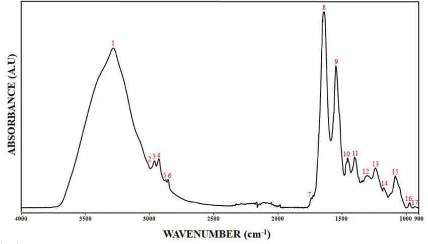
288
Investigation of the Toxic Effects of ...

in different regions of the world, this honey is widely used      disorders; however, there is no study reporting the effects
in the treatment of various disorders such as hypertension,       of RH on the structure and function of lipids in the cardiac
gastrointestinal complaints, sexual dysfunction toothache,        muscle. The ability of a tissue to function properly is
colds, etc.[2].                                                   related to its structure and ATR-FTIR spectroscopy gives
                                                                  information about the structure of the tissue at molecular
The effect of GTX is predominantly on the voltage-dependent       level. In this study, we aimed to reveal the molecular effects
sodium channel. It binds to the sodium channel in the             of two different doses of RH on the composition, structure
open position and the sodium channel becomes incapable            and function of the cardiac muscle lipids by using ATR-FTIR
of closing. As a result, action potential enters a prolonged      spectroscopy. To the best of our knowledge, this is the first
period of hyperpolarization process [3,4]. Through this action    study to investigate the effects of RH on cardiac muscle
mechanism, GTX affects the heart directly and causes a            lipids at the molecular level.
wide range of systemic effects including hypotension,
arrhythmia, nausea and a reduction in spontaneous motion
by affecting the central nervous system [1]. Most of the
                                                                  MATERIAL and METHODS
published studies on RH have presented case reports either        The RH that we used in our study was obtained from Düzce
on patient complaints after eating the honey or cases of          beekeepers. As a result of the palynological analysis, the
patients treated at emergency services [3]. In clinical studies   honey sample was confirmed to dominantly (≥45%) consist
examining the effects of RH on the cardiac system, it has         of the Rhododendron ponticum pollen [12].
been reported that hypotension and bradycardia were
seen in more than 90% of the patients [5].                        The amount of GTX-I and GTX-III in the RH used in this study
                                                                  was detected by liquid chromatography-mass/mass spectro-
In the literature, experimental studies on the effects            metry (LC-MS/MS) using the method developed by Kaplan
of RH on the cardiac system are extremely limited in              et al.[13]. The limit of detection and the limit of quantification
number, compared to the clinical trials on the subject.           values of the validated analysis method were 0.0033 mg/
For example, Onat et al.[6] observed obesity-related hypo-        kg and 0.01 mg/kg, respectively. The GTX-I and GTX-III
tension, bradycardia and respiratory rate depression in           amounts of the samples were found to be 32 and 8 μg/g,
RH-treated mice. In another study, blood pressure and             respectively, which were higher than the average values
heart rate were measured in rats with experimental hyper-         reported previously [13] and in parallel with the amounts of
tension and it was reported that RH reduced the blood             toxic substances in RH used in other studies [14-16].
pressure and heart rate in the hypertensive rats [7]. Zushi
et al.[8] have reported that the effect of GTX on the neuro-      All experimental procedures were approved by the Abant
muscular junction was achieved by increased permeability          İzzet Baysal University Medical Faculty Experimental Animals
of the membrane to sodium. In a recent experimental               Ethics Committee (2015/42). 18 male Mus musculus mice
study investigating the dose-related cardiovascular effects       (20-25 g), 8-12 weeks old, were used. The animals were
of GTX-III, it was reported that it could be lethal at high       housed in a 12 h light - 12 h dark cycle at a constant
doses due to cardiac arrest [5].                                  room temperature (22±2°C) and fed with mouse food
                                                                  and water. The mice were divided into three groups: the
Fourier transform infrared (FTIR) spectroscopy is a high-tech     control group (n=6), the 25 mg/kg RH-treated group (n=6)
tool that gathers valuable information about biological           and the 50 mg/kg RH-treated group (n=6). The different
tissues and membranes by measuring the vibrations of              concentrations of RH were prepared by dissolving in
molecules [9]. By using the attenuated total reflection (ATR)     water and they were administered via gavage to the
unit in FTIR spectroscopy, it is possible to detect spectral      animals in 0.01 mL per gram body weight ratio. 24 h after
changes in biological tissues faster and more accurately,         administration, the animals were decapitated and their
regardless of sample thickness [10]. The ATR-FTIR spectro-        heart tissues were removed and stored at -80°C for latter
scopy is a unique technique that enables the detection of         ATR-FTIR spectroscopy experimentation.
absorption bands of lipids, proteins, carbohydrates and
nucleic acids in biological systems in a single spectrum          The mouse cardiac muscle spectra were collected with
and at the same time, monitoring these molecules without          a Spectrum Two FTIR spectrometer equipped with an
using any labeling technique [10]. The main advantage of          ATR accessory (Perkin-Elmer Ltd., UK). 0.5 × 0.5 × 0.1 cm
this technique is that the samples can be investigated            sized sections were cut from the myocardial layer of the
without any preparation processes by placing them directly        heart for the ATR-FTIR studies and placed directly on the
on the ATR crystals.                                              diamond/zinc-selenide crystals of the ATR unit. For each
                                                                  sample, spectra were obtained at 4 cm-1 resolution and
Recent studies have revealed the importance of intra-             100 force gauge with 64 scans between the 4000-900
cellular myocardial and pericardial lipid deposits, showing       cm-1 wavenumbers. The analyses of spectral bands were
that even the smallest changes in these fat deposits              performed using Perkin Elmer software programme.
cause significant changes in cardiac performance [11]. It is      Bandwidth and band wavenumber values were calculated
known that RH leads to cardiac side effects and functional        at 75% of the band height [9,10].
289
CAKMAK-ARSLAN, EMIR
GOC-RASGELE, KEKECOGLU
For quantitative comparison between control and treated groups. The P values less than 0.05 were considered
samples, the areas under the lipid bands and the area ratios statistically significant.
of some specific infrared bands were calculated. To examine
the level of the lipid peroxidation of the system, the area RESULTS
under the olefinic=CH band and the unsaturated/saturated
lipid ratio (the area of the olefinic=CH stretching band/the Fig. 1 shows the ATR-FTIR spectrum of a control mouse
sum of the areas of the saturated lipid bands), which are the cardiac muscle in the wavenumber range of 4000-900 cm-1.
parameters used as an index for the determination of lipid The main bands on the figure are numbered and their
peroxidation in FTIR studies, were used [9,17]. To find out the definitions are given in Table 1 according to the literature.
changes in the amount of saturated lipids, the areas under
The cardiac muscle spectrum shown in Fig. 1 can be
the CH3 antisymmetric (antisym) stretching, CH2 antisym
examined in three different regions: The large band located
stretching and CH2 symmetric (sym) stretching bands and
at 3700-3030 cm-1 (band No. 1) mainly receives signals
the ratio of CH2 sym/CH2 sym + CH2 antisym stretching
from the N-H groups of proteins with the little contribution
vibrations were evaluated [9,18,19]. To have information
from O-H stretching of polysaccharides, carbohydrates
about the amount of triglycerides and cholesterol in the and water [20]. The 3025-2800 cm-1 region (band No’s. 2-6)
system, the area under the C=O ester stretching band is called the C-H stretching region and generally provides
was analyzed [20]. To determine the changes in the chain information about lipids [18]. The 1800-800 cm-1 region
length of the membrane phospholipids, the CH2/CH3 ratio (band No’s. 7-17), which is called the fingerprint region,
(the area of the CH2 antisym stretching/the area of the CH3 receives signals mostly from proteins and nucleic acids
antisym stretching) [21] and in the methyl concentration, and in small quantities from lipids and carbohydrates [22]. In
CH3/lipid (the area of the CH3 antisym stretching band/ this study, since we aimed to collect information about the
the sum of the areas of saturated lipid bands) ratio [20] were effects of different concentrations of RH on cardiac muscle
calculated. To compare the relative changes in lipid and lipids, the detailed analyses were performed mainly in the
protein concentrations in the system, the area ratios of the region of 3025-2800 cm-1. Fig. 2 shows the average ATR-
sum of the saturated lipid bands (CH3 antisym, CH2 antisym FTIR spectra in the 3025-2800 cm-1 region of the cardiac
and CH2 sym stretching bands) and proteins (Amide II muscle of control and 25 and 50 mg/kg RH-treated mice.
band) were obtained [21]. To have information about
membrane order and membrane fluidity, the wavenumber To obtain information about the changes in the concentra-
and bandwidth of the CH2 antisym stretching band were tions of lipid molecules after RH treatment, analyses of
analyzed, respectively [9]. the areas under the lipid bands in the FTIR spectra were
performed [18]. The changes in the band area values of the
Power analysis was performed to estimate the test power major functional groups are given in Table 2. As seen in
considering the sample size of each experimental group Table 2, the area under the olefinic=CH stretching band
(n=6) for a power of 80% to achieve significant statistical (band No. 2) increased significantly in the 50 mg/kg RH-
differences at the 5% significance level and the calculated treated group indicating an increase in the amount of
effect size value was 1.55. Mann-Whitney U test, which is unsaturated lipids. As also seen in Fig. 2 and Table 2, the
a non-parametric test used to compare two independent region under the saturated lipid bands (band No’s. 3, 4,
groups that do not require large normally distributed 6) increased significantly in the 50 mg/kg RH group. This
samples, was performed to test the significance of the result suggested that the 50 mg/kg RH administration
differences between the control and RH-treated groups caused an increase in the amount of saturated lipid in
two by two; that is, between the control and 25 mg/kg RH- the cardiac muscle. The result derived from the analysis
treated groups and the control and 50 mg/kg RH-treated of the lipid bands in the C-H region was supported by the
Fig 1. ATR-FTIR spectrum of control mouse cardiac muscle in
the 4000-900 cm-1 wavenumber region290
Investigation of the Toxic Effects of ...
Table 1. General band assignment of ATR-FTIR spectrum of cardiac muscle tissue based on literature [18,20,22]
Band No Wavenumber (cm-1) Definition of the Assignment
Amide A: Mainly N-H stretching of hydrogen-bonded amide groups of proteins with the little contribution from
1 3283
O-H stretching of polysaccharides, carbohdrates and water
2 3011 Olefinic=CH stretching: Unsaturated lipids
3 2959 CH3 antisymmetric stretching: Mainly lipids with little contribution from proteins, carbohydrates and nucleic acids
4 2924 CH2 antisymmetric stretching: Mainly lipids with little contribution from proteins, carbohydrates and nucleic acids
5 2874 CH3 symmetric stretching: Mainly proteins with little contribution from lipids, carbohydrates and nucleic acids
6 2855 CH2 symmetric stretching: Mainly lipids with little contribution from proteins, carbohydrates and nucleic acids
7 1738 C=O (carbonyl) ester stretching: Ester functional groups in phospholipids, triglycerides and cholesterol esters
8 1641 Amide I: C=O stretching in proteins (80%)
9 1545 Amide II: Proteins (60% N-H bending, 40% C-N stretching)
10 1454 CH2 bending: Mainly lipids with little contribution from proteins
11 1396 COO- symmetric stretching: Fatty acids and amino acid side groups
12 1302 Amide III: Proteins (40%C-N stretching, 30% N-H bending, 20% C-C stretching)
13 1238 PO-2 antisymmetric stretching: Mainly nucleic acids with some contribution from phospholipids
14 1172 CO-O-C antisymmetric stretching: Phospholipids, cholesteryl ester and nucleic acids
PO-2 symmetric stretching: Nucleic acids and phospholipids;
15 1080
C-O stretching: glycogen, polysaccarides and glycolipids
16 972 C-N+-C stretching: Nucleic acids, ribose-phosphate main chain vibrations of RNA
17 931 z-type DNA
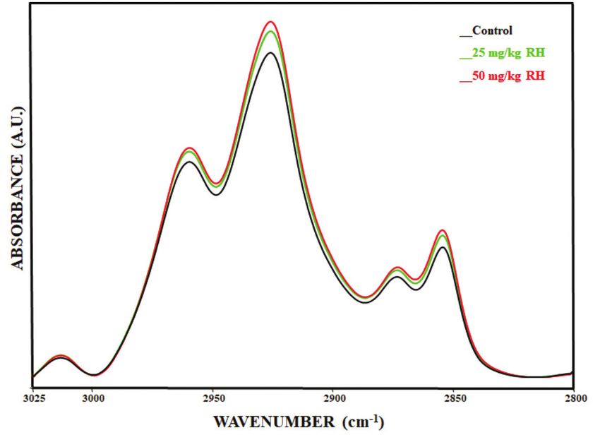
Fig 2. Average infrared spectra in the 3025-2800 cm-1 spectral
region of cardiac muscle of control, 25 mg/kg and 50 mg/kg
RH-treated mice (The spectra were normalized with regard to
the amide I band)
increase in the value of the C=O ester stretching band obtained from the analysis of the lipid band areas. The
located at 1738 cm-1 (band No. 7). As shown in Table 2, a unsaturated/saturated lipid ratio and the CH2/CH3 ratio
significant increase was observed in the area value of this increased significantly in 50 mg/kg RH-treated groups
band in the 50 mg/kg RH-treated group. compared to the control group. A significant decrease in
CH3/lipid ratio suggested a decrease in the amount of
In order to evaluate the effects of RH on the structure and methyl groups in the 50 mg/kg RH-treated group. The
composition of the cardiac muscle lipids, the area ratios of lipid/protein ratio increased significantly in the 50 mg/kg
some specific lipid functional groups were also evaluated [21]. RH-treated group.
The band area ratios calculated for this purpose are given
in Table 3. As shown in Table 3, the ratio of CH2 sym/CH2 sym The changes in the wavenumber and bandwidth values of
+ CH2 antisym stretching vibrations increased significantly the CH2 antisym stretching band are given in Table 3. As
in the 50 mg/kg RH-treated group. This increase confirmed seen from this table, the wavenumbers of the CH2 antisym
the increase in the amount of saturated lipids in the system stretching band shifted towards higher values in the 50291
CAKMAK-ARSLAN, EMIR
GOC-RASGELE, KEKECOGLU
Table 2. Changes in the band area values of the major lipid bands of the ATR-FTIR spectra of the cardiac muscle of control, 25 mg/kg and 50 mg/kg
RH-treated mice
Band No Functional Group Wavenumber (cm-1) Control 25 mg/kg RH P Valuea 50 mg/kg RH P Valueb
*
2 Olefinic=CH Stretch. 3011 0.021±0.004 0.030±0.006 0.100 0.037±0.005 0.034
*
3 CH3 Antisym. Stretch. 2959 0.433±0.018 0.448±0.038 0.807 0.461±0.018 0.034
*
4 CH2 Antisym. Stretch. 2924 0.815±0.025 0.855±0.072 0.376 0.896±0.063 0.043
*
6 CH2 Sym. Stretch. 2855 0.125±0.008 0.136±0.015 0.167 0.143±0.010 0.015
**
7 Carbonyl Ester Stretch. 1738 0.623±0.012 0.655±0.037 0.165 0.666±0.036 0.006
Values are given as “mean ± standard deviation” for each group. Degree of significance was denoted as * P292
Investigation of the Toxic Effects of ...
of the membrane [4]. The increase in intracellular sodium The physiological balance between lipid uptake and
concentration may affect the intracellular and extracellular oxidation prevents the accumulation of excess lipids. If
sodium/calcium exchange mechanisms [31]. In this way, the this balance is impaired for any reason, accumulation of
intracellular calcium concentration increases, while the myocardial lipid is observed and this situation leads to
sodium level decreases on the other side. The increase in various pathological responses [36]. Fatty acids significantly
intracellular calcium concentration is known to be one affect crucial membrane functions like membrane fluidity
of the main mechanisms triggering the formation of free and membrane structure stability, membranous ion and
radicals [32]. It is also known that exposure to GTX can lead substance transport and cardiac electrophysiology, which
to dysfunction in organs such as the liver and kidneys and is essential for cardiac function and excitability. In addition,
functional defects in organs and impaired balance in the they play a role as regulatory molecules in the formation of
biological system may lead to free radical formation [16]. On oxidative and ischemic damage, as a secondary messenger
the other hand, it is known that GTX plays an important in cell signaling and transduction and as an effector in
role in cardiotoxicity by binding to the muscarinic M2 apoptosis [37]. For this reason, abnormalities in fatty acid
receptors [33]. The increased vagal tone of GTX leads to metabolism affect the structure and function of the cardiac
the condensation of the cholinergic effect, especially in system adversely. In a study conducted by Oztasan et al.[25],
the cardiovascular system and to the impairment of the it has been demonstrated that RH caused changes in the
physiological balance of other systems [33]. These may lipid metabolism. In that study, diabetic rats were found to
indirectly lead to the degradation of the oxidant/anti- have reduced blood lipid levels after RH administration [25].
oxidant balance at the cellular level, the weakening of the RH may cause a decrease in the lipid content in the blood
cellular antioxidant defense system and the formation of due to this effect, which may in turn lead to an increase
free radicals. As a result, the increase in the area values of in the amount of lipids in tissues such as cardiac muscle.
olefinic=CH and carbonyl ester stretching bands and the Most of these lipids that accumulate in the cardiac muscle
ratio of unsaturated/saturated lipids observed in this study tissue affect cardiac function in the worst way and cause
could be attributed to lipid peroxidation as a consequence myocardial structural damage, such as cardiac fibrosis,
of the attack of free radicals to the fatty acids in the system. myocyte apoptosis and decreased contraction thought to
Our results showed that the mechanism of action of GTX be caused by frequent mitochondrial disorders [36]. Since
was related to the potential for oxidative stress formation. consumption of RH in high amounts is known to cause
In a study conducted by Eraslan et al.[34], in accordance with cardiac side effects and functional disorders [38], the lipids
our results, oxidative stress induced by different doses of accumulated in the tissue may contribute to all these
RH caused increases in the oxidative stress markers such functional disorders.
as MDA, NO (nitric oxide) and HNE (4-hydroxynonenal)
along with changes in enzyme levels. Similarly, lipid In the current study, the lipid/protein ratio in the 50 mg/kg
peroxidation induced by GTX in various cells and tissues RH-treated group was significantly increased. This finding
determined via increase in MDA level was reported by pointed out a change in the lipid asymmetry in the cardiac
Silici et al.[35] In the same study, an increase in antioxidant cell membranes and supported the conclusion that there
enzyme levels in the plasma and in various tissues was has been a change in lipid metabolism in the system
also observed. Since antioxidant enzymes play an active after the 50 mg/kg RH administration. It is known that
role in converting harmful free radicals into less harmful changes in lipid asymmetry cause significant alterations
or harmless compounds, this increase can be regarded as in intracellular and intercellular ion concentrations and
an indication of lipid peroxidation due to oxidative stress. ultimately in membrane function [39]. The wavenumber
of the CH2 antisym stretching band in the FTIR spectrum
According to the results of the present study, the amount of gives information about the order and disorder status of
saturated lipids increased significantly in the 50 mg/kg RH- the lipids in the membrane. In the present study, the
treated group. This result was supported by the increase wavenumber of the CH2 antisym stretching band of the 50
observed in the chain length, which is determined by mg/kg RH-treated group showed a significant shift towards
the increase in the CH2/CH3 ratio, in 50 mg/kg RH-treated higher values. This finding suggested that the 50 mg/kg
group [20]. The increase in lipid chain length may have been RH administration caused a reduction in the membrane
due to an increase in lipid content in the cardiac muscle order [9]. The reduction in membrane order may have been
tissue after 50 mg/kg RH administration. As systems with related to lipid peroxidation caused by oxidative stress
longer chained lipids contain relatively fewer methyl induced by RH [40]. In addition, the bandwidth of the CH2
groups, the CH3/lipid ratio decreased when the lipid chain antisym stretching band was significantly increased in the
length increased [21]. Thus, the observed reduction in the 50 mg/kg RH group. This increase in bandwidth indicated
CH3/lipid ratio following the administration of 50 mg/kg an increase in membrane fluidity [9]. The observed changes
RH confirmed the conclusion that the lipid chain length in lipid fluidity may be due to the changes in the lipid
had increased. These results suggested that the dose of composition, lipid concentrations and their changes
50 mg/kg RH caused changes in lipid metabolism in the relative to each other and to a change in the lipid/
cardiac muscle, resulting in the accumulation of lipids. protein ratio [9]. It is known that the effects of GTX on the293
CAKMAK-ARSLAN, EMIR
GOC-RASGELE, KEKECOGLU
skeleton and cardiac muscle are all due to changes in the 8. Zushi S, Miyagawa J, Yamamoto M, Kataoka K, Seyama I: Effect of
cell membrane [41]. As GTX is a fat-soluble toxin, the order grayanotoxin on the frog neuromuscular junction. J Pharmacol Exp Ther,
226 (1): 269-275, 1983.
and fluidity of the cell membrane is very important for
9. Cakmak G, Severcan M, Zorlu F, Severcan F: Structural and functional
permeability to GTX. As a result of the action mechanism damages of whole body ionizing radiation on rat brain homogenate
of GTX, some changes may occur in the cytosolic calcium membranes and protective effect of amifostine. Int J Radiat Biol, 92 (12):
concentration which plays important roles in events 837-848, 2016. DOI: 10.1080/09553002.2016.1230237
such as muscle contraction, cell division, apoptosis, and 10. Ozek NS, Bal IB, Sara Y, Onur R, Severcan F: Structural and functional
neurotransmitter release [4,42]. In our study, the decrease in characterization of simvastatin-induced myotoxicity in different skeletal
muscles. Biochim Biophys Acta, 1840 (1): 406-415, 2014. DOI: 10.1016/j.
the membrane order and the increase in the membrane bbagen.2013.09.010
fluidity were factors that increased the membrane 11. Wolf P, Winhofer Y, Krssak M, Krebs M: Heart, lipids and hormones.
permeability. Therefore, our results are consistent with Endocr Connect, 6 (4): R59-R69, 2017. DOI: 10.1530/EC-17-0031
the findings that RH affects the cell membrane, resulting 12. Maurizio A, Hodges FED: Pollen analysis of honey. Bee World, 32 (1):
in changes in intracellular ion concentration. Our findings 1-5, 1951. DOI: 10.1080/0005772x.1951.11094660
showed that the 50 mg/kg RH dose disrupted the normal 13. Kaplan M, Olgun EO, Karaoglu O: Determination of grayanotoxins
functioning of the cell membrane due to significant in honey by liquid chromatography tandem mass spectrometry using
dilute-and-shoot sample preparation approach. J Agric Food Chem, 62
changes in membrane order and fluidity. These adverse (24): 5485-5491, 2014. DOI: 10.1021/jf501560t
effects on the order and fluidity of the cell membrane 14. Eraslan G, Kanbur M, Karabacak M, Arslan K, Silig Y, Soyer Sarica Z,
could be one of the toxic mechanisms of RH on the cardiac Tekeli MY, Tas A: Effect on oxidative stress, hepatic chemical metabolizing
muscle. parameters, and genotoxic damage of mad honey intake in rats. Hum Exp
Toxicol, 37 (9): 991-1004, 2018. DOI: 10.1177/0960327117745691
Our results revealed that administration of 25 mg/kg of RH 15. Silici S, Yonar ME, Sahin H, Atayoglu TA, Ozkok D: Analysis
did not cause any significant change in the mouse cardiac of grayanatoxin in Rhododendron honey and effect on antioxidant
parameters in rats. J Ethnopharmacol, 156, 155-161, 2014. DOI: 10.1016/j.
muscle lipids except for an increase in the unsaturated/
jep.2014.08.027
saturated fatty acid ratio, which is an indication of lipid
16. Silici S, Dogan Z, Sahin H, Atayoglu T, Yakan B: Acute effects of
peroxidation. However, 50 mg/kg RH induced significant grayanotoxin in rhododendron honey on kidney functions in rats. Environ
changes on the tissue lipids at molecular level together with Sci Pollut Res, 23 (4): 3300-3309, 2016. DOI: 10.1007/s11356-015-5534-z
lipid peroxidation. The results of our study demonstrating 17. Severcan F, Gorgulu G, Gorgulu ST, Guray T: Rapid monitoring
the toxic effects 50 mg/kg RH on the structure, composition of diabetes-induced lipid peroxidation by Fourier transform infrared
and dynamics of cardiac muscle lipids are crucial to reveal spectroscopy: Evidence from rat liver microsomal membranes. Anal
Biochem, 339 (1): 36-40, 2005. DOI: 10.1016/j.ab.2005.01.011
the action mechanism of RH on the functions of the
18. Elibol-Can B, Jakubowska-Dogru E, Severcan M, Severcan F:
cardiac muscle and have been reported for the first time. The effects of short-term chronic ethanol intoxication and ethanol
The global changes in lipids and membranes observed in withdrawal on the molecular composition of the rat hippocampus by
this study may reflect one of the main mechanisms of the FT-IR spectroscopy. Alcohol Clin Exp Res, 35 (11): 2050-2062, 2011. DOI:
toxicity of GTX in RH. In addition, the results of this study 10.1111/j.1530-0277.2011.01556.x
show that the amount of RH is important and the induced 19. Gurbanov R, Bilgin M, Severcan F: Restoring effect of selenium on
the molecular content, structure and fluidity of diabetic rat kidney brush
effects depend on the dose consumed. border cell membrane. Biochim Biophys Acta, 1858 (4): 845-854, 2016. DOI:
10.1016/j.bbamem.2016.02.001
REFERENCES 20. Cakmak G, Miller LM, Zorlu F, Severcan F: Amifostine, a radioprotectant
agent, protects rat brain tissue lipids against ionizing radiation induced
1. Ullah S, Khan SU, Saleh TA, Fahad S: Mad honey: Uses, intoxicating/ damage: An FTIR microspectroscopic imaging study. Arch Biochem
poisoning effects, diagnosis, and treatment. RSC Adv, 8 (33): 18635-18646, Biophys, 520 (2): 67-73, 2012. DOI: 10.1016/j.abb.2012.02.012
2018. DOI: 10.1039/C8RA01924J 21. Cakmak G, Zorlu F, Severcan M, Severcan F: Screening of protective
2. Kukner A, Ilter G, Soyler G, Goc Rasgele P, Kekecoglu M, Kambur M: effect of amifostine on radiation-induced structural and functional
The Effect of rhododendron honey on mice liver tissue. Int J Morphol, 34 variations in rat liver microsomal membranes by FT-IR spectroscopy. Anal
(3): 842-847, 2016. DOI: 10.4067/S0717-95022016000300004 Chem, 83 (7): 2438-2444, 2011. DOI: 10.1021/ac102043p
3. Gunduz A, Turedi S, Russell RM, Ayaz FA: Clinical review of grayanotoxin 22. Elibol-Can B, Simsek-Ozek N, Severcan F, Severcan M, Jakubowska-
/mad honey poisoning past and present. Clin Toxicol, 46 (5): 437-442, Dogru E: Vitamin A deficiency induces structural and functional
2008. DOI: 10.1080/15563650701666306 alterations in the molecular constituents of the rat hippocampus. Br J
4. Kim SE, Shin MC, Akaike N, Kim CJ: Presynaptic effects of grayanotoxin Nutr, 113 (1): 45-55, 2015. DOI: 10.1017/s0007114514003432
III on excitatory and inhibitory nerve terminals in rat ventromedial 23. Viccellio P: Systemic poisonous plant intoxication. Handbook of
hypothalamic neurons. Neurotoxicology, 31 (2): 230-238, 2010. DOI: medical toxicology. Washington: Library of congress cataloging. 718, 1993.
10.1016/j.neuro.2009.12.006 24. Popescu R, Kopp B: The genus Rhododendron: An ethno-
5. Yılmaz İ, Kaya E, Yaykaşlı KO, Türker Y: Sıçanlarda intravenöz uygulanan pharmacological and toxicological review. J Ethnopharmacol, 147 (1): 42-
deli bal toksini grayanotoksin-III’ün doza bağımlı kardiyak etkileri. Behcet 62, 2013. DOI: 10.1016/j.jep.2013.02.022
Uz Cocuk Hast Derg, 7 (2): 121-128, 2017. DOI: 10.5222/buchd.2017.121 25. Öztaşan N, Altinkaynak K, Akçay F, Göçer F, Dane Ş: Effects of mad
6. Onat FY, Yegen BC, Lawrence R, Oktay A, Oklay S: Mad honey honey on blood glucose and lipid levels in rats with streptozocin-induced
poisoning in man and rat. Rev Environ Health, 9 (1): 3-10, 1991. DOI: diabetes. Turk J Vet Anim Sci, 29 (5): 1093-1096, 2005.
10.1515/reveh.1991.9.1.3 26. Cakmak-Arslan G, Haksoy H, Goc-Rasgele P, Kekecoglu M:
7. Oztasan N, Songur A: The use of “mad honey” as an antihypertensive Determination of the dose-dependent toxic effects of mad honey on
agent in rats-a preliminary study. Kocatepe Tıp Derg, 8 (1): 55-58, 2007. mouse liver using ATR-FTIR spectroscopy. Spectrochim Acta A Mol Biomol294 Investigation of the Toxic Effects of ... Spectrosc, (Article in Press) 2019. DOI: 10.1016/j.saa.2019.117719 34. Eraslan G, Kanbur M, Karabacak M, Arslan K, Silig Y, Soyer Sarica Z, 27. de Zwart LL, Meerman JHN, Commandeur JNM, Vermeulen NPE: Tekeli MY, Tas A: Effect on oxidative stress, hepatic chemical metabolizing Biomarkers of free radical damage applications in experimental animals parameters, and genotoxic damage of mad honey intake in rats. Hum Exp and in humans. Free Radic Biol Med, 26 (1-2): 202-226, 1999. DOI: 10.1016/ Toxicol, 37, 991-1004, 2018. DOI: 10.1177/0960327117745691 s0891-5849(98)00196-8 35. Sibel S, Enis YM, Huseyin S, Timucin AA, Duran O: Analysis 28. Moore DJ, Sills RH, Mendelsohn R: Peroxidation of erythrocytes: FTIR of grayanatoxin in Rhododendron honey and effect on antioxidant spectroscopy studies of extracted lipids, isolated membranes, and intact parameters in rats. J Ethnopharmacol, 156, 155-161, 2014. DOI: 10.1016/j. cells. Biospectroscopy, 1 (2): 133-140, 1995. DOI: 10.1002/bspy.350010207 jep.2014.08.027 29. Lamba OP, Borchman D, Garner WH: Spectral characterization 36. Schulze PC, Drosatos K, Goldberg IJ: Lipid use and misuse by of lipid peroxidation in rabbit lens membranes induced by hydrogen the heart. Circ Res, 118 (11): 1736-1751, 2016. DOI: 10.1161/circresaha. peroxide in the presence of Fe2+/Fe3+ cations: A site-specific catalyzed 116.306842 oxidation. Free Radic Biol Med, 16 (5): 591-601, 1994. DOI: 10.1016/0891- 37. Marin-Garcia J, Goldenthal MJ: Fatty acid metabolism in cardiac 5849(94)90059-0 failure: Biochemical, genetic and cellular analysis. Cardiovasc Res, 54 (3): 30. Barlas S, Tireli E, Dayıoğlu E, Barlas C: Miyokard korunması-II: 516-527, 2002. DOI: 10.1016/s0008-6363(01)00552-1 Miyokard metabolizması ve harabiyeti. GKD Cer Dergisi, 2, 313-317, 38. Gunduz A, Turedi S, Uzun H, Topbas M: Mad honey poisoning. Am J 1994. Emerg Med, 24 (5): 595-598, 2006. DOI: 10.1016/j.ajem.2006.01.022 31. Manzl C, Enrich J, Ebner H, Dallinger R, Krumschnabel G: Copper- 39. Becker W, Kleinsmith L, Hardin J, Wilbur B: Membranes: their induced formation of reactive oxygen species causes cell death and structure, function, and chemistry. 158-195, 2003. disruption of calcium homeostasis in trout hepatocytes. Toxicology, 196 40. Megli FM, Sabatini K: Mitochondrial phospholipid bilayer structure (1-2): 57-64, 2004. DOI: 10.1016/j.tox.2003.11.001 is ruined after liver oxidative injury in vivo. FEBS Lett, 573 (1-3): 68-72, 32. Adam-Vizi V, Starkov AA: Calcium and mitochondrial reactive 2004. DOI: 10.1016/j.febslet.2004.07.057 oxygen species generation: How to read the facts. J Alzheimers Dis, 20 41. Eken C: Grayanotoksin zehirlenmesi. Turk J Emerg Med, 4 (2): 76-77, (Suppl 2): S413-S426, 2010. DOI: 10.3233/JAD-2010-100465 2004. 33. Onat F, Yegen BC, Lawrence R, Oktay A, Oktay S: Site of action of 42. Bers DM, Eisner DA, Valdivia HH: Sarcoplasmic reticulum Ca2+ and grayanotoxins in mad honey in rats. J Appl Toxicol, 11 (3): 199-201, 1991. heart failure: Roles of diastolic leak and Ca2+ transport. Circ Res, 93 (6): 487- DOI: 10.1002/jat.2550110308 490, 2003. DOI: 10.1161/01.Res.0000091871.54907.6b
You can also read