Curcumin promotes cell cycle arrest and apoptosis of acute myeloid leukemia cells by inactivating AKT
←
→
Page content transcription
If your browser does not render page correctly, please read the page content below
ONCOLOGY REPORTS 45: 11, 2021

              Curcumin promotes cell cycle arrest and apoptosis of
               acute myeloid leukemia cells by inactivating AKT
             HAO ZHOU1,2, YICHONG NING3, GUIRONG ZENG4, CHANG ZHOU1 and XIAOFENG DING2

                              1
                             State Key Laboratory of Developmental Biology of Freshwater Fish,
    2
     The National and Local Joint Engineering Laboratory of Animal Peptide Drug Development, College of Life Sciences,
    Hunan Normal University, Changsha, Hunan 410081; 3Department of Clinical Laboratory, Chongzuo People's Hospital,
    Chongzuo, Guangxi 532200; 4Hunan Key Laboratory of Pharmacodynamics and Safety Evaluation of New Drugs and
          Hunan Provincial Research Center for Safety Evaluation of Drugs, Changsha, Hunan 410331, P.R. China

                                    Received September 29, 2020; Accepted January 19, 2021

                                                    DOI: 10.3892/or.2021.7962

Abstract. Curcumin, a phytochemical from rhizomes of the            by the AKT inhibitor afuresertib but were suppressed by the
plant Curcuma longa, has been reported to exert potential           AKT activator SC‑79, indicating that curcumin functions
anticancer properties in various cancer types, including            via AKT. In the AML xenograft mouse model, curcumin
acute myeloid leukemia (AML). However, the underlying               and afuresertib synergistically suppressed the engraftment,
mechanism remains poorly understood. The present study              proliferation and survival of AML cells. Collectively, the
demonstrated that curcumin had a stronger cytotoxic                 present study demonstrated that curcumin exerted anti‑AML
activity against AML cells compared with three other types          roles by inactivating AKT and these findings may aid in the
of phytochemicals (epigallocatechin gallate, genistein and          treatment of AML.
resveratrol). Protein phosphorylation profiling using an anti‑
body array identified that curcumin treatment increased the         Introduction
phosphorylation levels of 14 proteins and decreased those
of four proteins. A protein‑protein interaction network was         Acute myeloid leukemia (AML) is a hematological cancer type
constructed using the STRING database, in which AKT                 that is characterized by the clonal expansion and differentiation
was identified as a hub protein with the highest connectivity       arrest of myeloid progenitor cells (1). The standard treatment
(PRAS40, 4E‑BP1, P70S6K, RAF‑1 and p27). Western                    for AML is induction chemotherapy, which is based on a back‑
blotting results indicated that curcumin dose‑dependently           bone of cytarabine plus anthracycline treatment (2). However,
suppressed the phosphorylation of AKT, PRAS40, 4E‑BP1,              the survival time for most patients who receive conventional
P70S6K, RAF‑1 and p27 in AML cell lines (ML‑2 and                   therapy is short. Especially, the median survival of patients
OCI‑AML5). It was also demonstrated that curcumin                   aged ≥65 years is only 6 months (3). The 5‑year survival rate
regulated the cell cycle‑ and apoptosis‑related proteins            of patients with AML has not significantly increased despite
(cyclin D1, p21, Bcl2, cleaved‑caspase‑3 and cleaved‑PARP),         significant advances in targeted therapy and immunotherapy
leading to cell cycle arrest and apoptosis in both ML‑2 and         over recent years (4). Therefore, there is an urgent requirement
OCI‑AML5 cells. These effects of curcumin were enhanced             for the further identification of novel agents and therapeutics
                                                                    for AML.
                                                                        Phytochemicals, which are natural compounds from
                                                                    plants, have been recognized as vital resources for novel
                                                                    drugs (5). For example, curcumin (6), epigallocatechin
Correspondence to: Professor Chang Zhou, State Key Laboratory       gallate (EGCG) (7), genistein (8) and resveratrol (9) have
of Developmental Biology of Freshwater Fish, College of Life
                                                                    been reported to possess anti‑AML properties. Curcumin
Science, Hunan Normal University, Changsha, Hunan 410081,
P.R. China                                                          is the main polyphenol component extracted from rhizomes
E‑mail: zhouc@hunnu.edu.cn                                          of the plant Curcuma longa, and its therapeutic benefit
                                                                    has been demonstrated in various cancer types, including
Professor Xiaofeng Ding, The National and Local Joint Engineering
                                                                    AML (10). However, the underlying mechanism is complex
Laboratory of Animal Peptide Drug Development, College of Life
                                                                    and remains poorly understood, as curcumin has multiple
Sciences, Hunan Normal University, Changsha, Hunan 410081,
P.R. China                                                          targets and is involved in various signaling pathways (11).
E‑mail: dingxiaofeng@hunnu.edu.cn                                   Previous studies have reported that curcumin can exert its
                                                                    antitumor effects by acting as an inhibitor of kinases, such
Key words: acute myeloid leukemia, curcumin, AKT, cell cycle,       as protein kinase B (AKT/PKB) in head and neck cancer
apoptosis, afuresertib                                              cells (12), JAK1 in retinoblastoma cells (13) and p38MAPK
                                                                    in endothelial cells (14). In the present study, protein phos‑
                                                                    phorylation profiling using an antibody array demonstrated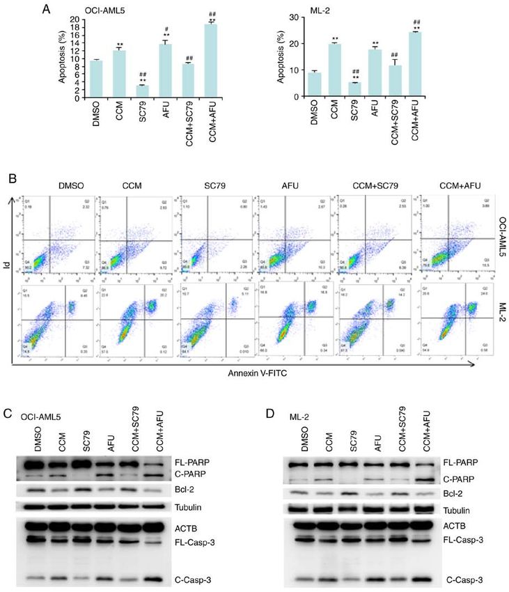
2                            ZHOU et al: CURCUMIN SUPPRESSES AML CELL PROLIFERATION VIA AKT

that curcumin treatment increased the phosphorylation              Phosphorylation profiling. A human phosphorylation pathway
levels of 14 proteins but decreased those of four proteins.        profiling array (cat. no. AAH‑PPP‑1‑4) was purchased from
Among the 18 proteins, AKT/PKB was found to be the                 RayBiotech, Inc., which can detect 55 phosphorylated proteins
main target of curcumin. Moreover, it was identified that          in five signaling pathways: MAPK, AKT, JAK/STAT, NF‑κ B
curcumin promoted cell cycle arrest and apoptosis of AML           and TGF‑β. ML‑2 cells were cultured in a 10‑cm dish until
cells by inactivating AKT.                                         cells reached 90% confluence, and then cells were treated with
                                                                   or without curcumin (25 µM) for 6 h. After treatment, the cells
Materials and methods                                              were harvested and lysed using the cell lysis buffer with a
                                                                   protease inhibitor cocktail and a phosphatase inhibitor cock‑
Chemicals and antibodies. Curcumin, genistein, epigal‑             tail. Phosphorylation array analysis was performed according
locatechin gallate (EGCG), resveratrol and decitabine              to the manufacturer's protocol. The array was sequentially
were purchased from Target Molecule Corp. Afuresertib              incubated with the sample and horseradish peroxidase‑conju‑
(GSK2110183) and SC79 were purchased from Selleck                  gated antibodies (provided within the kit), and then scanned
Chemicals. Antibodies against phosphorylated (p)‑P70S6             with ImageQuant LAS4000 Scanner (Cytiva). In total, two
kinase (P70S6K; T421/S424; cat. no. AP0540), p‑AKT1(S473;          biological replicates were performed, and the average expres‑
cat. no. A P 014 0), total A KT1 (cat. no. A11016),                sion levels were compared between the treatment and control
poly(ADP‑ribose) polymerase (PARP; cat. no. A11010),               samples.
ACTB (cat. no. AF0198) and caspase 3 (cat. no. A2156)
were obtained from ABclonal Biotech Co., Ltd. Antibodies           Western blotting. Cells were cultured in a 12‑well plate until
against p‑RAF‑1 (S301; cat. no. AF0047), p‑proline‑rich            the cell confluence reached ~90%, and then cells were treated
Akt substrate, 40 kDa (PRAS40; T246; cat. no. AF2387),             with the indicated chemicals. Cell lysates were prepared using
p‑p27/Kip1 (T198; cat. no. AF3325), p‑eukaryotic transla‑          the cell lysis buffer with a protease inhibitor cocktail and a
tion initiation factor 4E‑binding protein 1 (4E‑BP1; T36;          phosphatase inhibitor cocktail (Cell Signaling Technology).
cat. no. AF3431), β ‑tubulin (cat. no. AF7011) and Ki67            After quantification of the protein concentration, cell lysates
(cat. no. AF0198) were obtained from Affinity Biosciences.         containing equal amounts of total protein were denatured and
PE‑conjugated (clone HI30; cat. no. 560975; BD Bioscience)         separated on 10‑12% SDS‑PAGE. Following separation, the
and unconjugated mouse anti‑human CD45 antibody (clone             proteins were blotted onto PVDF membranes and blocked.
HI30; cat. no. 555480; BD Bioscience) were used for flow           After sequentially incubated with primary antibodies and
cytometry and immunohistochemistry (IHC), respectively.            appropriate secondary antibodies, the membranes were exposed
A FITC TUNEL cell apoptosis detection kit was purchased            to Pierce ECL Western Blotting Substrate (Thermo Fisher
from Wuhan Servicebio Technology Co., Ltd.                         Scientific, Inc.) and were imaged using a gel imaging system
                                                                   (Tanon 4600SF; Tanon Science and Technology Co., Ltd.).
Cell lines and culture. AML cell lines (HL‑60, ML‑2,
MOLM‑13, OCI‑AML3, OCI‑AML5 and U937) were                         Xenograft mouse models of AML. A total of 20 male
obtained from the American Type Culture collection, and            NOD/SCID mice (age, 5‑6 weeks; average weight, 23 mg)
were cultured according to the manufacturer's instructions.        were purchased from Hunan Slaccas Jingda Laboratory
All cell lines were mycoplasma‑free and were authenticated         Animal Co. Ltd., and housed in groups of 5 per cage with
by Yubo Biological Technology Co., Ltd. using short tandem         water and food ad libitum, in a specific‑pathogen‑free room
repeat analysis.                                                   with filtered air and controlled light/dark cycle (12/12 h),
                                                                   temperature (24±2˚C) and relative humidity (45‑65%).
Cytotoxicity assay. Cells were cultured in a 96‑well plate until   All mice were pretreated with an intraperitoneal injection
the cell confluence reached ~70%, and then cells were treated      of 20 mg/kg busulfan (APExBIO Technology LLC) 24 h
with different concentrations (0, 5, 10, 20, 40 and 80 µM) of      before inoculation, and were then injected intravenously
curcumin, genistein, EGCG, resveratrol or decitabine. After        with 1x10⁶ ML2 cells. At 15 days after inoculation, the mice
48 h, cell viability was determined using a MTT assay as           were randomly divided into four groups (5 mice per group),
described previously (15). Based on the results of the MTT         and then treated with vehicle, curcumin (2 mg/mouse),
assay, the half maximal inhibitory concentration (IC50) of each    afuresertib (1 mg/mouse) or curcumin (2 mg/mouse) +
chemical was calculated.                                           afuresertib (1 mg/mouse) via oral gavage every other day
                                                                   for 16 days. Curcumin and afuresertib were dissolved with
Cell cycle and apoptosis analyses. As reviewed by                  5% DMSO + 10% PEG300 + 5% Tween‑80. The humane
Kouhpeikar et al (16), in vitro examination of the efficacy of     endpoints were defined by body weight loss of 20%. All
curcumin against AML cells was conducted using 10‑50 µM            mice were euthanized by asphyxiation (CO2 displacement
curcumin to treat cells for 24‑48 h. In the present study, cells   rate was ~20% vol/min) 4 days after the last treatment, and
were treated with 25 µM curcumin for 24 h. After treatment,        the death was verified by respiratory arrest and cardiac
cell cycle and apoptosis were analyzed using a PI staining kit     arrest for >10 min. The experiments were performed
[Hangzhou Multi Sciences (Lianke) Biotech Co., Ltd.] and an        from to July 10 to August 12. The spleens were fixed in
Annexin V‑FITC/PI staining kit (Invitrogen; Thermo Fisher          10% formalin and processed for hematoxylin and eosin
Scientific, Inc.), respectively, according to the manufacturer's   (H&E) staining, immunohistochemistry (IHC) analysis
instructions. After staining, cells were analyzed using a flow     and TUNEL assay, as described previously (17,18). Bone
cytometer (CytoFlex; Beckman Coulter, Inc.).                       marrow (obtained from tibias and femurs) was crushed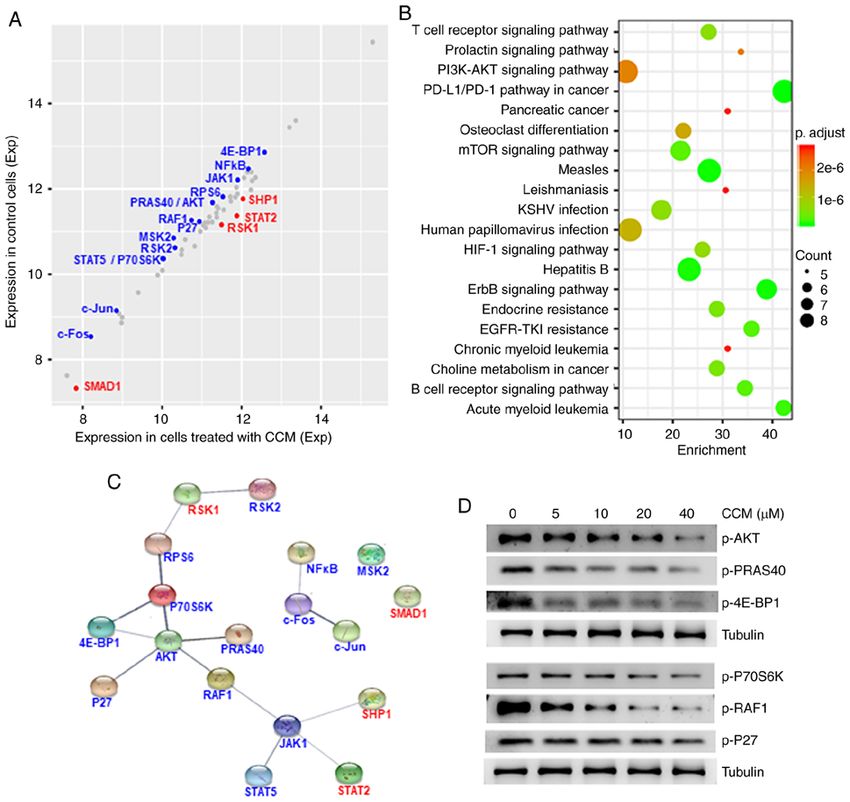
ONCOLOGY REPORTS 45: 11, 2021                                                     3

Table I. IC50 values (mean ± SD) of four phytochemicals and decitabine/curcumin against AML cell lines.

Cell line              Decitabine              Curcumin                 EGCG                    Genistein              Resveratrol

HL‑60                 69.13±13.65             46.98±0.79            111.94±34.72d             90.24±15.65             60.55±3.67
ML‑2                  33.67±1.57d             21.51±0.46b            34.65±1.81d              40.10±2.13b,d           28.70±1.29a,d
MOLM‑13               54.02±11.89             53.18±5.87            102.11±2.77b,d            59.65±10.02             64.93±5.01
OCI‑AML3              55.39±8.37              71.43±10.12            78.65±18.07             100.01±20.15a            48.01±8.11
OCI‑AML5             126.76±8.54d             38.45±0.38b           120.17±21.37d             64.59±2.13b,d           70.26±0.32b,c
U937                  56.10±2.17              59.80±1.34             74.44±1.40b,d            95.32±5.40b,d           74.32±4.20b,d

Differences between each phytochemical and decitabine (curcumin) were analyzed by Tukey test; aadjust P≤0.05 and badjust P≤0.01 vs.
decitabine; cadjust P≤0.05 and dadjust P≤0.01 vs. curcumin. IC50, half maximal inhibitory concentration; AML, acute myeloid leukemia;
EGCG, epigallocatechin gallate.

in PBS and created into single cell suspensions for flow            ‘AML’, ‘ErbB signaling pathway’, ‘EGFR tyrosine kinase
cytometry analysis.                                                 inhibitor (TKI) resistance’ and ‘B cell receptor signaling
                                                                    pathway’ (Fig. 1B; Table SII).
Statistical analysis. RStudio (https://rstudio.com) was used            To understand the association between these 18 proteins,
for statistical analysis. ANOVA and Tukey's post hoc test           a protein‑protein interaction (PPI) network was constructed
were performed to evaluate the significance of difference           using STRING software (https://string‑db.org). In a PPI
between samples, adjust P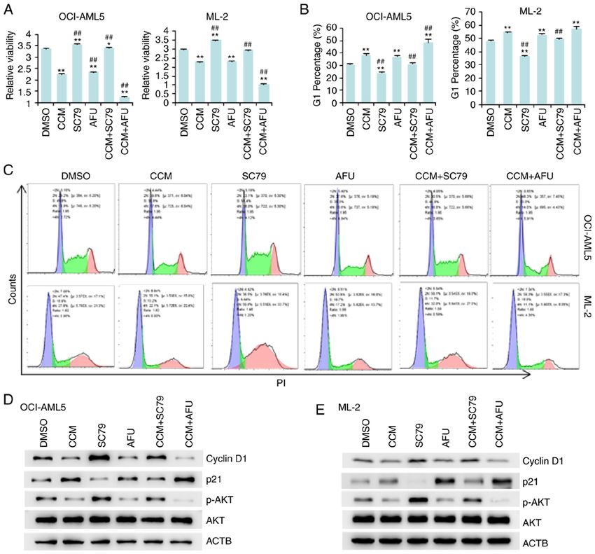
4                                  ZHOU et al: CURCUMIN SUPPRESSES AML CELL PROLIFERATION VIA AKT

Figure 1. Influence of curcumin (CCM) on protein phosphorylation, as detected via antibody array and western blotting. (A) Scatter plot demonstrating
the relative expression levels of the 55 phosphorylated proteins. ML‑2 cells were treated with or without CCM for 6 h. After treatment, a phosphorylation
antibody array was used for detecting the relative levels of phosphorylation. The red dots represent upregulated proteins, the blue dots represent downregulated
proteins and the grey dots represent non‑differentially phosphorylated proteins. (B) Most enriched KEGG pathways of the 18 differentially phosphorylated
proteins. (C) PPI network of the 18 differentially phosphorylated proteins. (D) Western blot analysis of phosphorylation levels of AKT and its interacting
proteins in ML‑2 cells after treatment with increasing concentrations of CCM for 6 h. CCM, curcumin; KEGG, Kyoto Encyclopedia of Genes and Genomes;
PPI, protein‑protein interaction.

However, SC‑79 produced the opposite results. Thus, the effects                   the antiapoptotic Bcl‑2 protein expression but increased
of curcumin were enhanced by afuresertib but attenuated by                        the cleavage of caspase‑3 (C‑Casp3) and PARP (C‑PARP)
SC‑79.                                                                            (Fig. 3C and D). Furthermore, the influence of curcumin on
                                                                                  these three proteins could be enhanced by afuresertib, but
Curcumin promotes AML cell apoptosis by inactivating AKT.                         was abrogated by SC‑79.
Annexin V and PI labeling followed by flow cytometry were                             The above results suggested that curcumin promoted AML
used to detect apoptotic cells. The results demonstrated that                     cell arrest and apoptosis by inactivating AKT. However, IC50
both curcumin and afuresertib promoted apoptosis, while                           values of curcumin were very weakly correlated with the
SC‑79 suppressed apoptosis. Moreover, curcumin‑induced                            levels of phosphorylated AKT in AML cell lines (Fig. S1).
apoptosis was stimulated by afuresertib, but diminished by                        Thus suggested that curcumin also exerted antitumor roles via
SC‑79 (Fig. 3A and B).                                                            other pathways, besides the AKT pathway.
   To identify the proteins involved in curcumin‑induced
apoptosis, the expression of three apoptosis‑related proteins,                    Curcumin and afuresertib synergistically reduce the leukemia
including Bcl‑2, caspase‑3 and PARP, were examined.                               burden in an AML xenograft mouse model. Next, the in vivo
The results indicated that curcumin treatment decreased                           efficacy of curcumin and afuresertib for the treatment of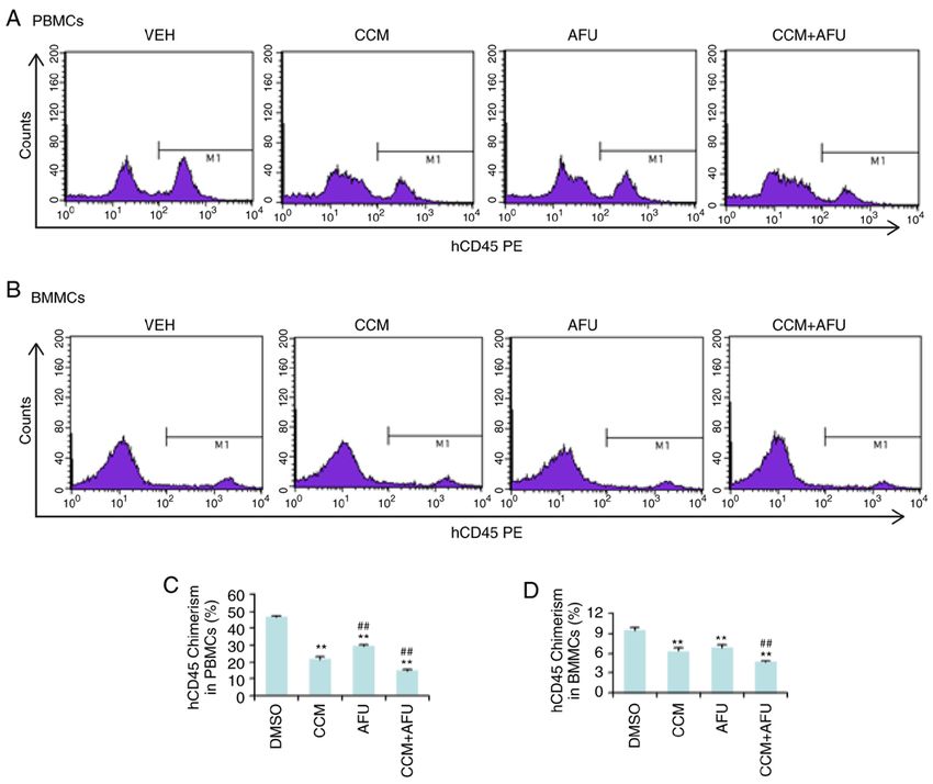
ONCOLOGY REPORTS 45: 11, 2021 5
Figure 2. Curcumin (CCM) suppresses AML cell proliferation by inactivating AKT. OCI‑AML5 and ML‑2 cells were treated with CCM (25 µM), SC‑79
(10 µM) or AFU (10 µM) alone or in combination for 24 h. After treatment, MTT assay, flow cytometry and western blotting were performed. (A) Cell viability
results from triplicate experiments. (B) Percentage of cells in the G1 phase. Data are presented as the mean ± SD of triplicate experiments. (C) Representative
images of flow cytometry. (D and E) Expression levels of cell cycle‑related proteins were detected via western blotting in OCI‑AML5 and ML‑2 cells. *P≤0.05
and **P≤0.01 vs. DMSO; ##P≤0.01 vs. CCM. AFU, afuresertib; CCM, curcumin; AML, acute myeloid leukemia.
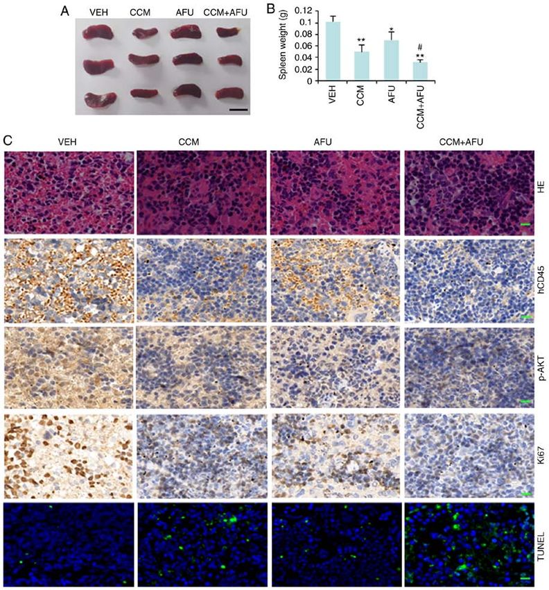
AML was evaluated. NOD/SCID mice were intravenously that treatment with curcumin or afuresertib could decrease
injected with 1x10⁶ ML‑2 cells. Drug treatment began 15 days splenomegaly in AML mice. IHC using an anti‑hCD45 anti‑
after injection and continued every other day for 16 days. body demonstrated that, compared with control mice, the mice
After treatment, peripheral blood mononuclear cells (PBMCs) treated with curcumin or afuresertib had decreased dissemina‑
and bone marrow mononuclear cells (BMMCs) were isolated tion of AML cells in the spleen, and the combinational use of
and evaluated for human hematopoietic (hCD45) chimerism curcumin and afuresertib was more effective compared with
via flow cytometry (Fig. 4). Compared with the control the use of a single drug (Fig. 5C).
group (VEH), the mice treated with curcumin (CCM) or afure‑ Subsequently, Ki‑67 staining and TUNEL assay were
sertib (AFU) either alone or in combination (CCM+AFU) had conducted to evaluate cell proliferation and apoptosis, respec‑
fewer human CD45+ cells in the bone marrow and peripheral tively. The results (Fig. 5C) demonstrated that treatment with
blood. Moreover, combination drug therapy was more effec‑ curcumin or afuresertib significantly increased apoptosis but
tive than single drug therapy in reducing the chimerism of decreased AKT phosphorylation and the cell proliferation
hCD45 (Fig. 4). These results indicated that curcumin and rate in spleen, while treatment with both drugs had stronger
afuresertib synergistically suppressed the engraftment of effects compared with treatment with a single drug. These
AML cells. findings suggested that treatment with curcumin or afure‑
The mice treated with curcumin and afuresertib either alone sertib suppressed the engraftment, proliferation and survival
or in combination had a smaller and lighter spleen compared of AML cells, and that combination therapy had increased
with the control mice (Fig. 5A and B). Thus, it was suggested efficacy compared with monotherapy.6 ZHOU et al: CURCUMIN SUPPRESSES AML CELL PROLIFERATION VIA AKT
Figure 3. Curcumin (CCM) promotes AML cell apoptosis by inactivating AKT. ML‑2 and OCI‑AML5 cells were treated with CCM (25 µM), SC‑79 (10 µM) or
AFU (10 µM) alone or in combination for 24 h. After treatment, flow cytometry and western blotting were performed. (A) Percentage of apoptosis is presented
as the mean ± SD of triplicate experiments. (B) Representative images of flow cytometry. (C and D) Western blot analysis of FL‑PARP, C‑PARP, FL‑casp3 and
C‑Casp3. **P≤0.01 vs. DMSO; #P≤0.05 and ##P≤0.01 vs. CCM. FL‑PARP, full length PARP; AFU, afuresertib; CCM, curcumin; AML, acute myeloid leukemia;
C‑PARP, cleaved‑PARP; FL‑casp3, full length caspase‑3; C‑Casp3, cleaved caspase‑3; PARP, poly(ADP‑ribose) polymerase.
Discussion and p38MAPK (14). To identifying the targets of curcumin
in AML, the present study performed a phosphorylation
The present study compared the cytotoxicity of four phyto‑ antibody array to detect the influence of curcumin on 55
chemicals (curcumin, EGCG, genistein and resveratrol) and phosphorylated proteins in five signaling pathways (MAPK,
identified that curcumin had the strongest anti‑acute myeloid AKT, JAK/STAT, NF‑κ B and TGF‑ β). The present results
leukemia (AML) efficacy. It has been reported that curcumin suggested that curcumin decreased the phosphorylation levels
has multiple targets and exerts its role via different molecular of 14 proteins but increased the phosphorylation levels of four
mechanism in various cancer types (11). Recently, several proteins. Then, a protein‑protein interaction (PPI) network of
studies have revealed that curcumin can inhibit the phos‑ these 18 proteins was conducted, in which AKT was a hub,
phorylation of certain kinases, such as AKT (12), JAK1 (13) indicating that AKT was a main target of curcumin.ONCOLOGY REPORTS 45: 11, 2021 7
Figure 4. Curcumin (CCM) and afuresertib (AFU) synergistically inhibit engraftment of AML cells in PB and BM of mice. NOD/SCID mice were pretreated
with intraperitoneal injection of 20 mg/kg busulfan 24 h before inoculation, and were then injected intravenously with 1x10⁶ ML‑2 cells. At 15 days after inocu‑
lation, the mice were randomly divided into four groups (5 mice per group), and were treated with VEH, CCM, AFU or CCM+AFU via oral gavage every other
day for 16 days. PBMCs and BMMCs were isolated and evaluated for human hematopoietic (hCD45) chimerism via flow cytometry. (A and B) Representative
images from flow cytometry. (C and D) Data are presented as the mean ± SD of three mice. **P≤0.01 vs. VEH; ##P≤0.01 vs. CCM. PB, peripheral blood;
BM, bone marrow; VEH, vehicle; PBMCs, peripheral blood mononuclear cells; BMMCs, bone marrow mononuclear cells; AFU, afuresertib; CCM, curcumin;
AML, acute myeloid leukemia.
Protein kinase B (AKT/PKB) is frequently overactivated by the AKT activator. Therefore, it was indicated that
in AML, and its phosphorylation is an independent poor prog‑ curcumin may function via AKT. However, the sensitivities
nostic factor of overall survival in adult de novo AML (23). to curcumin of AML cell lines were not significantly corre‑
AKT is a serine threonine kinase that contains three lated with their levels of AKT phosphorylation, suggesting
isoforms: AKT1, AKT2 and AKT3. It has been reported to that curcumin still functioned via other pathways, besides
serve roles in various cellular pathways, including prolifera‑ tbe AKT pathway.
tion, apoptosis and angiogenesis. Cyclin D1, which regulates The present study demonstrated the anti‑AML effect
the G1/S check point of the cell cycle, has been reported to of curcumin both in vitro and in vivo, and this effect was
be upregulated by the AKT/glycogen synthase kinase 3 β increased by the combination with afuresertib. Afuresertib has
axis (24). However, p21, a negative regulator of the cell been reported to exert antitumor effects in ovarian cancer (27),
cycle G1/S transition, is negatively regulated by AKT (25). malignant pleural mesothelioma (28) and chronic lymphocytic
AKT also promotes leukemia T cells by enhancing the leukemia (29). However, to the best of our knowledge, its role
transcription of Bcl‑2 (26). The present results suggested in AML has not been previously reported. The present study
that curcumin treatment increased AKT phosphorylation was the first report that afuresertib could potentially be used
and p21 expression but decreased the expression levels of for the treatment of AML.
cyclin D1 and Bcl‑2 in AML cells. Moreover, the effects of In conclusion, the present study demonstrated that curcumin
curcumin on the expression levels of p21, cyclin D1 and Bcl‑2 decreased the survival and proliferation of AML cells in vitro,
were enhanced by the AKT inhibitor but were suppressed as well as AML cell proliferation in hematopoietic tissue and8 ZHOU et al: CURCUMIN SUPPRESSES AML CELL PROLIFERATION VIA AKT
Figure 5. Curcumin (CCM) and afuresertib (AFU) synergistically inhibit engraftment, proliferation and survival of AML cells in the spleens of mice.
NOD/SCID mice were pretreated with intraperitoneal injection of 20 mg/kg busulfan 24 h before inoculation and were then injected intravenously with
1x10⁶ ML‑2 cells. At 15 days after inoculation, the mice were randomly divided into four groups (5 mice per group), and were treated with VEH, CCM, AFU
or CCM+AFU via oral gavage every other day for 16 days. (A) Gross appearance of spleen. Scale bar, 1 cm. (B) Average weight of spleen. (C) H&E, IHC
and TUNEL assays of the spleen. Scale bar, 50 µm. *P≤0.05 and **P≤0.01 vs. VEH; #P≤0.05 vs. CCM. VEH, vehicle; AFU, afuresertib; CCM, curcumin;
AML, acute myeloid leukemia; H&E, hematoxylin and eosin; IHC, immunohistochemistry.
dissemination into non‑hematopoietic tissues. Mechanistically, and Hunan Key Laboratory of Pharmacodynamics and Safety
curcumin treatment suppressed AKT activation, leading to Evaluation of New Drugs.
cell cycle arrest and apoptosis.
Availability of data and materials
Acknowledgements
All data generated and/or analyzed during the study are
We would like to thank RayBiotech Inc. (Guangzhou, China) available from the corresponding author on reasonable
for the assistance in the phosphorylation array analysis. request.
Funding Authors' contributions
This study was supported by the National Natural Science CZ and XD conceived and designed the study. HZ, YN and
Foundation of China (grant nos. 81872256 and 82070155) GZ performed the experiments. HZ, YN, CZ and XD analyzedONCOLOGY REPORTS 45: 11, 2021 9
and interpreted the data. HZ, CZ and XD wrote the manuscript. 13. Li Y, Sun W, Han N, Zou Y and Yin D: Curcumin inhibits
proliferation, migration, invasion and promotes apoptosis of
All authors read and approved the manuscript and agree to be retinoblastoma cell lines through modulation of miR‑99a and
accountable for all aspects of the research in ensuring that the JAK/STAT pathway. BMC Cancer 18: 1230, 2018.
accuracy or integrity of any part of the work are appropriately 14. Hosseini A, Rasmi Y, Rahbarghazi R, Aramwit P, Daeihassani B
and Saboory E: Curcumin modulates the angiogenic potential of
investigated and resolved. human endothelial cells via FAK/P‑38 MAPK signaling pathway.
Gene 688: 7‑12, 2019.
Ethics approval and consent to participate 15. Zhou J, Duan H, Xie Y, Ning Y, Zhang X, Hui N, Wang C,
Zhang J and Zhou J: MiR‑193a‑5p targets the coding region of
AP‑2alpha mRNA and induces cisplatin resistance in bladder
All animal experiments were approved by the Animal cancers. J Cancer 7: 1740‑1746, 2016.
Ethics Committee of Hunan Normal University and 16. Kouhpeikar H, Butler AE, Bamian F, Barreto GE, Majeed M and
Sahebkar A: Curcumin as a therapeutic agent in leukemia. J Cell
performed according to institutional animal care guidelines Physiol 234: 12404‑12414, 2019.
(no. 2018‑037). 17. Zhou C, Zhao XM, Li XF, Wang C, Zhang XT, Liu XZ, Ding XF,
Xiang SL and Zhang J: Curcumin inhibits AP‑2γ‑induced apop‑
tosis in the human malignant testicular germ cells in vitro. Acta
Patient consent for publication Pharmacol Sin 34: 1192‑1200, 2013.
18. Yang L, Qiu J, Xiao Y, Hu X, Liu Q, Chen L, Huang W, Li X,
Not applicable. Li L, Zhang J, et al: AP‑2βinhibits hepatocellular carcinoma
invasion and metastasis through Slug and Snail to suppress
epithelial‑mesenchymal transition. Theranostics 8: 3707‑3721,
Competing interests 2018.
19. Carlos‑Reyes A, Lopez‑Gonzalez JS, Meneses‑Flores M, Gallardo-
Rincón D, Ruíz‑García E, Marchat LA, Astudillo‑de la Vega H,
The authors declare that they have no competing interests. de la Cruz ON and López‑Camarillo C: Dietary compounds as
epigenetic modulating agents in cancer. Front Genet 10: 79, 2019.
References 20. He X and Zhang J: Why do hubs tend to be essential in protein
networks? PLoS Genet 2: e88, 2006.
21. Masamha CP and Benbrook DM: Cyclin D1 degradation is suffi‑
1. Döhner H, Weisdorf DJ and Bloomfield CD: Acute myeloid cient to induce G1 cell cycle arrest despite constitutive expression
leukemia. N Engl J Med 373: 1136‑1152, 2015. of cyclin E2 in ovarian cancer cells. Cancer Res 69: 6565‑6572,
2. Tallman MS, Wang ES, Altman JK, Appelbaum FR, Bhatt VR, 2009.
Bixby D, Coutre SE, De Lima M, Fathi AT, Fiorella M, et al: Acute 22. Georgakilas AG, Martin OA and Bonner WM: p21: A two‑faced
myeloid leukemia, version 3.2019, NCCN clinical practice guide‑ genome guardian. Trends Mol Med 23: 310‑319, 2017.
lines in oncology. J Natl Compr Cancer Netw 17: 721‑749, 2019. 23. Prijic S, Ugrina I, Labar B, Nemet D, Batinić J, Zadro R, Ries S,
3. Oran B and Weisdorf DJ: Survival for older patients with acute Gjadrov‑Kuvedžić K, Davidović S and Batinić D: Prognostic
myeloid leukemia: A population‑based study. Haematologica 97: significance of constitutive phosphatidylinositol 3‑kinase/akt
1916‑1924, 2012. and mitogen‑activated protein kinase phosphorylation in acute
4. Sami SA, Darwish NHE, Barile ANM and Mousa SA: Current myeloid leukemia. Leuk Lymphoma 56: 2281‑2288, 2015.
and future molecular targets for acute myeloid leukemia therapy. 24. Qin Z, Li Y, Li Y and Liu G: Tumor necrosis factor alpha
Curr Treat Options Oncol 21: 3, 2020. stimulates proliferation of dental pulp stem cells via akt/glycogen
5. Wang XJ, Chen JY, Fu LQ and Yan MJ: Recent advances in synthase kinase‑3β/cyclin D1 signaling pathway. J Endod 41:
natural therapeutic approaches for the treatment of cancer. 1066‑1072, 2015.
J Chemother 32: 53‑65, 2020. 25. Hu Z, Long T, Ma Y, Zhu J, Gao L, Zhong Y, Wang X, Wang X
6. Kian MM, Salemi M, Bahadoran M, Haghi A, Dashti N, and Li Z: Downregulation of GLYR1 contributes to microsatellite
Mohammadi S, Rostami S, Chahardouli B, Babakhani D and instability colorectal cancer by targeting p21 via the p38MAPK
Nikbakht M: Curcumin combined with thalidomide reduces and PI3K/AKT pathways. J Exp Clin Cancer Res 39: 76, 2020.
expression of STAT3 and Bcl‑xL, leading to apoptosis in acute 26. Wan YJ, Yang Y, Leng QL, Lan B, Jia HY, Liu YH, Zhang CZ
myeloid leukemia cell lines. Drug Des Devel Ther 14: 185‑194, and Cao Y: Vav1 increases bcl‑2 expression by selective activa‑
2020. tion of rac2‑akt in leukemia T cells. Cell Signal 26: 2202‑2209,
7. Liang K, Bae KH, Nambu A, Dutta B, Chung JE, Osato M 2014.
and Kurisawa M: A two‑pronged anti‑leukemic agent based 27. Blagden SP, Hamilton AL, Mileshkin L, Wong S, Michael A,
on a hyaluronic acid‑green tea catechin conjugate for inducing Hall M, Goh JC, Lisyanskaya AS, DeSilvio M, Frangou E, et al:
targeted cell death and terminal differentiation. Biomater Sci 8: Phase IB dose escalation and expansion study of AKT inhibitor
497‑505, 2019. afuresertib with carboplatin and paclitaxel in recurrent plat‑
8. de Blas E, Estañ MC, Del Carmen Gomez de Frutos M, Ramos J, inum‑resistant ovarian cancer. Clin Cancer Res 25: 1472‑1478,
Del Carmen Boyano‑Adánez M and Aller P: Selected polyphe‑ 2019.
nols potentiate the apoptotic efficacy of glycolytic inhibitors in 28. Yamaji M, Ota A, Wahiduzzaman M, Karnan S, Hyodo T,
human acute myeloid leukemia cell lines. Regulation by protein Konishi H, Tsuzuki S, Hosokawa Y and Haniuda M: Novel
kinase activities. Cancer Cell Int 16: 70, 2016. ATP‑competitive Akt inhibitor afuresertib suppresses the prolif‑
9. Li Y, Guo Y, Feng Z, Bergan R, Li B, Qin Y, Zhao L, Zhang Z and eration of malignant pleural mesothelioma cells. Cancer Med 6:
Shi M: Involvement of the PI3K/Akt/Nrf2 signaling pathway in 2646‑2659, 2017.
resveratrol‑mediated reversal of drug resistance in HL‑60/ADR 29. Chen CI, Paul H, Le LW, Wei EN, Snitzler S, Wang T, Levina O,
cells. Nutr Cancer 71: 1007‑1018, 2019. Kakar S, Lau A, Queau M, et al: A phase 2 study of ofatumumab
10. Giordano A and Tommonaro G: Curcumin and cancer. (Arzerra®) in combination with a pan‑AKT inhibitor (afure‑
Nutrients 11:2376, 2019. sertib) in previously treated patients with chronic lymphocytic
11. Liczbinski P, Michałowicz J and Bukowska B: Molecular mecha‑ leukemia (CLL). Leuk Lymphoma 60: 92‑100, 2019.
nism of curcumin action in signaling pathways: Review of the
latest research. Phytother Res 34: 1992‑2005, 2020.
12. Borges GA, Elias ST, Amorim B, de Lima CL, Coletta RD, This work is licensed under a Creative Commons
Castilho RM, Squarize CH and Guerra EN: Curcumin down‑ Attribution-NonCommercial-NoDerivatives 4.0
regulates the PI3K‑AKT‑mTOR pathway and inhibits growth
International (CC BY-NC-ND 4.0) License.
and progression in head and neck cancer cells. Phytother Res 34:
3311‑3324, 2020.You can also read