Promoter hypermethylation influences the suppressive role of long non coding RNA MEG3 in the development of multiple myeloma
←
→
Page content transcription
If your browser does not render page correctly, please read the page content below
EXPERIMENTAL AND THERAPEUTIC MEDICINE 20: 637-645, 2020

    Promoter hypermethylation influences the suppressive role of long
    non‑coding RNA MEG3 in the development of multiple myeloma
           WENJUN YU1,2*, QINGLIN SHI2*, CHAO WU2, XUXING SHEN2, LIJUAN CHEN2* and JIAREN XU1*

     1
      Department of Geriatric Medicine, Geriatric Hospital of Nanjing Medical University, Jiangsu Province Geriatric Institute;
         2
           Department of Hematology, First Affiliated Hospital of Nanjing Medical University, Jiangsu Province Hospital,
                                               Nanjing, Jiangsu 210000, P.R. China

                                      Received January 12, 2019; Accepted December 31, 2019

                                                     DOI: 10.3892/etm.2020.8723

Abstract. Methylation is a fundamental regulator of gene               Introduction
transcription. Long non‑coding RNA maternally expressed 3
(MEG3) inhibits cell proliferation in various types of cancer.         Multiple myeloma (MM) is one of the most common types of
However, the molecular mechanisms of MEG3 methylation in               haematological cancer, which is becoming more common in
the regulation of multiple myeloma (MM) are unknown. In the            the ageing population. It is also the cause of a number of fatal
present study, MEG3 upregulation was negatively associated             outcomes (1). As a haematological cancer that originates from
with the International Staging System (ISS) status of the bone         the malignant transformation of plasma cells, MM maintains
marrow samples of 39 patients with MM. MEG3 overexpres-                pathophysiologic heterogeneity due to its complex genetic
sion in an MM cell line resulted in elevated p53 expression.           background (2). Several distinct clinical phases of MM have
Furthermore, the results of methylation‑specific PCR revealed          been identified, including monoclonal gammopathy of unde-
that the abnormal methylation status of the MEG3 promoter              termined significance and smoldering multiple myeloma (3).
region was present in eight of the 39 bone marrow samples              With the development and progression of MM, several distinct
collected. Treatment of the MM cell line with the DNA                  patterns of genetic aberration are recognized, including cyto-
methylation inhibitor 5‑Aza‑2'‑deoxycytidine (5‑Aza‑CdR)               genetic abnormalities, chromosomal aberration and signaling
resulted in tumor cell proliferation inhibition, apoptosis induc-      pathway disorders (4).
tion and G 0/G1 cell cycle arrest. Furthermore, 5‑Aza‑CdR                  DNA methylation, a form of epigenetic control of gene
decreased aberrant hypermethylation of the MEG3 promoter               transcription, refers to cytosine methylation at position 5 in the
and increased the expression of MEG3. However, 5‑Aza‑CdR               pyrimidine ring, which can result in inappropriate silencing of
exerted no effect on p53 expression. To the best of our                genes involved in diverse biological processes, including cell
knowledge, the present study is the first to report that the           proliferation, apoptosis, migration and cell cycle arrest (5).
demethylation reagent 5‑Aza‑CdR may serve as a therapeutic             In normal cells, unmethylated CpG islands (a cluster of
agent in MM by upregulating MEG3 expression. However, the              CpG dinucleotides) are usually observed; however, human
mechanism of action was independent of p53 expression.                 malignancies are characterized by the gain of methylation at
                                                                       promoter associated CpG islands (6). The role of DNA meth-
                                                                       ylation in the mediation of multiple tumor suppressor gene and
                                                                       microRNA silencing has been implicated in the development
                                                                       and progression of MM (7).
                                                                           Long non‑coding RNAs (lncRNAs) are a class of non‑coding
Correspondence to: Dr Lijuan Chen, Department of Hematology,
                                                                       RNA with a length of >200 nucleotides, which possess little to
First Affiliated Hospital of Nanjing Medical University, Jiangsu
Province Hospital, 300 Guangzhou Road, Nanjing, Jiangsu 210000,        no capacity for protein synthesis (8). Numerous studies have
P.R. China                                                             reported that lncRNAs are deregulated in various types of
E‑mail: chenljb@126.com                                                cancer and are implicated in carcinogenesis and antitumor
                                                                       pathways (9). Wang et al (10) reported that protein tyrosine
Dr Jiaren Xu, Department of Geriatric Medicine, Geriatric Hospital
                                                                       phosphatase L1 could be epigenetically regulated in MM;
of Nanjing Medical University, Jiangsu Province Geriatric Institute,
18 Luojia Road, Nanjing, Jiangsu 210000, P.R. China                    a process which can be reversed by 5‑Aza‑2'‑deoxycytidine
E‑mail: xujiarencn@sina.com                                            (5‑Aza‑CdR), suggesting a potential therapeutic agent for
                                                                       MM. The maternally expressed 3 (MEG3) imprinted gene is
*
    Contributed equally                                                located on chromosome 14q32, which produces a non‑coding
                                                                       RNA transcript (11). lncRNA MEG3 has been identified
Key words: long non‑coding RNA, maternally expressed 3,                as a tumor suppressor in various types of cancer, including
promoter hypermethylation, multiple myeloma                            meningioma (12), breast cancer (13), bladder cancer (14)
                                                                       and hepatocellular carcinoma (15). A previous study also
                                                                       demonstrated the anticancer effect of MEG3 in MM (16).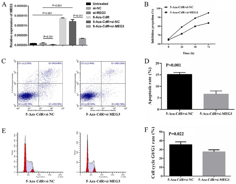
638 YU et al: PROMOTER HYPERMETHYLATION OF MEG3 IN MULTIPLE MYELOMA
The promoter region of MEG3 is rich in CpG islands, and the at 95˚C for 5 sec, annealing at 60˚C for 30 sec, followed by a
specific methylated and unmethylated CpG islands of differ- final extension at 72˚C for 5 min. MEG3 mRNA levels were
entially methylated regions (DMRs) are located upstream quantified according to the standard curve of MEG3 and
of the MEG3 gene (IG‑DMR and MEG3‑DMR) (17,18). β‑actin using the 2‑∆∆Cq method (24). β‑actin was used as the
Furthermore, lncRNA MEG3 expression has been reported internal reference gene.
to be induced following epigenetic modification of DNA
methylation in diverse malignancies, including gliomas (19), DNA isolation and methylation‑specific PCR (MSP). Genomic
ovarian cancer (20) and leukemia (21,22). (g)DNA was extracted from ARP1 cells and patient derived
In MM, Benetatos et al (21) observed MEG3 promoter MM cells using a TIANamp Genomic DNA kit according
hypermethylation in both bone marrow and peripheral blood to the manufacturer's protocol (Tiangen Biotech Co., Ltd.).
samples. This hypermethylation was correlated with MM Subsequently, DNA bisulfite conversion was performed on the
stage and subtype. Therefore, it was hypothesized that MEG3 gDNA using the EpiTect Plus Bisulfite kit (Qiagen GmbH),
expression could be epigenetically controlled by MEG3 according to the manufacturer's protocol.
promoter hypermethylation, which may ultimately influence The methylation status of MEG3 was determined by MSP,
the biological behavior of MM. using a Veriti96 PCR thermocycler (Applied Biosystems;
Thermo Fisher Scientific, Inc.) with Taq PCR MasterMix
Materials and methods (Tiangen Biotech Co., Ltd.). The following primers obtained
from previous studies (25,26) were used for MSP: Methylated
Study subjects. The present study was approved by the primer pair forward, 5'‑GTTAGTAATCGGGTTTGTCGGC‑3'
Institutional Review Board of the First Affiliated Hospital and reverse, 5'‑AATCATAACTCCGAACACCCGCG‑3'; and
of Nanjing Medical University. All participants provided unmethylated primer pair forward, 5'‑GAGGATGGTTAGTTA
written informed consent. Bone marrow biopsy samples TTGG GGT‑3' and reverse, 5'‑CCACCATAACCAACACCC
were collected from 39 patients with newly diagnosed TATA ATCACA‑3'. PCR was performed using the following
MM who were admitted to the First Affiliated Hospital thermocycling conditions: 94˚C for 3 min; 5 cycles of 94˚C
of Nanjing Medical University between January 2009 and for 30 sec, 70˚C for 30 sec and 72˚C for 30 sec; 5 cycles of
May 2014. Patient information is listed in Table I. The 94˚C for 30 sec, 65˚C for 30 sec and 72˚C for 30 sec; 30 cycles
diagnosis of MM was established according to the standard of 94˚C for 30 sec, 60˚C for 30 sec and 72˚C for 30 sec; and
morphological and immunophenotypical criteria (23). The a final extension at 72˚C for 7 min. The PCR products were
subtype of MM was classified according to the monoclonal run on a 3% agarose gel and were subsequently identified by
component. The stage of MM was classified according to the ethidium bromide staining at room temperature (27). The CpG
Durie‑Salmon staging system and the International Staging island usually localizes to the DMR which is located within
System (ISS) (24). ~4 kb of the DMR that contains the promoter of the MEG3
MM cells were isolated from bone marrow samples gene (28,29). A 160 bp product represented the methylated
using CD138 microbeads and MS‑columns (Miltenyi state and a 120 bp product represented the unmethylated state
Biotec; cat. no. 130‑051‑301) according to the manufacter's of MEG3 (26).
protocol. The MM cell line, ARP1 (American Type Culture
Collection), was cultured in RPMI 1640 medium (Gibco; Cell transfection and 5‑Aza‑CdR treatment. ARP‑1 cells were
Thermo Fisher Scientific, Inc.) supplemented with 10% fetal cultured in DMEM medium (Gibco; Thermo Fisher Scientific,
bovine serum (Gibco; Thermo Fisher Scientific, Inc.) and 1% Inc.) supplemented with 10% fetal bovine serum (Gibco;
penicillin‑streptomycin in an incubator at 37˚C with 5% CO2. Thermo Fisher Scientific, Inc.) and 1% penicillin‑strepto-
mycin in an incubator at 37˚C with 5% CO2. ARP1 cells
RNA isolation and reverse transcription‑quantitative PCR (2.0x10⁶/well) were plated into 6‑well plates and transfected
(RT‑qPCR). Total RNA was isolated from ARP1 cells and with the 4 µg pcDNA3.1‑MEG3 or 4 µg pcDNA3.1‑empty
patient derived MM cells using TRIzol® reagent (Thermo (provided by Professor Wei De, Nanjing Medical University)
Fisher Scientific, Inc.), according to the manufacturer's using Lipofectamine® 2000 (Thermo Fisher Scientific, Inc.),
protcol. Total RNA was reverse transcribed to cDNA using according to the manufacturer's protocol. For MEG3 knock-
the Primescipt RT Reagent kit with gDNA Eraser (Takara down, the following small interfering (si)RNA sequences
Biotechnology Co., Ltd.), according to the manufacturer's were used for Lipofectamine® 2000 transfection: si‑MEG3,
protocol. 5'‑GCUCAUACUU UGACUC UAU TT‑3'; and si‑negative
qPCR was subsequently performed on a StepOne Plus™ control (NC), 5'‑UUC U CC GAA C GU G UC ACG U TT‑3'.
Real‑Time PCR system (Applied Biosystems; Thermo Fisher Both sequences were designed and synthesized by Shanghai
Scientific, Inc.) using a SYBR Green qRT‑PCR assay according GenePharma Co., Ltd.
to the manufacturer's protocol (Takara Biotechnology Co., ARP1 cells were seeded at 2x10 4 cells/well in 96‑well
Ltd.). The following primer pairs, designed by Primer Premier culture plates and incubated with DMEM (200 µl) containing
5 (Premier Biosoft International), were used for qPCR: MEG3 0.1, 1, 5, 10, 50 or 100 µg/ml 5‑Aza‑CdR (Sigma‑Aldrich;
forward, 5'‑GGAG CTGTTGAGCCTTCAGT‑3' and reverse, Merck KGaA) for 72 h at 37˚C. Control cells were incubated
5'‑CAAGCCCTGTGCT TTGGAAC‑3'; and β‑actin forward, with DMEM containing PBS (20 µl). The Cell Counting
5'‑AGCGAGCATCCCCCAA AGT T‑3' and reverse, 5'‑GGG Kit‑8 (CCK‑8) assay (Selleck Chemicals) was used to analyze
CACGAAGGCTCATCATT‑3'. The following thermocycling cell proliferation according to the manufacturer's protocol.
conditions were used for the qPCR: 40 cycles of denaturation RT‑qPCR and MSP were performed to assess the expression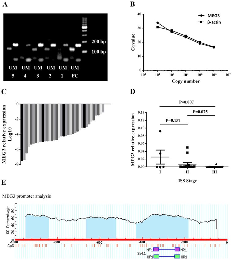
EXPERIMENTAL AND THERAPEUTIC MEDICINE 20: 637-645, 2020                                    639

Table I. Distribution of variables of patients with multiple    for 1.5 h with appropriate secondary antibodies (horseradish
myeloma.                                                        peroxidase conjugated goat anti‑rabbit IgG H&L; 1:4,000;
                                                                Abcam; cat. no. ab6721) at room temperature. Protein bands
Variable                                        Number (%)      were visualized using the Chemiluminescence horseradish
                                                                peroxidase substrate (cat. no. P90720; EMD Millipore) and
Median age (range)                               61 (36‑82)     the Molecular Imager ChemiDoc XRS+ chemiluminescence
Sex                                                             system (Bio‑Rad Laboratories, Inc.). Protein expression was
 Male                                             26 (66.7)     quantified using Image Lab software version 5.0 (Bio‑Rad
 Female                                           13 (33.3)     Laboratories, Inc.) with GAPDH as the loading control.
Subtypes
                                                                Flow cytometry. To analyze the cell cycle, ARP1 cells
 IgG                                              18 (46.2)
                                                                (2.0x10⁶/well) were plated in 6‑well plates and treated with a
 IgA                                              11 (28.2)
                                                                series of concentrations of 5‑Aza‑CdR (0, 5, 10 and 50 µg/ml).
 Light chain                                      10 (25.6)     After 48 h incubation at 37˚C with 5% CO2, cells were washed
Durie‑Salmon stage                                              with PBS and fixed with 75% cold ethanol for 24 h at ‑20˚C.
 I                                                 4 (10.3)     Subsequently, the cells were washed with PBS and stained
 II                                                5 (12.8)     using the Cell Cycle Detection kit according to the manufac-
 III                                              30 (76.9)     turer's protocol (Nanjing KeyGen Biotech Co., Ltd.) at room
International Staging System stage                              temperature for 30 min to analyze the cell cycle with FACS
                                                                (Becton, Dickinson and Company).
 I                                                 5 (12.8)
                                                                    To analyze apoptosis, ARP1 cells (2.0x10 6/well) were
 II                                               14 (35.9)
                                                                plated in 6‑well plates and treated with a series of concen-
 III                                              20 (51.3)     trations of 5‑Aza‑CdR (0, 5, 10 and 50 µg/ml). After 48 h
Serum creatinine (µmol/l)                                       treatment at 37˚C with 5% CO2, the cells were washed with
 >176.8                                              22         PBS. Subsequently, the cells were harvested and stained
 ≤176.8                                              12         using the Annexin V‑FITC Apoptosis Detection kit (Nanjing
Serum calcium (mmol/l)                                          KeyGen Biotech Co., Ltd.), according to the manufacturer's
 >2.98                                               31         protocol. Cells were stained with Annexin V and PI at 4˚C for
                                                                15 min in the dark, and subjected to FACS analysis (Becton,
 ≤2.98                                                3
                                                                Dickinson and Company).
 N/A                                                  5
                                                                    For restoration experiments, ARP1 cells were treated with
N/A, not detected.                                              5‑Aza‑CdR (50 µg/ml for 48 h at 37˚C with 5% CO2), followed
                                                                by MEG3 knockdown. At 48 h post‑transfection, cells were
                                                                used for cell cycle and apoptosis analyses.

                                                                Statistical analysis. The schematic diagram of CpG islands in
of MEG3 mRNA and the methylation status of the MEG3             the human MEG3 promoter was performed using MethPrimer
promoter, respectively.                                         software (Version 1.0; www.urogene.org/methprimer).
    For restoration experiments, ARP1 cells were treated with   Statistical analysis was performed using GraphPad Prism
5‑Aza‑CdR (50 µg/ml for 48 h at 37˚C), followed by MEG3         (version 5; GraphPad Software, Inc.). A chi‑squared test was
knockdown. MEG3 expression was detected by RT‑qPCR              used to compare categorical variables. Data were presented as
at 48 h post‑transfection. ARP1 cell proliferation following    the mean ± standard deviation from at least three repeats. Data
MEG3 knockdown was analyzed by the CCK‑8 assay at 0, 24,        were compared using a Student's t‑test or one‑way ANOVA
48 and 72 h post‑transfection.                                  followed by Tukey's post hoc test. P
640 YU et al: PROMOTER HYPERMETHYLATION OF MEG3 IN MULTIPLE MYELOMA
Figure 1. MEG3 methylation status and MEG3 expression in the bone marrow samples of patients with MM. (A) MEG3 methylation status. Samples 1 and 4
exhibited an abnormal methylation status. Samples 2, 3 and 5 exhibited a normal methylation status. (B) A standard curve was used to calculate the expres-
sion of MEG3 and β‑actin following reverse transcription‑quantitative PCR. (C) Relative expression of MEG3 in 39 bone marrow samples of patients with
MM. MEG3 expression was detected in 36 samples. (D) MEG3 expression was negatively associated with International Staging System stage. (E) Schematic
diagram of CpG islands (blue area) in the human MEG3 promoter, identified using MethPrimer software. The position of CpG islands in relation to the MEG3
transcription starting site and MSP primers were depicted. MEG3, maternally expressed 3; MM, multiple myeloma; PC, positive control; M, methylated band;
U, unmethylated band; MF1, methylated forward 1; MR1, methylated reverse 1; UF1, unmethylated forward 1; UR1, unmethylated reverse 1.
MM (Fig. 1D), indicating that MEG3 may serve as a tumor or 50 µg/ml). Re‑expression of MEG3 (Fig. 2B) and reversed
suppressor in human MM. The schematic diagram of CpG abnormal methylation pattern of the MEG3 promoter (Fig. 2A)
islands in the human MEG3 promoter is presented in Fig. 1E. were observed following treatment with 5‑Aza‑CdR. These
results indicated that CpG methylation may downregulate
MEG3 methylation status and expression is restored after MEG3 mRNA expression in MM cells.
treatment with 5‑Aza‑CdR. In the 39 newly diagnosed MM
samples, an abnormal methylation pattern of the MEG3 5‑Aza‑CdR contributes to the inhibition of MM cells. To
DMRs was identified in eight of the bone marrow samples. investigate the role of 5‑Aza‑CdR in cell proliferation, ARP1
Chi‑squared test was used to examine the association between cells were treated with different concentrations of 5‑Aza‑CdR.
MEG3 methylation status and ISS stage (Table II); however, The results of the CCK‑8 assay revealed that proliferation was
a significant correlation was not observed. ARP1 cells were inhibited by 5‑Aza‑CdR in a dose‑dependent manner (mean
treated with different concentrations of 5‑Aza‑CdR (0, 5, 10 inhibition proportion of 0, 5, 10, 50 and 100 µg/ml for 24 h:EXPERIMENTAL AND THERAPEUTIC MEDICINE 20: 637-645, 2020 641
Table II. Distribution of methylation status among the Association between MEG3 and p53 expression. The
International Staging System stages in patients with multiple present study investigated whether MEG3 regulated the
myeloma. expression of p53. ARP1 cells were successfully transfected
with pcDNA3.1‑MEG3 or pcDNA3.1‑empty (Fig. 4A). The
International staging pcDNA3.1‑MEG3 group displayed significantly increased
system stage levels of p53 protein (Fig. 4B). Furthermore, whether
‑‑‑‑‑‑‑‑‑‑‑‑‑‑‑‑‑‑‑‑‑‑‑‑‑‑‑‑‑‑‑‑‑‑‑‑‑‑‑‑‑‑‑‑‑‑‑‑‑‑‑‑‑‑‑‑‑‑ 5‑Aza‑CdR influences the expression of p53 was investigated.
Methylation status I II III P‑value The results revealed that 5‑Aza‑CdR treatment did not alter
the p53 expression of ARP1 cells (Fig. 4C).
Unmethylated 4 (12.9) 12 (38.7) 15 (48.4) 0.748
Methylated 1 (12.5) 2 (25.0) 5 (62.5) Restoration experiments. ARP1 cells treated with 5‑Aza‑CdR
followed by MEG3 knockdown were used to investigate
whether the tumor suppressive role of 5‑Aza‑CdR in MM
cells may be due to the upregulation of MEG3. MEG3 expres-
sion levels were detected in untreated, si‑NC, si‑MEG3,
5‑Aza‑CdR, 5‑Aza‑CdR + si‑NC and 5‑Aza‑CdR + si‑MEG3
groups, which demonstrated that 5‑Aza‑CdR could upregu-
late MEG3 expression in ARP1 cells (Fig. 5A). In co‑treated
cells, the increased antiproliferation (Fig. 5B), proapoptotic
(Fig. 5C and D) and enhanced cell cycle arrest (Fig. 5E and F)
effects induced by 5‑Aza‑CdR treatment were partly inhibited
by MEG3 knockdown.
Discussion
In the present study, low MEG3 expressions were associated
with higher ISS stages. A previous study reported that MEG3
overexpression inhibited proliferation, promoted apoptosis
and blocked the cell cycle in ARP1 cells (16). Furthermore,
the demethylating agent, 5‑Aza‑CdR, reversed the hyper-
methylation status of the MEG3 promoter and increased
MEG3 expression in ARP1 cells. Additionally, 5‑Aza‑CdR
produced an antiproliferative effect on ARP1 cells, which was
reversed by MEG3 knockdown. Taken together, the results of
the present study suggested that MEG3 may serve as a tumor
suppressor gene in MM and that MEG3 may be epigenetically
modified by the hypermethylation status of MEG3 promoter.
MEG3 is a maternally expressed imprinted gene, which
encodes an lncRNA (30). Multiple tumor samples have been
tested for MEG3 expression and loss of MEG3 expression
Figure 2. 5‑Aza‑CdR restores the normal MEG3 methylation status and has been identified in the majority of tumor types, including
MEG3 expression of ARP1 cells. (A) MEG3 methylation status and bladder (14), epithelial ovarian (20) and gallbladder cancer (31),
(B) expression of ARP1 cells. 5‑Aza‑CdR, 5‑Aza‑2'‑deoxycytidine; MEG3, as well as glioma (19) and nasopharyngeal carcinoma (32). In
maternally expressed 3; NC, negative control; PC, positive control; M,
methylated band; U, unmethylated band; 1, 0 µg/ml 5‑Aza‑CdR; 2, 5 µg/ml
MM, ISS stage is the most important prognostic system (23).
5‑Aza‑CdR; 3, 10 µg/ml 5‑Aza‑CdR; 4, 50 µg/ml 5‑Aza‑CdR. In the present study, MEG3 expression was detected in bone
marrow samples derived from patients with MM. Loss of
MEG3 expression was associated with higher ISS stages,
indicating that MEG3 may serve as a prognostic biomarker in
3.0, 38.5, 42.8, 59.6 and 61.5%; 48 h: 7.3, 45.3, 53.9, 74.3 and MM. The tumor suppressor role of MEG3 in MM identified
79.8%; and 72 h: 6.0, 45.1, 69.5, 91.6 and 93.5%; Fig. 3A). in the present study is consistent with the aforementioned role
The results of the apoptosis assay revealed that the number of of MEG3 in other malignancies identified in previous studies.
apoptotic cells increased by 5‑Aza‑CdR in a dose‑dependent The tumor suppressor function of p53 has long been
manner (6.02% for 0 µg/ml, 19.45% for 5 µg/ml, 24.58% for recognized and p53 has been reported to be mutated,
10 µg/ml, and 51.49% for 50 µg/ml; Fig. 3B and D). Similarly, which may lead to the loss of wild‑type p53 activity in the
cell cycle analysis demonstrated that the number of ARP1 cells majority of human malignancies (33). p53 abnormalities are
arrested at the G0/G1 phase was increased by 5‑Aza‑CdR in a regarded as independent prognostic markers in MM (34,35).
dose‑dependent manner (27.30±0.74 for 0 µg/ml, 31.93±0.79 Previously, a study reported that MEG3 could stimulate
for 5 µg/ml, 43.27±1.02 for 10 µg/ml, and 48.00±0.36 for p53‑dependent transcription (12). p53 exhibits a relative
50 µg/ml; Fig. 3C and E). The aforementioned results indicated low expression level due to rapid degradation caused by
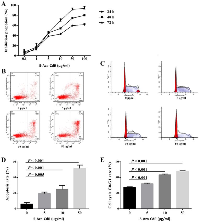
that 5‑Aza‑CdR inhibits the proliferation of MM cells. the ubiquitin‑proteasome pathway (36). The p53 protein642 YU et al: PROMOTER HYPERMETHYLATION OF MEG3 IN MULTIPLE MYELOMA Figure 3. 5‑Aza‑CdR functions as an anti‑tumor factor in the ARP1 cells. (A) 5‑Aza‑CdR inhibited the proliferation of the ARP1 cell line. (B) Representative images of the flow cytometry analysis of the (B) apoptosis and (C) cell cycle of ARP1 cells following 5‑Aza‑CdR treatment. (D) 5‑Aza‑CdR promoted the apoptosis of the ARP1 cell line. (E) 5‑Aza‑CdR arrested the cell cycle at G 0/G1 in the ARP1 cell line. 5‑Aza‑CdR, 5‑Aza‑2'‑deoxycytidine. is regulated by MDM2 proto‑oncogene (MDM2), which DNA methylation, an epigenetic regulation mechanism, is an E3 ubiquitin ligase that inhibits the function of p53 serves a role in silencing MEG3 gene expression in different and promotes its degradation (37). MEG3 has been iden- types of cancer (42). The role of the DNA methylation tified as a tumor suppressor, which exerts its effect by inhibitor 5‑Aza‑CdR is associated with the hypermethylation downregulating MDM2 expression, as well as activating regulation of various genes in MM (10). Downregulation of p53 (38). Furthermore, MDM2 suppression contributes to MEG3 expression is associated with the hypermethylation p53 accumulation induced by MEG3 (39). Previous studies status of the MEG3 promoter in a number of malignancies, have demonstrated that p53 serves as target of MEG3 in including glioma (19), ovarian cancer (20) and leukemia (21). multiple types of cancer, including breast (13), non‑small In MM, Benetatos et al (21) reported MEG3 promoter hyper- cell lung (38) and bladder cancer (40), as well as glioma (19) methylation status in 12 out of 21 bone marrow samples and and hepatoma (41). In the present study, p53 protein levels 9 out of 14 peripheral blood samples. In the present study, were upregulated in ARP1 cells overexpressing MEG3, MEG3 promoter methylation status was identified in the indicating that MEG3 may suppress MM cell proliferation bone marrow samples of 39 patients with MM and abnormal by upregulating p53. hypermethylation was identified in eight samples. The
EXPERIMENTAL AND THERAPEUTIC MEDICINE 20: 637-645, 2020                                                        643

Figure 4. Effect of MEG3 overexpression and 5‑Aza‑CdR on p53 expression in ARP1 cells. (A) MEG3 overexpression efficiency in ARP1 cells transfected
with pcDNA3.1‑empty and pcDNA3.1‑MEG3. (B) MEG3 overexpression increased the protein levels of p53 in the ARP1 cell line; (C) 5‑Aza‑CdR exhibited
no effect on p53 protein expression. MEG3, maternally expressed 3; 5‑Aza‑CdR, 5‑Aza‑2'‑deoxycytidine.

Figure 5. MEG3 knockdown reverses the antitumor effects of 5‑Aza‑CdR. Analysis of (A) MEG3 mRNA expression, (B) cell proliferation, (C) representative
plots and (D) quantification of apoptosis. (E) Representative plots and (F) quantification of the cell cycle in ARP1 cells treated with 5‑Aza‑CdR followed by
MEG3 knockdown. MEG3, maternally expressed 3; 5‑Aza‑CdR, 5‑Aza‑2'‑deoxycytidine; si, small interfering RNA; NC, negative control.
644                       YU et al: PROMOTER HYPERMETHYLATION OF MEG3 IN MULTIPLE MYELOMA

discrepancy between the present study and previous studies       Ethics approval and consent to participate
could be attributed to the small sample size and diverse
genetic backgrounds of the patients included in the current      The present study was approved by the Institutional Review
study. Furthermore, no significant association was identified    Board of the First Affiliated Hospital of Nanjing Medical
between MEG3 methylation status and ISS stage, which could       University. All participants provided written informed
be attributed to the small number of samples and the low         consent.
proportion of samples with an abnormal methylation status.
The functional role of 5‑Aza‑CdR in ARP1 cells was further       Patient consent for publication
explored. The results further demonstrated a demethylation
effect of 5‑Aza‑CdR on MEG3 promoter hypermethylation in         Not applicable.
ARP1 cells. In addition, MEG3 levels revealed a dose‑depen-
dent relationship with 5‑Aza‑Cdr concentration. Furthermore,     Competing interests
5‑Aza‑CdR inhibited proliferation, promoted apoptosis and
induced G 0/G1 cell cycle arrest in ARP1 cells. The effects      The authors declare that they have no competing interests.
of 5‑Aza‑CdR were reversed by MEG3 knockdown. The
results suggested that the demethylation reagent, 5‑Aza‑CdR,     References
might restore MEG3 expression by demethylating the MEG3
promoter and therefore, may exert an anticancer effect in         1. Kazandjian D: Multiple myeloma epidemiology and survival: A
MM. Furthermore, the effect of 5‑Aza‑CdR on p53 expres-              unique malignancy. Semin Oncol 43: 676‑681, 2016.
                                                                  2. Morgan GJ, Walker BA and Davies FE: The genetic architecture
sion was investigated; however, no alterations to the levels         of multiple myeloma. Nat Rev Cancer 12: 335‑348, 2012.
of p53 protein were observed. This may be attributed to the       3. Mateos MV and Landgren O: MGUS and smoldering multiple myeloma:
different effect of 5‑Aza‑CdR on different genes. Therefore,         Diagnosis and epidemiology. Cancer Treat Res 169: 3‑12, 2016.
                                                                  4. Glavey SV, Manier S, Sacco A, Salem K, Kawano Y, Bouyssou J,
the results indicated that the antitumor effect of 5‑Aza‑CdR         Ghobrial IM and Roccaro AM: Epigenetics in multiple myeloma.
involved MEG3 but was independent of p53.                            Cancer Treat Res 169: 35‑49, 2016.
    In conclusion, the present study suggested that MEG3          5. Wilkins JF: Genomic imprinting and methylation: Epigenetic
                                                                     canalization and conflict. Trends Genet 21: 356‑365, 2005.
may serve as a tumor suppressor by upregulating p53 levels        6. Liyanage VR, Jarmasz JS, Murugeshan N, Del Bigio MR,
in MM. Furthermore 5‑Aza‑CdR inhibited MM cell prolif-               Rastegar M and Davie JR: DNA modifications: Function and
eration by upregulating MEG3 expression. However, this was           applications in normal and disease States. Biology (Basel) 3:
                                                                     670‑723, 2014.
independent of p53 expression. Further investigation into how     7. Wong KY and Chim CS: DNA methylation of tumor suppressor
5‑Aza‑CdR affects MEG3 and why p53 expression is not                 protein‑coding and non‑coding genes in multiple myeloma.
altered in MM is required. Additionally, further investigation       Epigenomics 7: 985‑1001, 2015.
                                                                  8. Wilusz JE, Sunwoo H and Spector DL: Long noncoding RNAs:
into the mechanisms of MEG3 may provide novel therapeutic            Functional surprises from the RNA world. Genes Dev 23:
targets for MM.                                                      1494‑1504, 2009.
                                                                  9. Schmitt AM and Chang HY: Long noncoding RNAs in cancer
                                                                     pathways. Cancer Cell 29: 452‑463, 2016.
Acknowledgements                                                 10. Wang W, Wang J, Chen M, Liang Y, LI Z, Zhang Z and Jing H:
                                                                     5‑Azacitidine remolds the methylation status and inhibits growth
The authors would like to thank Professor Wei De (Nanjing            in multiple myeloma. Blood 126: 4817, 2015.
                                                                 11. Miyoshi N, Wagatsuma H, Wakana S, Shiroishi T, Nomura M,
Medical University) for providing the pcDNA3.1‑MEG3 and              Aisaka K, Kohda T, Surani MA, Kaneko‑Ishino T and Ishino F:
pcDNA3.1‑empty plasmid.                                              Identification of an imprinted gene, Meg3/Gtl2 and its human
                                                                     homologue MEG3, first mapped on mouse distal chromosome
                                                                     12 and human chromosome 14q. Genes Cells 5: 211‑220, 2000.
Funding                                                          12. Zhang X, Gejman R, Mahta A, Zhong Y, Rice KA, Zhou Y,
                                                                     Cheunsuchon P, Louis DN and Klibanski A: Maternally
The present study was supported by the National Natural              expressed gene 3, an imprinted noncoding RNA gene, is asso-
                                                                     ciated with meningioma pathogenesis and progression. Cancer
Science Foundation of China (grant nos. 81800200 and                 Res 70: 2350‑2358, 2010.
81670199) and the Jiangsu Province's Medical Elite Program       13. Sun L, Li Y and Yang B: Downregulated long non‑coding RNA
(grant no. ZDRCA2016015).                                            MEG3 in breast cancer regulates proliferation, migration and
                                                                     invasion by depending on p53's transcriptional activity. Biochem
                                                                     Biophys Res Commun 478: 323‑329, 2016.
Availability of data and materials                               14. Ying L, Huang Y, Chen H, Wang Y, Xia L, Chen Y, Liu Y and
                                                                     Qiu F: Downregulated MEG3 activates autophagy and increases
                                                                     cell proliferation in bladder cancer. Mol Biosyst 9: 407‑411, 2013.
The datasets used and/or analyzed during the current study       15. Zhuo H, Tang J, Lin Z, Jiang R, Zhang X, Ji J, Wang P and Sun B:
are available from the corresponding author on reasonable            The aberrant expression of MEG3 regulated by UHRF1 predicts
request.                                                             the prognosis of hepatocellular carcinoma. Mol Carcinog 55:
                                                                     209‑219, 2016.
                                                                 16. Shen X, Bai H, Zhu H, Yan Q, Yang Y, Yu W, Shi Q, Wang J,
Authors' contributions                                               Li J and Chen L: Long non‑coding RNA MEG3 functions as
                                                                     a competing endogenous RNA to regulate HOXA11 expres-
                                                                     sion by sponging miR‑181a in multiple myeloma. Cell Physiol
WY and QS assisted with all experiments and wrote the manu-          Biochem 49: 87‑100, 2018.
script. CW and XS were responsible for the flow cytometry        17. Astuti D, Latif F, Wagner K, Gentle D, Cooper WN, Catchpoole D,
experiments and statistical analysis. JX and LC designed             Grundy R, Ferguson‑Smith AC and Maher ER: Epigenetic altera-
                                                                     tion at the DLK1‑GTL2 imprinted domain in human neoplasia:
the current study and critically reviewed the manuscript. All        Analysis of neuroblastoma, phaeochromocytoma and Wilms'
authors read and approved the final manuscript.                      tumour. Br J Cancer 92: 1574‑1580, 2005.
EXPERIMENTAL AND THERAPEUTIC MEDICINE 20: 637-645, 2020 645
18. Kagami M, O'Sullivan MJ, Green AJ, Watabe Y, Arisaka O, 31. Jin L, Cai Q, Wang S, Wang S, Mondal T, Wang J and Quan Z:
Masawa N, Matsuoka K, Fukami M, Matsubara K, Kato F, et al: Long noncoding RNA MEG3 regulates LATS2 by promoting
The IG‑DMR and the MEG3‑DMR at human chromosome the ubiquitination of EZH2 and inhibits proliferation
14q32.2: Hierarchical interaction and distinct functional proper- and invasion in gallbladder cancer. Cell Death Dis 9: 1017,
ties as imprinting control centers. PLoS Genet 6: e1000992, 2010. 2018.
19. Li J, Bian EB, He XJ, Ma CC, Zong G, Wang HL and Zhao B: 32. Chak WP, Lung RW, Tong JH, Chan SY, Lun SW, Tsao SW,
Epigenetic repression of long non‑coding RNA MEG3 mediated Lo KW and To KF: Downregulation of long non‑coding
by DNMT1 represses the p53 pathway in gliomas. Int J Oncol 48: RNA MEG3 in nasopharyngeal carcinoma. Mol Carcinog 56:
723‑733, 2016. 1041‑1054, 2017.
20. Sheng X, Li J, Yang L, Chen Z, Zhao Q, Tan L, Zhou Y and Li J: 33. Muller PA and Vousden KH: Mutant p53 in cancer: New func-
Promoter hypermethylation influences the suppressive role of tions and therapeutic opportunities. Cancer Cell 25: 304‑317,
maternally expressed 3, a long non‑coding RNA, in the develop- 2014.
ment of epithelial ovarian cancer. Oncol Rep 32: 277‑285, 2014. 34. Chng WJ, Price‑Troska T, Gonzalez‑Paz N, Van Wier S,
21. Benetatos L, Hatzimichael E, Dasoula A, Dranitsaris G, Tsiara S, Jacobus S, Blood E, Henderson K, Oken M, Van Ness B,
Syrrou M, Georgiou I and Bourantas KL: CpG methylation Greipp P, et al: Clinical significance of TP53 mutation in
analysis of the MEG3 and SNRPN imprinted genes in acute myeloma. Leukemia 21: 582‑584, 2007.
myeloid leukemia and myelodysplastic syndromes. Leuk Res 34: 35. Drach J, Ackermann J, Fritz E, Krömer E, Schuster R,
148‑153, 2010. Gisslinger H, DeSantis M, Zojer N, Fiegl M, Roka S, et al:
22. Li ZY, Yang L, Liu XJ, Wang XZ, Pan YX and Luo JM: The Presence of a p53 gene deletion in patients with multiple myeloma
long noncoding RNA MEG3 and its target miR‑147 regulate predicts for short survival after conventional‑dose chemotherapy.
JAK/STAT pathway in advanced chronic myeloid leukemia. Blood 92: 802‑809, 1998.
EBioMedicine 34: 61‑75, 2018. 36. Brooks CL and Gu W: p53 regulation by ubiquitin. FEBS
23. Greipp PR, San Miguel J, Durie BG, Crowley JJ, Barlogie B, Lett 585: 2803‑2809, 2011.
Bladé J, Boccadoro M, Child JA, Avet‑Loiseau H, Kyle RA, et al: 37. Harris SL and Levine AJ: The p53 pathway: Positive and nega-
International staging system for multiple myeloma. J Clin tive feedback loops. Oncogene 24: 2899‑2908, 2005.
Oncol 23: 3412‑3420, 2005. 38. Lu KH, Li W, Liu XH, Sun M, Zhang ML, Wu WQ, Xie WP and
24. Livak KJ and Schmittgen TD: Analysis of relative gene expres- Hou YY: Long non‑coding RNA MEG3 inhibits NSCLC cells
sion data using real‑time quantitative PCR and the 2(‑Delta Delta proliferation and induces apoptosis by affecting p53 expression.
C(T)) method. Methods 25: 402‑408, 2001. BMC Cancer 13: 461, 2013.
25. Murphy SK, Wylie AA, Coveler KJ, Cotter PD, Papenhausen PR, 39. Zhou Y, Zhong Y, Wang Y, Zhang X, Batista DL, Gejman R,
Sutton VR, Shaffer LG and Jirtle RL: Epigenetic detection of Ansell PJ, Zhao J, Weng C and Klibanski A: Activation of p53
human chromosome 14 uniparental disomy. Hum Mutat 22: by MEG3 non‑coding RNA. J Biol Chem 282: 24731‑24742,
92‑97, 2003. 2007.
26. Benetatos L, Dasoula A, Hatzimichael E, Georgiou I, Syrrou M 40. Feng SQ, Zhang XY, Fan HT, Sun QJ and Zhang M: Upregulation
and Bourantas KL: Promoter hypermethylation of the MEG3 of LncRNA MEG3 inhibits cell migration and invasion and
(DLK1/MEG3) imprinted gene in multiple myeloma. Clin enhances cisplatin chemosensitivity in bladder cancer cells.
Lymphoma Myeloma 8: 171‑175, 2008. Neoplasma 65: 925-932, 2018.
27. Sigmon J and Larcom LL: The effect of ethidium bromide on 41. Zhu J, Liu S, Ye F, Shen Y, Tie Y, Zhu J, Wei L, Jin Y, Fu H, Wu Y
mobility of DNA fragments in agarose gel electrophoresis. and Zheng X: Long noncoding RNA MEG3 interacts with p53
Electrophoresis 17: 1524‑1527, 1996. protein and regulates partial p53 target genes in hepatoma cells.
28. Robertson KD: DNA methylation and human disease. Nat Rev PLoS One 10: e0139790, 2015.
Genet 6: 597‑610, 2005. 42. Zhou Y, Zhang X and Klibanski A: MEG3 noncoding RNA: A
29. Schmidt M, Dehne S and Feierabend J: Post‑transcriptional tumor suppressor. J Mol Endocrinol 48: R45‑53, 2012.
mechanisms control catalase synthesis during its light‑induced
turnover in rye leaves through the availability of the hemin
cofactor and reversible changes of the translation efficiency of
mRNA. Plant J 31: 601‑613, 2002.
30. Zhang X, Rice K, Wang Y, Chen W, Zhong Y, Nakayama Y,
Zhou Y and Klibanski A: Maternally expressed gene 3 (MEG3)
noncoding ribonucleic acid: Isoform structure, expression, and
functions. Endocrinology 151: 939‑947, 2010.You can also read