Expression of tropomyosin in relation to myofibrillogenesis in axolotl hearts
←
→
Page content transcription
If your browser does not render page correctly, please read the page content below
Zajdel et al. Regenerative Medicine Research 2013, 1:8
http://www.regenmedres.com/content/1/1/8

 REVIEW                                                                                                                                        Open Access

Expression of tropomyosin in relation to
myofibrillogenesis in axolotl hearts
Robert W Zajdel2, Matthew D McLean2, Syamalima Dube1 and Dipak K Dube1,2*

  Abstract
  The anatomy, function and embryonic development of the heart have been of interest to clinicians and researchers
  alike for centuries. A beating heart is one of the key criteria in defining life or death in humans. An understanding
  of the multitude of genetic and functional elements that interplay to form such a complex organ is slowly evolving
  with new genetic, molecular and experimental techniques. Despite the need for ever more complex molecular
  techniques some of our biggest leaps in knowledge come from nature itself through observations of mutations
  that create natural defects in function. Such a natural mutation is found in the Mexican axolotl, Ambystoma
  mexicanum. It is a facultative neotenous salamander well studied for its ability to regenerate severed limbs and tail.
  Interestingly it also well suited to studying segmental heart development and differential sarcomere protein
  expression due to a naturally occurring mendelian recessive mutation in cardiac mutant gene “c”. The resultant
  mutants are identified by their failure to beat and can be studied for extended periods before they finally die due
  to lack of circulation. Studies have shown a differential expression of tropomyosin between the conus and the
  ventricle indicating two different cardiac segments. Tropomyosin protein, but not its transcript have been found to
  be deficient in mutant ventricles and sarcomere formation can be rescued by the addition of TM protein or cDNA.
  Although once thought to be due to endoderm induction our findings indicate a translational regulatory
  mechanism that may ultimately control the level of tropomyosin protein in axolotl hearts.
  Keywords: Ambystoma mexicanum, Cardiac lethal mutation, Non-beating ventricle, Conus, Ectopic expression,
  Translational repression

Introduction                                                                        fortnight post-hatching which is ideal for studying this
The Mexican axolotl, a facultative neotenous salamander,                            process before the lack of circulation, secondary to
provides a valuable model to study heart development due                            abnormal sarcomere formation is lethal. The mutant
to a cardiac lethal mutation (gene c) that affects only heart                       axolotl heart provides a unique opportunity for studying
muscle [1,2]. It has also been used extensively for organ                           the intricate process of cardiac development and for
regeneration research, particularly of its limbs and tails [3]                      examining the specific functional role of each tropomyosin
but have included initial studies into the regeneration of                          (TM) isoform in this process. Ultimately the protein level
the heart [4]. The axolotl cardiac gene c mutation is a                             of TM is profoundly diminished in the ventricle of c/c
Mendelian, autosomal recessive lethal mutation with                                 mutant hearts, resulting in an absence of organized
significant effects on tropomyosin protein levels in the                            myofibrils and subsequently the inability to beat [5-8].
cardiac tissue. Morphological studies of the abnormal                               It is important to note that the conus is not deficient in
cardiomyogenesis in mutants have shown they lack or-                                tropomyosin protein, retains organized myofibrils and is
ganized myofibrils, have large collections of amorphous                             capable of beating independently, unlike the atria and
material, but still retain normal electrophysiological                              ventricle in mutant hearts [8]. Notably, the mutant
properties [5-7]. The embryos can survive for up to a                               hearts can be rescued in situ by supplying exogenous
                                                                                    TM protein or TM cDNA in an expression construct
* Correspondence: dubed@upstate.edu                                                 under the control of an appropriate promoter(s) [9,10].
1
 Department of Medicine, SUNY Upstate Medical University, 750 East Adams
Street, Syracuse, NY 13210, USA                                                     Mutant hearts can also be rescued in situ by a specific
2
 Department of Cell and Developmental Biology, SUNY Upstate Medical                 non-coding RNA that is unrelated to TM [11-13].
University, 750 East Adams Street, Syracuse, NY 13210, USA

                                      © 2013 Zajdel et al.; licensee BioMed Central Ltd. This is an open access article distributed under the terms of the Creative
                                      Commons Attribution License (http://creativecommons.org/licenses/by/2.0), which permits unrestricted use, distribution, and
                                      reproduction in any medium, provided the original work is properly cited.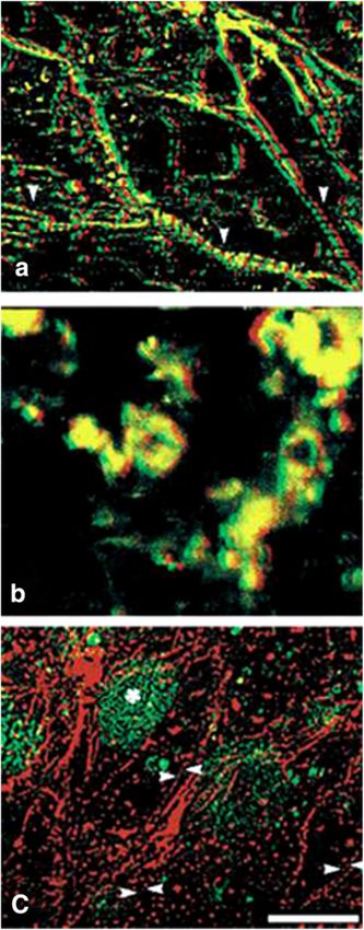
Zajdel et al. Regenerative Medicine Research 2013, 1:8                                                                         Page 2 of 10
http://www.regenmedres.com/content/1/1/8

However, the exact mechanism by which this RNA                        type isoform (TPM1β) of the TPM1 gene (Figure 1 and
modulates the expression of tropomyosin is yet to be                  Table 1). TPM1κ transcripts and its corresponding protein
elucidated. To better understand the mechanism(s)                     are expressed in both axolotl hearts and skeletal muscle
effecting tropomyosin expression in mutant hearts, we                 there appears to be a differential translation of the tran-
undertook an extensive molecular characterization of                  script. Using qRT-PCR the expression level of TPM1κ
the various isoforms of tropomyosin in the Mexican                    transcripts is higher than TPM1α (ratio α:κ is 0.32) in
axolotl.                                                              adult axolotl hearts although TPM1κ protein is less than
                                                                      10% of the total sarcomeric TM, as determined by CH1
Isoform diversity of tropomyosin in vertebrates                       antibody [24]. The opposite is true in adult skeletal muscle
The thin filaments of striated muscle in vertebrate consist           where the level of TPM1κ transcript is significantly lower
of actin, tropomyosin, the troponin (Tn) complex (Tn-I,               compared to TPM1α (ratio of α:κ > 13) but the level of
Tn-C and Tn-T), tropomodulin, and a few other proteins                TPM1κ protein constitutes ~30% of the total sarcomeric
[14]. Actin filaments interaction with Ca +2 governs                  TM [24] Similarly, the levels of expression of TPM1α and
muscle contraction and relaxation. Tropomyosin is a                   TPM1κ transcripts in human hearts are comparable but
coiled coil actin-binding protein found along the length of           the actual TPM1κ protein level is only ~5% of the total
seven actin monomers. A set of four known genes (TPM1,                sarcomeric TM [25]. Comparatively, TPM1α protein con-
TPM2, TPM3 and TPM4) that encode tropomyosin in ver-                  stitutes ~90-95% of the total TM in human hearts [24,25]
tebrates [15-19] give rise to various tropomyosin protein             while TPM1κ is not expressed in human skeletal muscle
isoforms that play important roles in striated, smooth and            at all [26]. TPM1κ transcripts are also expressed in embry-
non-muscle cells. There remains even further diversity in             onic chicken heart but not in adult heart and skeletal
other models such as zebrafish where six tropomyosin                  muscle [27]. It remains unknown if the protein is expressed
genes have been identified [15]. The creation of different            since human TPM1κ antibody may not cross-react with
tropomyosin isoforms occurs through various mecha-                    chicken TPM1κ protein [25]. This discrepancy between
nisms, including the use of different promoters, alternative          transcript and protein levels in the two isoforms in heart
mRNA splicing, different 3’ end mRNA processing and                   tissue suggests that TPM1κ transcripts may undergo
tissue specific translation control [20].                             translational repression.
   We have cloned and sequenced the cDNA of three                        Accounting for the other tropomyosin genes other
sarcomeric TM isoforms from cardiac tissues. These iso-               than TPM1, TPM2α (sarcomeric isoform of the TPM2
forms are designated as TPM1α, TPM1κ, and TPM4α                       gene) is also expressed in mammalian hearts in addition
[21-23] (Table 1). TPM1α, one of nine alternatively                   to the previously described TPM1α and TPM1κ The
spliced isoforms of the TPM1 gene, is known to be the                 sarcomeric isoform of the TPM3 gene, TPM3α, is only
major sarcomeric isoform in mammalian hearts [15-19].                 expressed in slow-twitch skeletal muscle. No sarcomeric
We first identified and characterized another alternatively           isoform of the TPM4 gene is expressed in mammalian
spliced sarcomeric isoform of the TPM1 gene in axolotl                striated muscles because the TPM4 gene is truncated in
hearts [22], designated TPM1κ. TPM1α and TPM1κ have                   mammals [15-17]. On the contrary, TPM4α is a major
an identical exon composition except for exon 2 where                 TM isoform in amphibian cardiac tissues [23,31] and is
TPM1κ contains exon 2a instead of exon 2b (Figure 1 and               the only isoform for sarcomeric TM in adult avian
Table 1). Exon 2a is characteristic of the smooth muscle              hearts [27,30,32].

Table 1 Exon compostion of various high molecular weight TM isoforms with old & new nomenclature [15,17,19,28,29]
Nomenclature of various isoforms TPM Gene encoding the       Various isoforms of TM        Exon composition          Nomenclature used in
of TM referred to in this article Isoforms: New Nomenclature currently known as                                      previous publications
                                  (Old Nomenclature)                                                                 on axolotl
TPM1α                               TPM1(α–TM)               Striated Muscle               1a,2b,3,4,5.6b,7,8,9a/b ATmC-1/α-Tm-1
TPM1β                               TPM1(α-TM)               Smooth Muscle                 1a,2a,3,4,5,6b,7,8,9d     Sm α-Tm
TPM1γ                               TPM1(α-TM)               TM-2Fibroblast                1a,2b,3,4,5,6b,7,8,9d
TPM1δ                               TPM1(α-TM)               TM-3Fibroblast                1a,2b,3,4,5,6a,7,8,9d
TPM1ε                               TPM1(α-TM)               TM-5aFibroblast               1b,3,4,5,6b,7,8,9d
TPM1κ                               TPM1(α-TM)               Novel Striated/Card           1a,2a,3,4,5,6b,7,8,9a/b ATmC-2/α-Tm-2
TPM2α                               TPM2(β-TM)               Striated/Sk Muscle            1a,2b,3,4,5,6b,7,8,9a/b
TPM3α                               TPM3(hTMnm)              Sk.Muscle                     1a,2b,3,4,5,6b,7,8,9a/b
TPM4α                               TPM4(TM4)                StrTM4                        1a,2b,3,4.5,6b,7,8,9a/b ATmC-3/Str.TM-4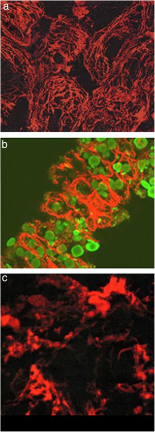
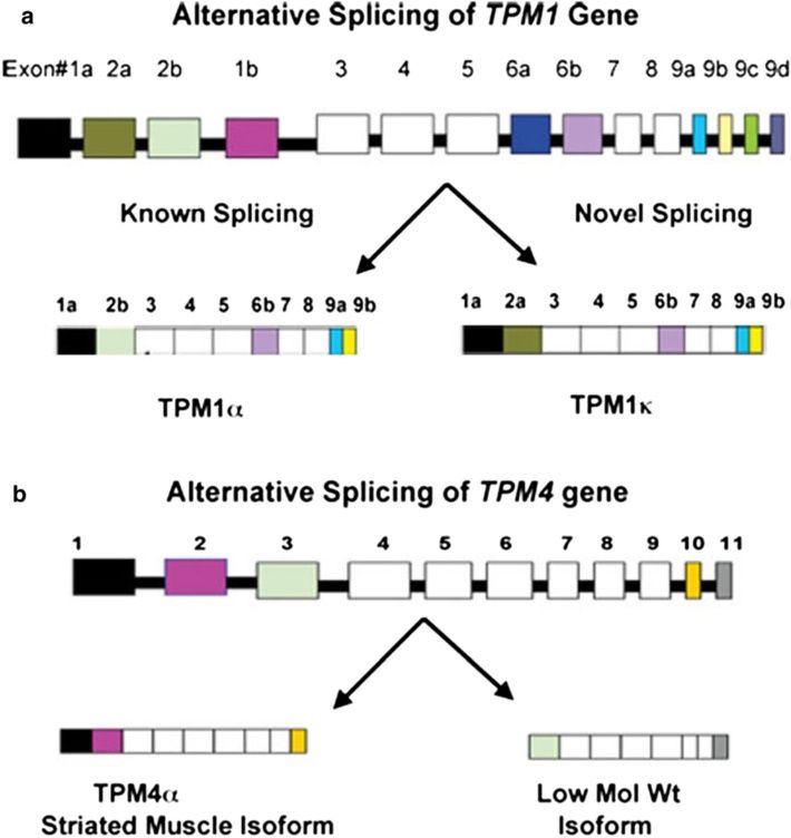
Zajdel et al. Regenerative Medicine Research 2013, 1:8                                                                                Page 3 of 10
http://www.regenmedres.com/content/1/1/8

 Figure 1 Alternative splicing patterns of TPM1 and TPM4 genes. (a) Exon composition of the TPM1 gene and alternative splicing that
 generates two striated muscle specific isoforms -TPM1α and TPM1κ [26]. (b) Exon composition of TPM4 gene (adapted from avian species as
 proposed by Fleenor et al. [30] and alternative splicing generate two isoforms in axolotl, TPM4α and a low-molecular weight TPM4 transcript.

Sarcomeric tm protein in cardiac mutant axolotl hearts                     mutation is the deficiency in tropomyosin protein al-
Among the various myofibril proteins, tropomyosin has                      though the underlying cause of this functional deficit is
been shown by a variety of experiments to be drastically                   less clear. Interestingly, other myofibril structural proteins
reduced in cardiac mutant hearts [2,6,9,12]. Increasing                    such as actin, myosin and myosin binding protein C
the intracellular levels of TM in cardiac mutant heart                     (MyBp-C) were found to be at or near normal levels in
cells via introduction of FITC-labeled exogenous TM pro-                   the mutant hearts [5,28] while one protein, tropomodulin
tein by itself or an expression construct allowing in-vivo                 which is intricately related functionally with sarcomere
TM production subsequently promoted myofibrillogenesis                     maturation is increased [33].
(Figure 2) [9]. Control mutant heart stained with CH1                        The most comprehensive study on the analysis of
monoclonal antibody specific for sarcomeric TM, dem-                       sarcomeric tropomyosin protein expression in normal
onstrated minimal staining when examined by confocal                       and mutant axolotl hearts were reported by Zhang et al.
microscopy (Figure 2b). However, examination of mutant                     [12]. To determine whether multiple isoforms of tropomy-
heart transfected with an expression construct of murine                   osin exist in embryonic axolotl hearts and to verify if they
TPM1α cDNA under the control of mouse α-MYHC pro-                          are differentially regulated in mutant hearts, 2D western
moter demonstrated the formation of organized myofibrils                   blot with the monoclonal antibody (CH1), was performed.
(Figure 2c). The results prove mutant hearts are capable                   Five different protein spots (tropomyosin isoforms)
of forming cardiac myofibrils when provided with suffi-                    from both normal and mutant embryonic hearts at
cient levels of tropomyosin protein. This unequivocally                    stages 36 to 42 were detected (Figure 3). All isoforms of
demonstrated the functional defect in the gene “c”                         tropomyosin detected by the sarcomer specific CH1
Zajdel et al. Regenerative Medicine Research 2013, 1:8                                                                                 Page 4 of 10
http://www.regenmedres.com/content/1/1/8

 Figure 2 Sarcomeric tropomyosin expression in normal, mutant, and TPM1α -transfected mutant hearts. a. Confocal micrograph of
 stage-39 normal hearts stained with CH1 anti-tropomyosin antibdody (and rabbit anti-mouse lissamine rhodamine secondary antibody),
 well-organized sarcomeric myofibrils can be seen throughout the ventricle of the heart (arrow). b. Heart from stage-39 mutant embryo stained
 with CH1 does not show any organized myofibril, Only small areas of amorphous staining can be visualized within the ventricle (arrow).
 c. Stage-36 mutant heart lipofected with an expression construct containing a murine TPM1α cDNA under the control of α-Myosin Heavy Chain
 promoter, which subsequently induced TM and promoted myofibrillogenesis. Mutant heart stained with α-actinin primary antibody.
  Well-organized sarcomeric myofibrils can be seen throughout the heart (arrows). Staining of the Z-lines confirmed the sarcomeric organization
 seen in TPM1α transfected hearts that were stained with tropomyosin primary antibody (results not shown here) [ref]. Interestingly, statge-36
 mutant heart tansfected with a murine TPM2α cDNA under the same promoter did express some TM protein but sarcomeric myofibrils did not
 form throughout the heart in contrast to TPM1α transfected hearts [7].

antibody (30) were located between pI 4 to 5 with mo-                       insufficient transcription of the cardiac specific TPM4α
lecular weight of ~38 kD. The results showed protein                        isoform is highly unlikely. The most plausible explanation
levels of the 4 CH1 recognized TM isoforms were de-                         based on available evidence of TM deficiency in mutant
creased significantly in mutant hearts compared to normal                   hearts is a translational insufficiency of the tropomyosin
hearts (Figure 3a and 3b) [12].                                             transcripts in mutant hearts [12].
   Although mutant axolotl hearts are deficient in sarco-
mere specific TM proteins, mRNA levels of each of                           Molecular analysis and manipulation of tropomyosin
three striated muscle isoforms (TPM1α, TPM1κ, and                           isoforms in normal and mutant axolotl hearts
TPM4α) are comparable in normal and mutant hearts                           As stated earlier, there are at least three striated muscle
[12,23]. Hence, the tropomyosin deficiency in mutant                        isoforms of tropomyosin present in the axolotl. Two iso-
heart is not due to an insufficiency in transcription or                    forms of tropomyosin cDNA have been identified which
post-transcriptional splicing [23]. We cloned and se-                       apparently are derived from the single alpha-tropomyosin
quenced cDNAs of three isoforms from mutant hearts;                         gene (TPM1) through alternative splicing [21,22]. Spinner
no mutation(s) was detected in any of these cDNAs that                      et al. [23] cloned another tropomyosin cDNA, which is
may cause truncated non-functional TM isoform(s). Add-                      the product of a TM4 type tropomyosin gene from axolotl
itionally, we have cloned and sequenced the promoter re-                    heart. An expression construct with each of these isoforms
gion of the TPM4 gene from the DNA isolated from                            upon transfection into mutant hearts canaugment tropo-
normal and mutant axolotl hearts and again, no differ-                      myosin proteinlevels and promotes myofibrillogenesis.
ences were observed [34]. Hence, the possibility of                         The important question is whether or not any one of these

 Figure 3 2-D Western blot analysis of normal and mutant axolotl hearts using CH1 monoclonal antibody. a. Stage-36 normal hearts show
 4 different CH1-recognizable TM isoforms #1, #2, #3, and #4). b. Mutant heart at stage-36 all show 4 CH1-recognizable TM isoforms as in normal
 hearts at much lower levels along with an extra isoform (#5). #5 isoform is detectable in normal hearts at later developmental stages (results not
 shown). The top right corner represents an overexposed blot B [12]. The figure was adapted from Zhang et al. [12].
Zajdel et al. Regenerative Medicine Research 2013, 1:8 Page 5 of 10
http://www.regenmedres.com/content/1/1/8
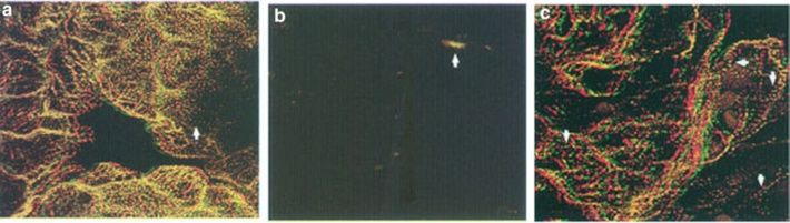
Figure 4 Transfection of isoform-specific sense and anti-sense
oligonucleotides in the ventricle of normal hearts. a. Confocal
microscopy of normal axolotl hearts transfected with TPM1a anti-sense
oligonucleoted and subsequently stained with CH1 monoclonal
antibody. TPM1a anti-sense oligonucleotide did not result in a
drastic disruption of organized myofibrils in the ventricle, which is
comparable with the normal untreated control hearts (figure not
shown). Sarcomeric TM can be seen in most of the cells. Contractility of
the anti-sense treated hearts were not affected. b. TPM1κ anti-sense
transfection disrupted the myofibril organization in normal axolotl heart
compared to TPM1κ sense transfection. Very little organized structure is
seen when examined by the tropomyosin staining. The secondary
antibody is contained within amorphous areas in the cells. c. It
shows that the TPM1κ sense oligonucleoides did not affect the
structure. Since no effect on myofibril structure with TPM1κ sense
was observed, we tagged the oligonucleotide with FITC (green) to
verify its presence and found it to be within the myocytes. Double
staining of the nucleus (green) and the myofibrils at the periphery
of the cells (red) can be seen. The green staining is ovoid in shape
and primarily located at the center of the cells [10]. Confocal z-series
images of stage ~38 embryonic axolotl hearts transfected with either
TPM1κ anti-sense or sense oligonucleotides. Immunodetection of
sarcomeric tropomyosin using CH1 monoclonal antibody is shown
in red. The results depicted by this figure suggests that TPM1κ plays
a critical role in maintaining the myofibrillar structure in embryonic
axolotl hearts.
isoforms alone and/or in various combination(s) is neces-
sary for myofibrllogenesis in axolotl hearts in vivo. In
order to address this issue we developed procedures for
disruption of myofibrils in normal axolotl hearts mim-
icking the mutant hearts by lipofecting antibodies against
sarcomeric TM into normal hearts in situ. Myofibrils in
lipofected normal hearts indeed became greatly disorga-
nized [10,35]. As CH1 antibodies react with all three
sarcomeric tropomyosins, it is not possible to evaluate
the requirement of a particular isoform that is involved
in cardiac myofibrillogenesis. Later we developed anti-
body against TPM1κ in rabbits using a 15-mer peptide
sequence (LDELHKSEESLLTAD) derived from axolotl
exon 2a [36]. Recall, TPM1κ is unique as a sarcomeric
TM in that it contains exon 2a instead of exon 2b which
is found in TPM1α. The affinity purified anti-TPM1κ
antibody upon transfection could disarray the organized
myofibrils in axolotl hearts [37]. The results strongly
suggest that TPM1κ plays a critical role in myofibril
formation in axolotl hearts.
Additionally, isoform specific sense and anti-sense
oligonucleotide was transfected into normal axolotl
hearts. TPM1κ expression was blocked in whole embry-
onic axolotl heart by transfection of exon 2a-specific
anti-sense oligonucleotide (Figure 4b). In contrast, myofi-
brils were unaffected in normal control heart when trans-
fected with FITC label sense oligonucleotides (Figure 4c).
RNA was isolated from treated and untreated hearts and
subsequently RT-PCR was carried out with isoform spe-
cific primer-pairs. The results confirmed the lowerZajdel et al. Regenerative Medicine Research 2013, 1:8 Page 6 of 10
http://www.regenmedres.com/content/1/1/8
Figure 5 Effect of transfection of TM isoform-specific sense- and
anti-sense oligonucleotides on myofibril organization in hearts
from normal axolotl hearts. a. Stereo anaglyph of a 24 section
confocal laser scanning microscope z-series of normal axolotl heart
stained with CH1 anti-tropomyosin antibody. This type of image is
shown because it demonstrates branching myofibrils in whole hearts.
b. Stereo anaglyph of normal hearts transfected with TPM4α exon
2- specific anti-sense, 5′-T*A*C*T*AGCTCGTCCTCAAGC*T*G*C*-3′,
where N* represents the phophothioate blocked oligonucleotide.
Myofibirl organization was disrupted in a majority of the cardio-
myocytes. Most of the tropomyosin appears to be in amorphous
areas when expressed. Some of the cells do not appear to have a
detectable level of tropomyosin. Gross morphology was normal
and the cells appeared to be intact although myofibril structure
was largely disrupted. The contractility was diminished significantly
[36]. c. Normal heart transfected with TPM4α exon 2-specific sense
chimeric olighonucleotide, 5′-fG*C*A*GCTTGAAGGCGAGC
TA*G*T*A*-3, where *N represents phosphothioate blocked
nucleotide, and fG represents G tagged with Fluorescein at the
5′end. This image is a compressed z-series of 2 sections that was
not stereo offset but used to demonstrate double staining. Isolated
pieces of myofibrils that were contained within these sections of
the cardiac cells were stained with tropomyosin antibody (re,
arrowheads). This image is primarily useful for demonstrating the
presence green fluorescence (GFP) within the nuclear area of the
cariomyocytes by five days (asterik). Hoescht staining of the nuclei
in the same heart coincided with the FITC staining that were
localized in a majority of nuclei (figure not shown). The figure was
adapted from Spinner et al. (2004) [36].
transcript expression of TPM1κ in anti-sense treated
hearts. The conclusion was substantiated by the in vitro
analysis of the specificity of the TPM1κ anti-sense oligo-
nucleotides used in this study. Confocal analysis of the
sense and anti-sense oligonucleotide transfected normal
axolotl hearts was carried out after staining with anti-
tropomyosin antibody (CH1). Immunohistochemical ana-
lysis unequivocally confirmed that the inhibition of the ex-
pression of TPM1κ disrupted myofibril structure of the
myofibrils in anti-sense transfected normal axolotl hearts.
In contrast, TPM1α anti-sense oligonucleotide did not
cause a disruption of the myofibrillar organization in axo-
lotl hearts (Figure 4a).
In a separate study, we found that the antisense TPM4α
oligonucleotide disrupted myofibril formation and inhib-
ited beating in normal axolotl hearts, while the sense
strands did not. A fluorescein-tagged sense oligonucleo-
tide clearly showed that the oligonucleotide was intro-
duced within the cells of intact hearts. The results
implicate the essential role of TPM4α in cardiac myofibril-
logenesis (Figure 5) [8,10,36].
Differential expression of tropomyosin in conus and the
ventricle
Despite the dramatic and lethal effects of the homozy-
gous cardiac gene “c” mutation in the axolotl ventricle,
the conus of the heart beats and has organized myofi-
brils (Figure 6). In order to understand whether variousZajdel et al. Regenerative Medicine Research 2013, 1:8 Page 7 of 10 http://www.regenmedres.com/content/1/1/8 Figure 6 (See legend on next page.)
Zajdel et al. Regenerative Medicine Research 2013, 1:8 Page 8 of 10
http://www.regenmedres.com/content/1/1/8
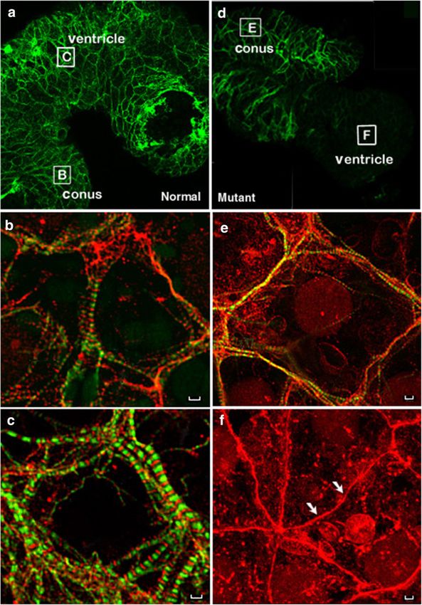
(See figure on previous page.)
Figure 6 Tropmodulin but not tropomyosin found in cardiac “c” mutant axolotl ventricle. CH1 anti-sarcomeric TM antibody labels the
conus but not the ventricle in stage 38/39 cardiac mutant axolotl hearts. Stage 38/39 normal (a–c) and mutant (d–f) axolotl hearts were double
stained for immunofluorescent microscopy with CH1 anti-sarcomeric TM antibody (FITC channel) and polyclonal anti-tropomodulin antibody,
R1749 (Rhodamine channel). Only the FITC channel is shown for whole heart images (a, d) demonstrating the lack of staining in the ventricle of
mutant hearts while all higher magnification images utilize a dual rhodamine-FITC filter. Letter labeled boxes in A and D correspond with the
area shown in their respective higher magnification image. TM labeled with CH1 antibody is found in a cross-striated pattern in the ventricle
of normal hearts (c) and in the conus of both normal (b) and mutant (e) hearts. Tropomyosin staining is markedly absent in mutant ventricle
(f) resulting in a failure of normal sarcomere formation with tropmodulin found in linear arrays near the surface of the membranes (Arrows, F).
Scale Bar = 100 μm.
TM isoforms are differentially expressed in different beating in the mutant (c/c) axolotl hearts in situ [11].
segments of the heart and whether the known TM iso- Zhang et al. [12] demonstrated that the MIR gene is es-
forms contribute to myofibril formation in a segment sential for tropomyosin (TM) expression in axolotl hearts
specific manner, we employed anti-sense oligonucleotides during development at the level of translation or post-
to separately knockdown post-transcriptional expression translation. qRT-PCR using isoform-specific primer-pairs
of TPM1α and TPM4α in axolotl heart segments. We showed that mRNA expression of three sarcomeric tropo-
evaluated the organization of myofibrils in the conus and myosin isoforms (TPM1α, TPM1κ, and TPM4α) in un-
ventricle of normal and cardiac mutant hearts using im- treated mutant hearts and in normal hearts knocked down
munohistochemical techniques. We concluded that the with double-stranded MIR (dsMIR) are similar to un-
TPM1α isoform, a product of the TPM1 gene, was essen- treated normal. However, at the protein level, sarcomeric
tial for myofibrillogenesis in the conus, whereas TPM4α, tropomyosin isoforms detected with CH1 monoclonal
the striated muscle isoform of the TPM4 gene, was essen- antibodies, are significantly reduced in mutant and dsMIR
tial for myofibrillogenesis in the ventricle. Our results sup- treated normal hearts. However, this study neither showed
port the segmental theory of vertebrate heart development the mechanism by which MIR may induce sarcomeric TM
and suggest the conus is a different transcriptional tissue synthesis in axolotl hearts nor addressed the role of spe-
unit. Since the conus is an outflow tract structure and in cific tropomyosin isoforms in cardiac myofibrillogenesis.
humans is a conical pouch of the right ventricle from Recently, Kochegarovr et al. [40] randomly cloned RNAs
which the pulmonary artery arises it will be interesting to from fetal human heart. RNA from one of the clones (clone
examine tropomyosin isoform diversity in these two seg- #291) was found to promote myofibril formation in mutant
ments in other systems including humans [8]. Develop- axolotl in situ. This RNA induced expression of cardiac
ment of the conus appears to be unaffected in the mutant markers in mutant hearts: tropomyosin, troponin and α-
heart and is comparable to the normal heart. The different syntrophin. The nucleotide sequences of the cloned RNA
functions of the heart segments would suggest that differ- matches in partial with the human microRNA-499a and b,
ent isoforms could be needed in accordance with that although it differs in length. qRT-PCR data suggest this
function. The physiologic characteristics necessary for the RNA may induce the TPM4α (ATmC-3) isoform in mutant
ventricle versus the conus and subsequent outflow tract heart, producing more sarcomeric tropomyosin protein
are different [38]. Isoform diversity in specific heart seg- and subsequently promote myofibril formation. The mech-
ments includes tropomyosin but also could include other anism by which this non-coding RNA induces tropomyosin
sarcomeric proteins such as myosin heavy chain [39] Fur- in mutant hearts may well be different from that of MIR,
ther studies could examine the localization and function which acts at the post transcriptional level [12].
of specific isoforms in adult heart segments. There has
also been research on isoform diversity and the relation- Review and conclusions
ship to diseases such as dilated cardiomyopathy [25]. Although our complete understanding of the mechanism
These studies suggest that with changing physiologic pa- of tropomyosin expression in mutant axolotl hearts as
rameters, the isoforms can also be changed. Ultimately, well as the nature and function of gene “c” is far from
the study of segment specific tropomyosin isoforms may over, we would like to end this review with a positive
help in the understanding of time and function specific note. Our finding of sarcomeric TM isoform TPM1κ in
sarcomeric proteins and their relationship to regeneration axolotl led to the discovery of this isoform in human
of heart function in damaged hearts. hearts [26]. Unlike in axolotl, it is not expressed in hu-
man skeletal muscle. Most importantly, an upregulation
Promotion of myofibrillogenesis in mutant hearts in situ of TPM1κ protein has been reported in hearts from
by a non-coding rna human dilated cardiomyopathy patients [25]. However, it
A noncoding RNA, Myofibril-Inducing RNA (MIR) is is not yet known whether the upregulation of TPM1κ is
capable of promoting myofibrillogenesis and heart the cause or a consequence of cardiomyopathy in thisZajdel et al. Regenerative Medicine Research 2013, 1:8 Page 9 of 10
http://www.regenmedres.com/content/1/1/8
patient. Our anti-sense experiments suggest strongly the 2. Lemanski LF: Morphology of developing heart in cardiac lethal mutant
functional significance of TPM1κ in axolotl hearts. In Mexican axolotls, ambystoma mexicanum. Devel Biol 1973, 33:312–333.
3. Voss GJ, Kump DK, Walker JA, Voss SR: Variation of Salamender tail
addition, the lower expression level of TPM1κ protein in regeneration is associated with genetic factors that determine tail
axolotl heart and skeletal muscle [24] and also in human morphology. PLos ONE 2013, 8(7):e67274. doi:10,1371/journal.pone.0067274.
hearts [25] points towards translational repression of 4. Cano-Martinez A, Vargas-Gonzaleez A, Guarner-Lans V, Prado-Zayago E,
Leon-Oleda M, Nieto-Lima B: Functional and structural regeneration in the
TPM1κ. Further, upon injection intraperitoneally into axolotl heart (Ambystom mexicanum) after partial ventricular
juvenile axolotl, Shz-1, a cardiogenic small molecule, aug- amputation. Arch Cariol Mexi 2010, 80:79–86.
mented the expression levels of transcripts of TPM1α, 5. Lemanski LF: Role of tropomyosin in actin filament formation in
embryonic salamander heart cells. J Cell Biol 1979, 82:227–238.
TPM1κ, and TPM4α in hearts. But the increased tran- 6. LaFrance SM, Lemanski LF: Imunofluorescent confocal analysis of
script level did not resulted into increased sarcomeric TM tropomyosin in developing hearts of normal and cardiac mutant
protein expression [41]. Finally, although the transcript axolotls. Int J Devel Biol 1994, 38:695–700.
7. Zajdel RW, Dube DK, Lemanski LF: The cardiac mutant axolotl is a unique
levels of all TPM isoforms in normal and mutant axolotl animal modelfor evaluation of cardiac myofibrillogenesis. Exp Cell Res
heart ventricles are comparable, the proteins of all three 1999, 248:557–566.
isoforms are diminished significantly. This observation 8. Zajdel RW, MeLean MD, Denz CR, Dube S, Thurston H, Poiesz BJ, Dube DK:
Differential expression of tropomyosin during segmental heart
also point towards the translational repression of TM in development in Mexican axolotl. J Cell Biochem 2006, 99:952–965.
cardiac tissues [42]. The evidence for translational control 9. Zajdel RW, McLean MD, Lemanski SL, Muthuchamy M, Wieczorek DF,
of sarcomeric TM in mammalian hearts was originally Lemanski LF, Dube DK: Ectopic expression of tropomyosin promotes
myofibrillogenesis in mutant axolotl hearts. Dev Dynamics 1998,
came from the works from the laboratory of Dr. David 213:412–420.
Wieczorek, University of Cincinnati, Cincinnati, OH 10. Zajdel RW, McLean MD, Lemanski LF, Dube DK: Alteration of cardiac
[29,43]. Rethinesamy et al. [29] and Blanchard et al. [44] myofibrillogenesis by lipofectin-mediated delivery of exogenous proteins
and nucleic acids into whole embryonic hearts. Anat Embryol 2000,
independently ablated one of the two alleles of the TPM1
210:217–228.
gene in mice that resulted in half of the TPM1α transcripts 11. Lemanski LF, Nakatsugawa M, Bhatia R, Erginel-Unaltuna N, Spinner BJ, Dube
in ablated mice hearts compared to wild-type. However, DK: A specific synthetic RNA promotes cardiac myofibrillogenesis in the
TPM1α protein level was unchanged in ablated mice Mexican axolotl. Biochem Biophys Res Com 1996, 229:974–981.
12. Zhang C, Jia P, Huang X, Sferrazza GF, Athauda G, Achary MP, Wang J,
hearts suggesting a higher translational efficiency of Lemanski SL, Dube DK, Lemanski LF: Myofibril-inducing RNA (MIR) is
TPM1α transcripts in ablated mice hearts. Again, Rajan essential for tropomyosin expression and myofibrillogenesis in axolotl
et al. [25] reported that although the level of transcripts hearts. J Biomed Sci 2009, 16:81.
13. Zhang C, Dube DK, Huang X, Zajdel RW, Bhatia R, Foster D, Lemanski SL,
of TPM1α and TPM1κ human hearts is parallel, TPM1κ Lemanski LF: A point mutation in bioactive RNA results in the failure of
protein is only ~5% of the total sarcomeric TM whereas mutant heart correction in Mexican axolotls. Anat Embryol 2003,
TPM1α protein constitutes about 90-95% of the total 206:495–506.
14. Perry SV: Vertebrate tropomyosin, properties and function. J Muscle Res
sarcomeric TM. The results strongly suggest the trans- Cell Motil 2001, 22:5–49.
lational repression of TPM1κ transcripts in human 15. Schevzov G, Whittaker SP, Fath T, Lin JJ, Gunning PW: Tropomyosin
hearts. The immediate future goal of our laboratory is isoforms and reagents. Bioarchitecture 2011, 1:135–164.
16. Gunning P, O’Neill G, Hardmen E: Tropomyosin-based regulation of actin
to explore further the translational repression of tropo- cytoskeleton in time and space. Physiol Rev 2008, 88:1–35.
myosin expression in vertebrate hearts as well as to find 17. Lees-Miller JP, Helfman DM: The molecular basis for tropomyosin isoform
out the functional role of TPM1κ. diversity. Bioessays 1991, 13:429–437.
18. Wieczorek DF: Regulation of alternatively spliced alpha-tropomyosin gene
expression by nerve extract. J Bol Chem 1988, 263:10456–10463.
Competing interests
19. Pittenger MF, Kazzaz JA, Helfman DM: Functional properties of non-muscle
The authors declare that they have no competing interests.
tropomyosin isoforms. Curr Opin Cell Biol 1994, 1:96–104.
20. Piples K, Wieczorek DF: Tropomyosin 3 increases striated muscle isoform
Authors’ contributions diversity. Biochemistry 2000, 39:8291–8297.
As evident from the cited literature, each of the authors has been involved 21. Luque EA, Lemanski LF, Dube DK: Molecular cloning, sequencing and
in this research for a long time and each of us contributed equally for expression of atropomyosin form cardiac muscle of the Mexican axolotl,
writing this review article. All authors have read and approved the final ambystoma mexicanum. Biochem Biophys Res Comm 1994, 203:319–325.
version. 22. Luque EA, Spinner BJ, Dube S, Dube DK, Lemanski LF: Differential expression
of a novel isoform of alpha-tropomyosin in cardiac and skeletal muscle of
Acknowledgement the Mexican axolotl (Ambystoma mexicanum). Gene 1997, 185:175–180.
The work in this laboratory was supported by grants from American Heart 23. Spinner BJ, Lemanski LF, Dobbins N, Dube DK: Transient expression of the
Association (both National & New York State affiliate), CNY Children’s Miracle cardiac specific TM4-type tropomyosin suggest a unique role in heart
Network, Syracuse, NY, grants from Golisano Children’s Hospital, Syracuse, NY development in the Mexican axolotl. J Cell Biochem 2002, 85:747–761.
to DKD, an AHA (NY Affiliate) grant to RW. 24. Thomas A, Rajan S, Thurston H, Masineni L, Sreeharsha N, Dube P, Bose A,
Muthu V, Dube T, Wieczorek DF, Dube DK: Expression of a novel
Received: 9 August 2013 Accepted: 8 October 2013 tropomyosin isoform in axoltl heart and skeltel muscle. J Cell Biochem
Published: 4 December 2013 2010, 110:875–881.
25. Rajan S, Jagatheesan G, Karam CN, Alves ML, Bodi I, Schwartz A, Buclago CF,
References D’Souza KM, Akhter SA, Boivin GP, Dube DK, Petrasheveskaya N, Herr AB,
1. Humphrey RR: Genetic and experimental studies on a mutant gene (c) Hullin R, Liggett SB, Beata MW, Solaro JR, Wieczorek DF: Molecular and
determining absence of heart action in embryos of the Mexican axolotl functional characterization of a novel cardiac-specific human
(Ambystoma mexicanum). Dev Biol 1972, 27:365–375. tropomyosin isoform. Circulation 2010, 121:410–418.Zajdel et al. Regenerative Medicine Research 2013, 1:8 Page 10 of 10
http://www.regenmedres.com/content/1/1/8
26. Denz CR, Narshi A, Zajdel RW, Dube DK: Expression of a novel cardiac-
specific tropomyosin isoform in humnas. Biochem Biophys Res Comm
2004, 320:1291–1297.
27. Zajdel RW, Denz CR, Lee S, Dube S, Ehler E, Perriard E, Periard J-C, Dube DK:
Identification, characterization, and expression of a novel alpha
tropomyosin isoform in cardiac tissue in developing chicken. J Cell
Biochem 2003, 89:427–439.
28. Ward SM, Dube DK, Fransen ME, Lemanski LF: Differential expression of
C-protein isoforms in the developing heart of normal and cardiac lethal
mutant axolotls (Ambystoma mexicanum). Devel Dynamics 1996,
205:93–103.
29. Rethinasamy P, Muthuchamy M, Hewett T, Boivin G, Wolska BM, Evans C,
Solaro RJ, Wieczorek DF: Molecular and physiological effects of
alpha-tropomyosin ablation in the mouse. Circ Res 1998, 82:116–123.
30. Fleenor DE, Hickman KH, Lindquester GJ, Devlin RB: Avian cardiac
tropomyosin gene produces tissue-specific isoforms through alternative
TNA splicing. J Muscle Res Cell Motel 1992, 13:55–63.
31. Hardy S, Theze N, Lepetit D, Allo MR, Thiebaud P: The Xenopus laevis TM-4
gene encodes non-muscle and cardiac tropomyosin isoforms through
alternative splicing. Gene 1995, 156:265–270.
32. Forry-Schaudies S, Gruber CE, Hughes SH: Chicken tropomyosin and a low
molecular weight non-muscle tropomyosin are related by alternative
splicing. Cell Growth Diff 1990, 1:473–481.
33. McLean MD, Zajdel RW, Dube S, Thurston H, Dube DK: Tropomodulin
expression in developing hearts of normal and cardiac mutant Mexican
axolotl. Cardiovasc Toxicol 2006, 6:85–98.
34. Denz CR, Zhang C, Jia P, Du J, Huang X, Dube S, Thomas A, Poiesz BJ, Dube
DK: Absence of mutation at the 5’-upstream promoter region of the
TPM4 gene from cardiac mutant axolotl. (Ambystoma mexicanum)
Cardiovasc Toxicol 2011, 11:235–243.
35. Zajdel RW, Denz CR, Narshi A, Dube S, Dube DK: Anti-sense-mediated
inhibition of expression of the novel striated tropomyosin isoform
TPM1κ disrupts myofibril organization in embryonic axolotl hearts. J Cell
Biochem 2005, 95:840–848.
36. Spinner BJ: Molecular analysis of isoform diversity of cardiac tropomyosin in
the Mexican axolotl. Upstate Medical University, Department of Anatomy
and Cell Biology: Ph.D. thesis; 1998.
37. Zajdel RW, McLean MD, Denz CR, Dube S, Lemanski LF, Dube DK:
Manipulation of myofibrillogenesis in whole heart. In Myofibrillogenesis.
Edited by Dube DK. Boston: Birkhauser; 2002:87–100.
38. Satin J, Fujii S, DeHaan RI: Devekopment of cardiac beat rate in early
chick embryos is regulated by regional cues. Dev Biol 1988, 129:103–113.
39. Zhang R, Xu X: Transient and transgenic analysis of the zebrafish
ventricular myosin hearvy chain (vmhc) promoter: an ingibitory
mechanism of ventricle-specific gene expression. Dev Dyn 2009,
238:1564–1573.
40. Kochegarov A, Moses A, Lian W, Meyer J, Michael C, Hanna MC, Lemanski
LF: A new unique form of microRNA from human heart, microRNA-499c,
promotes myofibril formation and rescues cardiac development in
mutant axolotl embryos. J Biomed Sci 2013, 20:20.
41. Pinnamaneni S, Dube S, Welch C, Shrestha R, Benz PM, Lynn Abbott L, Poiesz
BJ, Dube DK: Effect of Shz-1, a cardiogenic small molecule, on expression of
tropomyosin in axolotl heart. American Based Res J 2013, 2:24–40.
42. Dube DK, Benz PM, Dube S, Poiesz BJ: Do we know the complete story of
TPM1κ expression in vertebrate hearts? J Cytol Histol 2012, 3:3.
43. Schevzov G, Fath T, Vrhovski B, Vlahovich N, Rajan S, Hook J, Joya JE,
Lemckert F, Puttur F, Lin JJ, Hardeman EC, Wieczorek DF, O’Neill GM,
Gunning PW: Divergent regulation of the sarcomere and the
cytoskeleton. J Biol Chem 2008, 283:275–283. Submit your next manuscript to BioMed Central
44. Blanchard EM, Iizuka K, Christe M, Conner DA, Geisterfer-Lowrance A, and take full advantage of:
Schoen FJ, Maughan DW, Seidman CE, Seidman JG: Targeted ablation of
the murine alpha-tropomyosin gene. Circ Res 1997, 81:1005–1010. • Convenient online submission
• Thorough peer review
doi:10.1186/2050-490X-1-8
Cite this article as: Zajdel et al.: Expression of tropomyosin in relation to • No space constraints or color figure charges
myofibrillogenesis in axolotl hearts. Regenerative Medicine Research • Immediate publication on acceptance
2013 1:8.
• Inclusion in PubMed, CAS, Scopus and Google Scholar
• Research which is freely available for redistribution
Submit your manuscript at
www.biomedcentral.com/submitYou can also read