Hereditary multiple osteochondromas in Jordanian patients: Mutational and immunohistochemical analysis of EXT1 and EXT2 genes
←
→
Page content transcription
If your browser does not render page correctly, please read the page content below
ONCOLOGY LETTERS 21: 151, 2021

        Hereditary multiple osteochondromas in Jordanian patients:
              Mutational and immunohistochemical analysis
                         of EXT1 and EXT2 genes
          ZIYAD MOHAIDAT1, KHALDON BODOOR2, ROWIDA ALMOMANI3, MOHAMMED ALORJANI4,
             MOHAMMAD‑AKRAM AWWAD5, AUDAI BANY‑KHALAF6 and KHALID AL‑BATAYNEH7

    1
     Orthopedic Division, Special Surgery Department, Faculty of Medicine, Jordan University of Science and Technology,
     King Abdullah University Hospital, Irbid 22110; 2Department of Applied Biology, Faculty of Science; 3Department of
      Laboratory Medical Sciences, Faculty of Science; 4Department of Pathology, Faculty of Medicine, Jordan University
     of Science and Technology, Irbid 22110; 5Department of Clinical Sciences, Faculty of Medicine, Yarmouk University,
    Irbid 21110; 6Orthopedic Division, Special Surgery Department, Faculty of Medicine, Jordan University of Science and
        Technology, Irbid 22110; 7Department of Biology, Faculty of Sciences, Yarmouk University, Irbid 21110, Jordan

                                        Received May 28, 2020; Accepted November 26, 2020

                                                    DOI: 10.3892/ol.2020.12412

Abstract. The aim of the present study was to investigate            Jordanian patients with HMO, who may represent an ethnic
the molecular characteristics of hereditary multiple osteo‑          group that is infrequently investigated, were males and had
chondromas (HMO) in a subset of Jordanian patients with              a mild clinical disease course; whereas most patients with
a focus on the genetic variants of exostosin (EXT1)/(EXT2)           EXT1 gene mutations were not necessarily associated with a
and their protein expression. Patients with HMO and their            severe clinical disease course. The role of EXT2 gene remains
family members were included. Recorded clinical character‑           a subject of debate, since patients with EXT1 mutations alone
istics included age, sex, tumors number and location, joint          did not express the non‑mutated EXT2 gene.
deformities and associated functional limitations. Mutational
analysis of EXT1 and EXT2 exonic regions was performed.              Introduction
Immunohistochemical staining for EXT1 and EXT2 was
performed manually using two different commercially avail‑           Hereditary multiple osteochondromas (HMO) is characterized
able rabbit anti‑human EXT1 and EXT2 antibodies. A total             by multiple cartilage‑capped bony projections (exostoses) that
of 16 patients with HMO from nine unrelated families were            usually arise from the metaphysis of long bones (1). It is also
included, with a mean age of 13.9 years. A total of 75%              known as hereditary multiple exostoses, multiple cartilagi‑
(12/16) of the patients were male and (69%) (11/16) had a mild       nous exostoses, osteochondromas and diaphyseal aclasis (2).
disease (class I). EXT mutation analysis revealed only EXT1          The prevalence of HMO is ~1:50,000, and the male: Female
gene mutations in 13 patients. Seven variants were detected,         ratio is 1.5:1 (3,4). Although HMO can be asymptomatic and
among which three were novel: c.1019G>A, p. (Arg340His),             diagnosed incidentally, it can disrupt bone growth and cause
c.962+1G>A and c.1469del, p. (Leu490Argfs*9). Of the                 short stature, unequal limb lengths and joint deformities with
16 patients, 3 did not harbor any mutations for either EXT1 or       significant morbidity (5,6). The most serious complication of
EXT2. Immunohistochemical examination revealed decreased             HMO is the malignant transformation into chondrosarcoma,
expression of EXT1 protein in all patients with EXT1 muta‑           occurring in 0.5‑5% of the patients (6). Therefore, clinical
tion. Surprisingly, EXT2 protein was not detected in these           and radiological follow up is crucial for the management of
patients, although none had EXT2 mutations. The majority of          patients with HMO. However, there is currently no standard
                                                                     follow‑up protocol for HMO. Genetic analysis of EXT genes
                                                                     to identify patients with HMO at higher risk of developing
                                                                     severe disease or malignant transformation may contribute to
Correspondence to: Dr Ziyad Mohaidat, Orthopedic Division,           the future management of such patients.
Special Surgery Department, Faculty of Medicine, Jordan University       HMO is an autosomal dominant inherited disease with
of Science and Technology, King Abdullah University Hospital,        a penetrance of 100% (5). Genetic analysis of HMOs in
1 Alramtha Street, Irbid 22110, Jordan                               different populations identified two main causative genes,
E‑mail: zmmohaidat@just.edu.jo                                       namely exostosis 1 (EXT1) and exostosis 2 (EXT2) (5,7,8).
                                                                     Mutations in the EXT1 and EXT2 genes account for >90% of
Key words: hereditary multiple osteochondromas, exostosis,           all HMO cases (7,9). EXT1 and EXT2 both encode for a glyco‑
exostosin 1, exostosin 2, novel mutations                            syltransferase required for heparan sulfate (HS) synthesis and
                                                                     polymerization as HS proteoglycans (HSPGs) (8). The role
                                                                     of EXT1 and EXT2 proteins in HS synthesis involves the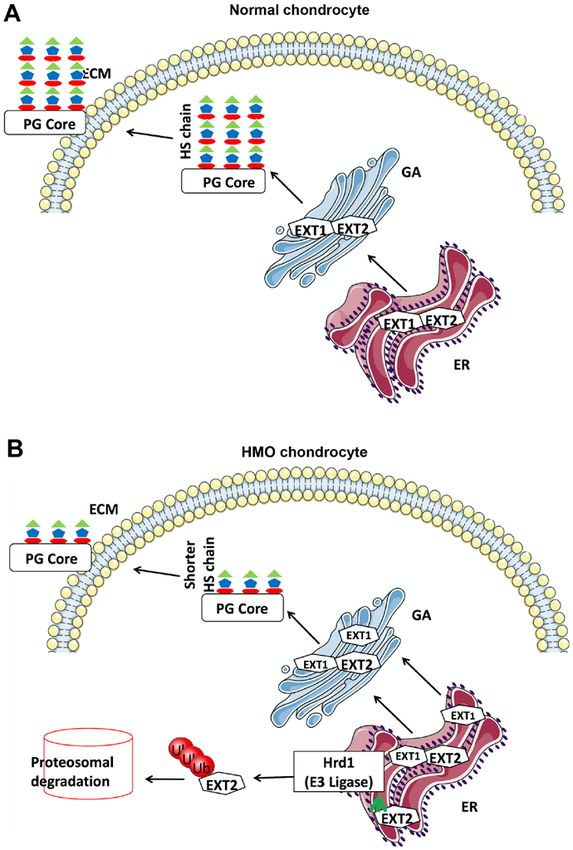
2                  MOHAIDAT et al: ROLE OF EXT1 AND EXT2 IN HEREDITARY MULIPLE OSTEOCHONDROMAS

formation of heterocomplex of both proteins (1). HSPGs play     Table I. HMO severity score described by Mordenti et al (10).
a key role in the regulation of different signaling pathways
involved in chondrocyte proliferation and differentiation in    Class                                      Subclass
the growth plate (8).
    The present study investigated 16 Jordanian index cases     I: No deformities‑no
from nine different unrelated families with confirmed           functional limitations
diagnosis of HMO. The different clinical characteristics in       IA                          ≤5 sites with exostosis
addition to the mutational spectrum of the EXT genes and the      IB                          >5 sites with exostosis
expression of their corresponding proteins were evaluated in    II: Deformities‑no
this group of patients.                                         functional limitations
                                                                  IA                          ≤5 sites with deformities
Materials and methods                                             IB                          >5 sites with deformities
                                                                III: Deformities‑functional
Patients with HMO. The present study was conducted at           limitations
several Orthopedic Surgery clinics over a 2‑year period           IIIA                        1 site with functional limitations
between January 2018 and December 2019. A total of
                                                                  IIIB                        >1 site with functional limitations
42 individuals were included in this study, among which
16 were diagnosed with HMO. Of the patients, 12 were
men and 4 were women who had a mean age of 13.9 years
(age range, 6‑27 years). HMO diagnosis was confirmed            an ABI 310 DNA sequencer (Applied Biosystems; Thermo
by either histopathological or radiological examinations.       Fisher Scientific, Inc.).
These patients were evaluated clinically, and their available       The obtained sequences were compared with the normal
radiological examinations were reviewed. Disease severity       EXT1 (NM_000127) and EXT2 (NM_000401) genes refer‑
was determined according to the severity score described by     ence sequences and chromatograms were visualized by
Mordenti et al (10) (Table I). Blood samples were collected     using the ChromasPro 1.34 (Technelysium Pty Ltd.) software
from all participants. Paraffin‑embedded tissues of patients    package or Mutation Surveyor (V4.07; SoftGenetics, LLC).
with HMO who underwent surgical excision were avail‑            Sequence nomenclatures for the coding and noncoding vari‑
able from the archives of Pathology Department. Informed        ants were described in accordance with the Human Genome
consent was obtained from all individuals who participated      Variation Society Nomenclature standards (http://www.hgvs.
in this study. The study protocol was approved by the Human     org/mutnomen). To assess and predict the impact of newly
Research Ethics Committee.                                      identified missense variants, the Mutation taster (http://www.
                                                                mutationtaster.org/) and Polyphen2 programs (http://genetics.
Molecular analysis. Genomic DNA from the patients and           bwh.harvard.edu/pph2/index.shtml) were used.
their available family members was extracted from peripheral
blood samples using a Gentra Puregene kit (Qiagen GmbH)         Immunohistochemistry. Formalin fixed, paraffin embedded
following the manufacturer's instructions. The quality and      HMO tissues were used in this study for immunohistochemical
concentration of the DNA was determined by NanoDrop             staining of EXT1 and EXT2. Tissues were processed following
2000 V7.3.1 (Thermo Fisher Scientific, Inc.). All exons and     the manufacturer's instructions. Immunostaining for EXT1
exon‑flanking intron sequences of EXT1 (NM_000127)              and EXT2 was performed manually on the sample sections
and EXT2 (NM_000401) genes were amplified by PCR                using two different commercially available rabbit anti‑human
(polymerase chain reaction). PCR was performed in a final       EXT1(dilution 1:200; cat. no. abx100786; Abbexa Ltd.; and
volume of 25 µl containing 40 ng genomic DNA, 1X Master         cat. no. HPA044394; Sigma‑Aldrich; Merck KGaA) and rabbit
Mix (GoTaq® Green Master Mix; Promega Corporation),             anti‑human EXT2 (dilution 1:200; cat. no. abx03435; Abbexa
and 5 pmol of forward and reverse primers (Table II). The       Ltd.; and cat. no. SAB2108124; Sigma‑Aldrich; Merck KGaA)
following thermocycling conditions were used: Initial           antibodies. Tissue sections were observed under a light micros‑
denaturing step (95˚C for 7 min) followed by 40 cycles of       copy at a magnification of x40. Tissues known to express
95˚C for 1 min, annealing at 60˚C for 90 sec, extension step    EXT1 and EXT2 (normal femoral head cartilage) were used
at 72˚C for 90 sec and final extension at 72˚C for 7 min. PCR   as positive control, and negative controls were created by omit‑
was performed using an iCycler (Bio‑Rad Laboratories,           ting the primary antibody step. The scoring criteria for EXT1
Inc.). The PCR products were separated by 2% agarose gel        and EXT2 were as follow: 0, 0‑10%; 1, 10‑30%; 2, 30‑85%;
electrophoresis, visualized by ethidium bromide and the         and 3, >85%. The intensity of the reaction was scored as: 0,
products were purified using the Norgen's PCR Purification      negative; 1, weak; 2, moderate; and 3, strong. The samples that
kit (cat. no. 45700; Norgen, Bioteck Corp.). GAPDH was          were scored as 1 or more were considered as positive.
used as the loading control and for normalization. Sanger
sequencing was performed in both sense and antisense direc‑     Results
tions by using the BigDye Terminator Cycle Sequencing
kit version 3.1 (Applied Biosystems; Thermo Fisher              Patients. A total of nine families (A‑I) with 42 members were
Scientific, Inc.), according to the manufacturer's protocol.    included in the present study. These families included a total
Sequencing reactions were purified using the NucleoSEQ kit      of 16 patients with HMO, and three families (B, E and F) only
(Macherey‑Nagel GmbH) and final analysis performed using        had one affected member (Table III).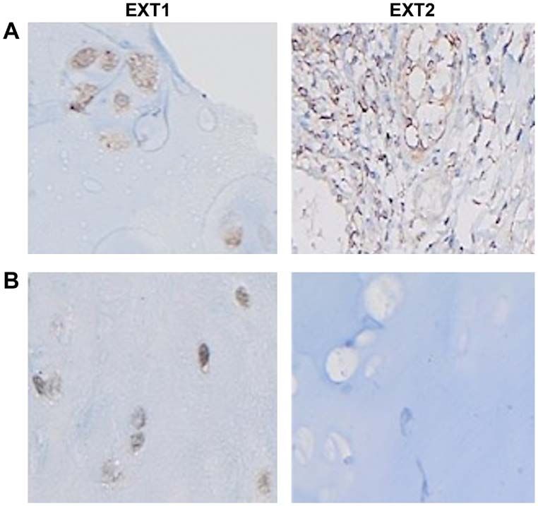
ONCOLOGY LETTERS 21: 151, 2021 3
Table II. Primer sequences used for PCR amplification and Sanger sequencing.
Gene Exon Forward (5'‑3') Reverse (5'‑3')
EXT1 Exon 1 CGAGCGCAGGAGTAAACACC CGTTTTTTGGCCTGCATGTG
Exon 1 GAGCTGAAAGTGTTGATTGG GAGACTCTGCACCTTTGGATC
Exon 1 CCTCTTTGTCCTGAGTCTGG CCATCCCCCAACTTCACACC
Exon 2 CCCCACATTCGCAATGAGTC GAGAGGTGATAATGTTAAACCC
Exon 3 CTGATTGGAACAGCTTCTGCTG TGAAAGTTTGGACGGGGGCAGC
Exon 4 GTGCATCTCTTTGTTTTACAG GCTGAGAGAAGTGTATAAAGG
Exon 5 CCTTTCCAAATATCATCAGG GGCCTTTAGTTCTGTATGAC
Exon 6 GAGCAAGGAGGAGTAATTTTC ATAACAGGTAAGGAGGGCGG
Exon 7 AAGAGGCTTTGGGTTGGAGG AAGTGCCCCATGGAGAAAC
Exon 8 GGGAGAATTGTCCTGAAAAC ATCGTGCAACATGAGGTGAC
Exon 9 TTAGTGGGGAGAAGGTAATG TTCCTATTTATGCAGCAGCC
Exon 10 GTCTCAGAAGTCCACTTGTC ACGTGAGTCCTCATTACCTG
Exon 11 CCTTGCACTTCTCTCATCATTATCC GAAGAGAGAGCAGCTTGACC
EXT2 Exon 1 GCCTGAATATAAGCACCTAC AAAAGCGGGCAGTCATTGTC
Exon 2 TCAAGTGTCATTTGCCATCC CCCTTCCCTTTAGTTCCCTG
Exon 3 GGCTTGGGGATCCTTGATAG ACTTCTAAATCTTCAGGAGG
Exon 4 ACTCTGTAAACGTTAGCTGG AGGACCCTACCCTGTAACTG
Exon 5 TCAGTGGAGGTGAAGACTGG CATAGGCCAAGCAGCTTTGC
Exon 6 GTATTGCTTGGCGTCAACCC GTAGTAGTTCTTGAACCAGG
Exon 7 GGATGTTGTTTCTGCTTGTG ACTCAGGCATTCAGCTCCTG
Exon 8 CCTGGAGTTGACTATGATAG TTATGCTGCCCTTATCAGGC
Exon 9 CATGTTTGGGTTTGCTGACG AAATGGAGGCATGCTGTCTC
Exon 10 GGATACAAGCTGATTCTCCC GCACACCTTTTGGACTCTAC
Exon 11 TGGAACATCTCCAGAATCCC AAGCCCTCTTGGCAGGTATG
Exon 12 TATGAGAGAAAGCTTGTCCC CCAATGTGACCGCATCAATC
Exon 13 CATGCAACATCTCAGCTTAC ACTATGGCTACCAGCTGCTG
Exon 14 CAGACTGTGGCTACTTGAGC AGTAGGTCAACCTTCCACCC
These patients had a mean age of 13.9 years at their initial variant [c.96dup, p.(Ser33Glufs*11)] and one splice site
diagnosis (range, 6‑27 years). A total of 75% (12/16) of the variant (c.962+1G>A) (11,12). Three of the seven variants:
patients were males. The total number of tumors was 135, with c.1019G>A, p.(Arg340His); c.962+1G>A; and c.1469del,
over half (58%) being located around the knee. According to p.(Leu490Argfs*9) were novel since they were not found in
HMO severity classification by Mordenti et al (10) (Table I), known databases such as ExAC (http://exac.broadinstitute.
most (69%) (11/16) of these patients had a mild disease org/), GenomeAD (https://gnomad.broadinstitute.org) and
(class I) (Table III). Moderate (class II) and severe (class III) dbSNP (https://www.ncbi.nlm.nih.gov/snp). The [c.1019G>A,
disease forms were recorded in 2 (13%) and 3 (19%) patients, p. (Arg340His] variant was identified in two unrelated fami‑
respectively (Table III). lies (B‑II.1, C‑II.2, C‑III.1 and C‑III.2; Table III).
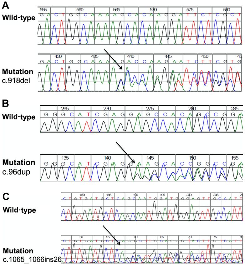
In family A, a novel heterozygous variant c.918del, p.
EXT1 and EXT2 genes mutational analysis. Mutational (Lys306Asnfs*53) was identified in three affected patients
analysis of the 16 patients from nine families (A‑I) and (A‑I.1, A‑II.1 and A‑II.3; Table III and Fig. 1). This deletion
their family members for both EXT1 and EXT2 genes variant creates a frameshift starting at codon Lys306 and a
revealed different heterozygous mutations in only EXT1 new reading frame ends in a new stop codon at position 53
gene. While 13 patients (77%) from seven unrelated families downstream of the mutation (Table III and Fig. 2A). Unaffected
harbored these EXT1 mutations, the remaining 3 patients individuals in this family were wild type for this variant.
(23%) from two families were negative for both EXT1 and A second novel variant [c.96dup; p. (Ser33Glufs*11)] was
EXT2 genetic variation in the targeted sequenced regions. detected in family D (D‑I.1 and D‑II.1; Table III and Fig. 2B).
Seven different genetic variants were identified in the EXT1 This one‑base‑pair duplication variant in exon 1 creates a
gene. These variants consisted of; two missense variants frameshift starting at codon Ser33 and the new reading frame
[c.1019G>A, p. (Arg340His); and c.82T>A, p. (Phe28Ile)], is predicated to end in a stop codon at position 11 (Table III
two deletions variants [c.918del, p.(Lys306Asnfs*53); and Fig. 2B).
and c.1469del, p.(Leu490Argfs*9)], one insertion variant A third novel heterozygous variant was identified in family
[c.1065_1066ins26, p.(Val356Cysfs*12)], one duplication F (F‑II.3) which was an insertion of 26 base pairs (bps) in4
Table III. Clinical severity class and pathogenic EXT1 gene variants identified.
EXT1 EXT2
Age at Clinical protein protein
diagnosis severity Nucleotide change Clinical Novel IHC IHC
Family Patient Sex (years) class (EXT1 gene) Genomic position Protein level significance mutation staining staining
A I.1 M 20 IB c.918del g.119122368del p. (Lys306Asnfs*53) Likely pathogenic Yes RE No
II.1 F 7 IB (Exon 1)
II.3 F 7 IB
B II.1 M 10 IB c.1019G>A g.118849384C>T p. (Arg340His) Pathogenic Noa RE No
(Exon 2) VUS
c.82T>A g.119123204A>T p. (Phe28Ile) Yes
(Exon 1)
C II.2 M 18 IIIB c.1019G>A g.118849384C>T p. (Arg340His) Pathogenic Noa RE No
III.1 M 12 IA (Exon 2)
III.2 M 15 IA
D I.1 M 6 IA c.96dup g.119123190dup p. (Ser33Glufs*11) Likely pathogenic Yes RE No
II.1 M 27 IB (Exon 1)
E II.1 M 6 IIA c.1469del g.118831982del p. (Leu490Argfs*9) Pathogenic Nob RE No
(Exon 6)
F II.3 M 7 IB c.1065_1066ins26 g.118847781_118847782ins26 p. (Val356Cysfs*12) Likely pathogenic Yes RE No
(Exon 3)
G I.1 F 10 IIIA c.962+1G>A g.119122323C>T p.? Pathogenic Noc RE No
II.4 M 25 IA (Donor splice site of intron 1)
H I.1 M 24 IB None
I 1.1 F 20 IIIA None
11.1 M 9 IIA None
a
MOHAIDAT et al: ROLE OF EXT1 AND EXT2 IN HEREDITARY MULIPLE OSTEOCHONDROMAS
Ref. (11). bRef. (12). chttps://www.ncbi.nlm.nih.gov/clinvar/variation/642916/. HMO, hereditary multiple osteochondromas; M, male; F, female; RE, reduced expression; VUS, variant of undetermined
significance; IHC, immunohistochemical.ONCOLOGY LETTERS 21: 151, 2021 5
Figure 1. Pedigrees of families (A-G) with EXT1 genetic variants leading to HMO. (A) Family A, (B) Family B, (C) Family C, (D) Family D, (E) Family E,
(F) Family F, (G) Family G. Squares, males; circles, females; solid symbols, affected individuals with heterozygous mutations; open symbols, unaffected
individuals. EXT1 genotypes: Mu, mutant allele; wt, wild‑type allele. EXT, exostosin; HMO; hereditary multiple osteochondromas.
exon 3 [c.(1065_1066ins26); p. (Val356Cysfs*12)] (Table III variant [c.96dup; p. (Ser33Glufs*11)] was detected in this
and Fig. 2C). This variant was predicted to create a frame shift family. With regards to the EXT2 gene, no protein expression
mutation starting at codon Val356 and a new reading frame was detected in any osteochondroma tissues from the 12 tested
ends in a new stop codon at downstream position 12. patients (Fig. 3B).
Careful medical evaluation of all family members
(affected and unaffected individuals) of families B, E and F, Discussion
who only had one affected member each (Table III and Fig. 1)
was performed. The tested mother and father of these families Several HMO studies investigated the associations of the
were clinically normal with no signs of HMO. Furthermore, different clinical characteristics with the genetic findings in
genetic testing for the presence of a causative genetic variant different populations. The clinical characteristics of patients
of the parents showed negative mutation results. The affected with HMO in the present study, including the mean age at
individuals (B‑II.1, E‑II.1 and F‑II.3) in these families carry diagnosis of 13.9 years, as well as male predilection, were
a de novo mutation and were the first ones who gained the similar to what was reported in the literature (1,13). The knee
mutant allele. joint was the most common tumor location, which is also
consistent with the findings of other studies (14‑16).
Immunohistochemical staining of EXT1 and EXT2. HMO severity was reported using various clinical
Osteochondroma tissue was available for immunohisto‑ classifications (10). These classifications were based on
chemical staining from 12 of the 13 patients with EXT1 the clinical parameters of HMO patients, including, age,
gene mutations. EXT1 protein expression was found to be tumor number, joint deformities, limb length discrepancy,
significantly decreased (weak staining pattern; Fig. 3B) in in addition to the morbidity associated with HMO tumors.
all examined tissues, apart from tissues obtained from the 2 The clinical classification, used in the present study,
proband patients of family D (D‑I.1 and D‑II.1). Their tissues revealed that the majority of Jordanian patients with HMO
exhibited moderate staining pattern when compared to EXT1 have a mild disease form (class 1). Since different HMO
protein expression in normal chondrocytes (Fig. 3A). A novel studies (14,17,18) used different scoring systems to assess6 MOHAIDAT et al: ROLE OF EXT1 AND EXT2 IN HEREDITARY MULIPLE OSTEOCHONDROMAS
Figure 2. Sanger sequencing data of (A-C) the three novel variants identified in the present study, patients (mutation) and controls (wild‑type). (A) Novel
Mutation c.918del, (B) Novel Mutation c.96dup, (C) Novel Mutation c.1065_1066ins26. EXT, exostosin; HMO; hereditary multiple osteochondromas.
HMO severity, comparing HMO severity among different in disease severity between these two gene mutations (19,24).
populations can be difficult. In the present study, only 2 of the 13 patients with EXT1 muta‑
HMO is not only clinically heterogenous, but it is also tions exhibited a severe form of the disease (class III). This can
genetically heterogenous (6,17,18). Mutational analysis be attributed to the variability of HMO severity, even among
studies reported variable frequencies of EXT1 and EXT2 patients with the same EXT gene mutations particularly
mutations in different ethnic groups (13). Several authors EXT1 gene (18,25). In addition, ethnicity can be considered
reported EXT1 mutations to be more common, particularly in as another influential factor. This is also consistent with the
Caucasians (7,19‑21). Although the present study investigated findings of a previous study investigating osteochondroma
a different ethnic group, it revealed a similar predominance in Jordanian patients in whom a milder form of HMO was
of EXT1 mutations in this group of Jordanian patients with observed compared with that of other populations (4).
HMO. On the other hand, no potential pathogenic genetic vari‑ With regards to the mutational analysis of the patients in the
ants of EXT2 gene were identified in the present study. This present study, EXT1 allelic heterogeneity was observed and the
was inconsistent with other studies which reported that EXT2 identified mutations were shown to be dispersed throughout the
mutations to be present in 20‑45% of the patients (7). This coding regions of the gene (Fig. 4). Furthermore, the truncated
inconsistency may be explained by the presence of mutations mutations occurred in 66.6% of the tested families. These
in noncoding parts of the genes. In addition, the small number results are similar to those reported from different studies in
of the included patients can be a contributing factor. the literature (9). A mutational database from these studies
Several phenotype‑genotype studies of HMO reported is assembled in the Multiple Osteochondromas Mutation
that EXT1 gene mutations were more likely than EXT2 gene Database (MOdb) (http://medgen.ua.ac.be/LOVDv.2.0/), with
mutations to be associated with a more severe form of the >600 and 200 different mutations in EXT1 and EXT2, respec‑
disease (14,15,18,20‑23). Other studies reported no difference tively. The majority of these mutations (80%) are nonsense,ONCOLOGY LETTERS 21: 151, 2021 7
Figure 3. Immunohistochemical staining of chondrocytes. (A) Immunohistochemical staining of EXT1 and EXT2 from control unaffected tissues (magni‑
fication, x40). (B) Immunohistochemical staining of EXT1 and EXT2 from a patient with hereditary multiple osteochondromas (magnification, x40). EXT,
exostosin.
Figure 4. Structure of the EXT1 gene, with the positions of the identified genetic variants indicated. Gray boxes indicate 5' and 3' untranslated region, and black
boxes indicate EXT1 exonic region. EXT, exostosin.
whereas the remaining 20% are frameshift mutations and mechanism of the disease, which involved a 80.7 kb intronic
splice‑junction mutations, causing an early termination of deletion of EXT1gene and a 68.9‑kb duplication proximal
translation or partial/complete deletion of the gene and loss of of EXT 1 (28). Furthermore, genetic variants in the 5' and
protein function (7). 3'UTRs, deep intronic causing variants or in the promoter
In the present study, 19% (3/16) of patients with HMO regions were not determined in the present study. In addition,
had no point mutations in the coding regions for either EXT1 several studies have reported that 10‑15% of patients with
or EXT2 genes. This may be explained by variants involving HMO have no mutations in either EXT1 or EXT2 genes (29‑31),
large rearrangements such as deletions, duplications, inver‑ suggesting that other genes may be involved in the pathogen‑
sions, translocations or somatic mosaicism, that include the esis of the disease. Therefore, testing this subgroup of patients
EXT1 and EXT2 genes. Deletion of a single or multiple exons by whole exome sequencing or whole genome next generation
were previously detected in ~10% of all HMO cases (7,26,27). sequencing will be an attractive approach to identify other
Another study reported a complex rearrangement as causative possible disease‑causing gene(s) (32,33).8 MOHAIDAT et al: ROLE OF EXT1 AND EXT2 IN HEREDITARY MULIPLE OSTEOCHONDROMAS Figure 5. Model of the function of the EXT1/EXT2 complex in HS synthesis in (A) normal chondrocytes and (B) HMO chondrocytes. (A) In normal chondrocytes, EXT1 and EXT2 form a complex, which is involved in the HS synthesis in the Golgi apparatus. The EXT1/EXT2 complex, through its galac‑ tosyltransferase activity, aids in the formation of HS proteoglycans HSPGs, which are next exported to the cell exterior. (B) In HMO chondrocytes, decreased levels of EXT1 disrupts the stoichiometry of the EXT1/EXT2 complex, resulting in low HS synthesis and, thus, diminished HSPGs on the cell exterior. Low levels of EXT1 render EXT2 unable to be transported into the Golgi complex, which is instead retained in the ER and thereby targeted for ER‑associated protein degradation. EXT2 is shown to be ubiquitinated at Lys 245 and is degraded though ubiquitin‑proteasome system. HS, heparan sulfate; HSPGs, HS proteoglycans; EXT, exostosin; HMO; hereditary multiple osteochondromas; ER, endoplasmic reticulum; ECM, extracellular matrix; GA, Golgi apparatus; PG, proteoglycans. To the best of our knowledge, the majority of pheno‑ EXT2 mutations. Previous studies demonstrated that the pres‑ type‑genotype research studies on HMO did not test for ence of fully functional EXT1 and EXT2 proteins is required the expression of EXT genes in the resected tumor tissues. for their correct localization in the Golgi complex (36,37). In Immunohistochemistry studies revealed a decreased expres‑ addition, the present study proposes a model (Fig. 5), in which sion of EXT1 and/or EXT2 corresponding to the EXT gene mutations in EXT1 result in a truncated product and/or inac‑ mutations status (3,34,35). In the present study, the patients tive form of the protein that can no longer bind to its EXT2 with EXT1 mutations exhibited a decreased expression of partner. The very low levels of EXT1, EXT2 and EXT1/2 EXT1 protein. Surprisingly, the same patients exhibited no alter the stoichiometry of the complex and greatly diminish its expression of EXT2 proteins, although none of them harbored glycosyltransferase activity. Low levels of EXT1 can no longer
ONCOLOGY LETTERS 21: 151, 2021                                                    9

associate with its requisite partner EXT2 and, thus, EXT2 is         Acknowledgements
retained in the endoplasmic reticulum (ER) and is targeted
through the ER‑associated protein degradation pathway                Not applicable.
for degradation. The identification of EXT2 protein as a
substrate for the Hrd1 E3 ligase and the identification of the       Funding
lysine involved in ubiquitin attachment to the protein (38,39)
is consistent with the aforementioned model, as explained            The present study was supported by the Jordan University of
in Fig. 5.                                                           Science and Technology (grant no. 20180318).
    Mutations in EXT1 and EXT2 are associated with the
pathogenesis of HMO; however, the mechanism through                  Availability of data and materials
which HS synthesis alteration leads to exostoses has yet to
be elucidated. Heterozygous EXT1 or EXT2 mutations are               Sanger sequencing data are available at https://www.ebi.
common molecular changes identified in >80% of the inves‑            ac.uk/eva with the following accessions: Project, PRJEB41290;
tigated exostoses (40). There remains the question of whether        analyses, ERZ1673834. The following links can also be
osteochondromas arise via loss of heterozygosity or haploinsuf‑      used: https://www.ebi.ac.uk/ena/data/view/PRJEB41290,
ficiency mechanism (40,41). The results of the present study and     https://www.ebi.ac.uk/eva/?eva‑study=PRJEB41290 and
early biochemical studies (36,37,42) of the EXT1/2 complex           https://wwwdev.ebi.ac.uk/eva/?eva‑study=PRJEB41290.
suggest that haploinsufficiency for either EXT1 or EXT2 affects
the ability of chondrocytes to synthesize HS, as explained in        Authors' contributions
Fig. 5. Although EXT1 and EXT2 are ubiquitously expressed,
mutations in these genes are only manifested in chondrocytes,        ZM performed the collection and analysis of data, study design,
suggesting that chondrocytes require two fully active EXT1 and       writing and editing of the manuscript. KB performed the data
EXT2 proteins. However, Reijnders et al (41) refuted the haplo‑      analysis, study design, writing and editing of the manuscript.
insufficiency theory and demonstrated that osteochondromas           MA was responsible for the study design and writing of the
arise via loss of heterozygosity and inactivation of both alleles    manuscript. RA performed the data collection, study design
(Knudson's two‑hit model) (43). Further genetic analysis studies     and writing of the manuscript. MAA performed the data
of large cohorts of patients with HMO are required to determine      collection, study design and writing of the manuscript. ABK
the contribution of LOH and haploinsufficiency in the molecular      cotributed to the data collection, study design and writing of
pathogenesis of the disease.                                         the manuscript. KAB performed the data analysis and writing
    The main limitation of the present study was the lack of         of the manuscript. All authors read and approved the final
DNA samples from some members of family C (unaffected                manuscript.
individuals I.1 and I.2), and so genetic testing or further inves‑
tigations for mosaic mutations (pyrosequencing or cloning of         Ethics approval and consent to participate
the suspected PCR products) could not be conducted.
    In conclusion, the present study conducted a pheno‑              Approval for this research was obtained from the Human
type‑genotype study of HMO in 16 Jordanian patients from             Research Ethics Committee and IRB at Jordan University of
nine families. These patients are representative of an ethnic        Science and Technology (approval no. 22/116/2018). All individ‑
group in which the genetic background of HMO is infre‑               uals included in this study and/or their legal guardians provided
quently investigated. The majority of these patients were            written informed consent for participating in this study.
males, diagnosed the age of ~14 years, and exhibited a mild
clinical disease form. Genetic analysis revealed mutations           Patient consent for publication
exclusively involving EXT1 gene and none involved EXT2
gene. These mutations were not necessarily associated with a         All participants and/or their legal guardians provided a written
severe clinical disease. Three of the identified mutations were      informed consent regarding the publication of case details and
novel. Three patients did not show any mutations for either          any associated images.
EXT1 or EXT2 genes. Upon immunohistochemical testing,
osteochondroma tissue resected form all patients with EXT1           Competing interests
mutations exhibited decreased expression of EXT1 protein.
Surprisingly, EXT2 protein was not detected in these patients        The authors declare that they have no competing interests.
although none had EXT2 mutations. Therefore, a model may
be suggested that questions the role of EXT2 gene in HMO             References
pathogenies. HMO continues to represent a clinically and
genetically heterogenous disease among different ethnic               1. D'arienzo A, Andreani L, Sacchetti F, Colangeli S and
                                                                         Capanna R: Hereditary multiple exostoses: Current insights.
groups. Therefore, further genetic and immunohistochemical               Orthop Res Rev 11: 199‑211, 2019.
studies are required to further elucidate the pathogenesis            2. Hakim DN, Pelly T, Kulendran M and Caris JA: Benign tumours
of HMO. In addition, mutational analysis studies can be                  of the bone: A review. J Bone Oncol 4: 37‑41, 2015.
                                                                      3. Bovée JV: Multiple osteochondromas. Orphanet J Rare Dis 3: 3,
helpful in screening for patients with HMO, particularly                 2008.
those who may be at a risk of developing a severe form of the         4. Mohaidat ZM, Saleh AA, Al‑Omari MH, Obeidat AA and
                                                                         Khasawneh RA: Osteochondroma in Jordanian Patients: Clinical
disease, which will have a significant impact on the clinical            manifestations and management. J Clin Diagnostic Res 12:
management and follow up of patients with HMO.                           RC11‑RC15, 2018.
10 MOHAIDAT et al: ROLE OF EXT1 AND EXT2 IN HEREDITARY MULIPLE OSTEOCHONDROMAS
5. Beltrami G, Ristori G, Scoccianti G, Tamburini A and 24. Jennes I, Entius MM, Van Hul E, Parra A, Sangiorgi L and
Capanna R: Hereditary multiple exostoses: A review of clinical Wuyts W: Mutation screening of EXT1 and EXT2 by denaturing
appearance and metabolic pattern. Clin Cases Miner Bone high‑performance liquid chromatography, direct sequencing
Metab 13: 110‑118, 2016. analysis, fluorescence in situ hybridization, and a new multiplex
6. de Souza AM and Bispo Júnior RZ: Osteochondroma: Ignore or ligation‑dependent probe amplification probe set in patients with
investigate? Rev Bras Ortop 49: 555‑564, 2014. multiple osteochondromas. J Mol Diagn 10: 85‑92, 2008.
7. Jennes I, Pedrini E, Zuntini M, Mordenti M, Balkassmi S, 25. Cousminer DL, Arkader A, Voight BF, Pacifici M and Grant SFA:
Asteggiano CG, Casey B, Bakker B, Sangiorgi L and Wuyts W: Assessing the general population frequency of rare coding vari‑
Multiple osteochondromas: Mutation update and description of ants in the EXT1 and EXT2 genes previously implicated in
the multiple osteochondromas mutation database (MOdb). Hum hereditary multiple exostoses. Bone 92: 196‑200, 2016.
Mutat 30: 1620‑1627, 2009. 26. Vink GR, White SJ, Gabelic S, Hogendoorn PC, Breuning MH
8. Zak BM, Crawford BE and Esko JD: Hereditary multiple exos‑ and Bakker E: Mutation screening of EXT1 and EXT2 by direct
sequence analysis and MLPA in patients with multiple osteochon‑
toses and heparan sulfate polymerization. Biochim Biophys dromas: Splice site mutations and exonic deletions account for more
Acta 1573: 346‑355, 2002. than half of the mutations. Eur J Hum Genet 13: 470‑474, 2005.
9. Busse‑Wicher M, Wicher KB and Kusche‑Gullberg M: The 27. Szuhai K, Jennes I, De Jong D, Bovée JV, Wiweger M, Wuyts W
extostosin family: Proteins with many functions. Matrix Biol 35: and Hogendoorn PC: Tiling resolution array‑CGH shows that
25‑33, 2014. somatic mosaic deletion of the EXT gene is causative in EXT
10. Mordenti M, Ferrari E, Pedrini E, Fabbri N, Campanacci L, gene mutation negative multiple osteochondromas patients. Hum
Muselli M and Sangiorgi L: Validation of a new multiple osteo‑ Mutat 32: E2036‑E2049, 2011.
chondromas classification through Switching Neural Networks. 28. Waaijer CJ, Winter MG, Reijnders CM, de Jong D, John Ham S,
Am J Med Genet Part A 161A: 556‑560, 2013. Bovée JV and Szuhai K: Intronic deletion and duplication prox‑
11. Fusco C, Nardella G, Fischetto R, Copetti M, Petracca A, imal of the EXT1 gene: A novel causative mechanism for multiple
Annunziata F, Augello B, D'Asdia MC, Petrucci S, Mattina T, et al: osteochondromas. Genes Chromosomes Cancer 52: 431‑436, 2013.
Mutational spectrum and clinical signatures in 114 families with 29. Hong G, Guo X, Yan W, Li Q, Zhao H, Ma P and Hu X:
hereditary multiple osteochondromas: Insights into molecular Identification of a novel mutation in the EXT1 gene from a
properties of selected exostosin variants. Hum Mol Genet 28: patient with multiple osteochondromas by exome sequencing.
2133‑2142, 2019. Mol Med Rep 15: 657‑664, 2017.
12. Bali DS, Goldstein JL, Banugaria S, Dai J, Mackey J, Rehder C 30. Zhang H, Ping XL, Lee PK, Wu XL, Yao YJ, Zhang MJ,
and Kishnani PS: Predicting cross‑reactive immunological Silvers DN, Ratner D, Malhotra R, Peacocke M and Tsou HC:
material (CRIM) status in Pompe disease using GAA mutations: Role of PTCH and p53 genes in early‑onset basal cell carcinoma.
Lessons learned from 10 years of clinical laboratory testing Am J Pathol 158: 381‑385, 2001.
experience. Am J Med Genet Part C Semin Med Genet 160C: 31. Wu Y, Xing X, Xu S, Ma H, Cao L, Wang S and Luo Y: Novel and
40‑49, 2012. recurrent mutations in the EXT1 and EXT2 genes in Chinese kindreds
with multiple osteochondromas. J Orthop Res 31: 1492‑1499, 2013.
13. Santos SCL, Rizzo IMPO, Takata RI, Speck‑Martins CE, 32. Liu H, Wu S, Duan L, Zhu W, Zhang S, Hu X, Jia W, Yang G,
Brum JM and Sollaci C: Analysis of mutations in EXT1 and Liu C, Li W, et al: Identification of a novel EXT1 mutation in
EXT2 in Brazilian patients with multiple osteochondromas. patients with hereditary multiple exostosis by exome sequencing.
Mol Genet Genomic Med 6: 382‑392, 2018. Oncol Rep 33: 547‑552, 2015.
14. Jäger M, Westhoff B, Portier S, Leube B, Hardt K, Royer‑Pokora B, 33. Zhang F, Liang J, Guo X, Zhang Y, Wen Y, Li Q, Zhang Z, Ma W,
Gossheger G and Krauspe R: Clinical outcome and genotype in Dai L, Liu X, et al: Exome sequencing and functional analysis
patients with hereditary multiple exostoses. J Orthop Res 25: identifies a novel mutation in EXT1 gene that causes multiple
1541‑1551, 2007. osteochondromas. PLoS One 8: e72316, 2013.
15. Porter DE, Lonie L, Fraser M, Dobson‑Stone C, Porter JR, 34. Cheung PK, McCormick C, Crawford BE, Esko JD, Tufaro F and
Monaco AP and Simpson AH: Severity of disease and risk Duncan G: Etiological point mutations in the hereditary multiple
of malignant change in hereditary multiple exostoses. A exostoses gene EXT1: A functional analysis of heparan sulfate
genotype‑phenotype study. J Bone Joint Surg Br 86: 1041‑1046, polymerase activity. Am J Hum Genet 69: 55‑66, 2001.
2004. 35. Hameetman L, David G, Yavas A, White SJ, Taminiau AH,
16. Saglik Y, Altay M, Unal VS, Basarir K and Yildiz Y: Cleton‑Jansen AM, Hogendoorn PC and Bovée JV: Decreased
Manifestations and management of osteochondromas: A retro‑ EXT expression and intracellular accumulation of heparan
spective analysis of 382 patients. Acta Orthop Belg 72: 748‑755, sulphate proteoglycan in osteochondromas and peripheral chon‑
2006. drosarcomas. J Pathol 211: 399‑409, 2007.
17. Li Y, Wang J, Wang Z, Tang J and Yu T: A genotype‑phenotype 36. McCormick C, Duncan G, Goutsos KT and Tufaro F: The putative
study of hereditary multiple exostoses in forty‑six Chinese tumor suppressors EXT1 and EXT2 form a stable complex that
patients. BMC Med Genet 18: 126, 2017. accumulates in the Golgi apparatus and catalyzes the synthesis
18. Francannet C, Cohen‑Tanugi A, Le Merrer M, Munnich A, of heparan sulfate. Proc Natl Acad Sci USA 97: 668‑673, 2000.
Bonaventure J and Legeai‑Mallet L: Genotype‑phenotype corre‑ 37. Kobayashi S, Morimoto K, Shimizu T, Takahashi M, Kurosawa H
lation in hereditary multiple exostoses. J Med Genet 38: 430‑434, and Shirasawa T: Association of EXT1 and EXT2, hereditary
2001. multiple exostoses gene products, in Golgi apparatus. Biochem
19. Delgado MA, Martinez‑Domenech G, Sarrión P, Urreizti R, Biophys Res Commun 268: 860‑867, 2000.
Zecchini L, Robledo HH, Segura F, De Kremer RD, Balcells S, 38. Lee KA, Hammerle LP, Andrews PS, Stokes MP, Mustelin T,
Silva JC, Black RA and Doedens JR: Ubiquitin ligase substrate
Grinberg D and Asteggiano CG: A broad spectrum of genomic identification through quantitative proteomics at both the protein
changes in latinamerican patients with EXT1/EXT2‑CDG. Sci and peptide levels. J Biol Chem 286: 41530‑41538, 2011.
Rep 4: 6407, 2014. 39. Ernst R, Claessen JH, Mueller B, Sanyal S, Spooner E,
20. Pedrini E, Jennes I, Tremosini M, Milanesi A, Mordenti M, van der Veen AG, Kirak O, Schlieker CD, Weihofen WA and
Parra A, Sgariglia F, Zuntini M, Campanacci L, Fabbri N, et al: Ploegh HL: Enzymatic blockade of the ubiquitin‑proteasome
Genotype‑phenotype correlation study in 529 patients with pathway. PLoS Biol 8: e1000605, 2011.
multiple hereditary exostoses: Identification of ‘protective’ and 40. Hall CR, Cole WG, Haynes R and Hecht JT: Reevaluation of
‘risk’ factors. J Bone Joint Surg Am 93: 2294‑2302, 2011. a genetic model for the development of exostosis in hereditary
21. Sarrión P, Sangorrin A, Urreizti R, Delgado A, Artuch R, multiple exostosis. Am J Med Genet 112: 1‑5, 2002.
Martorell L, Armstrong J, Anton J, Torner F, Vilaseca MA, et al: 41. Reijnders CM, Waaijer CJ, Hamilton A, Buddingh EP, Dijkstra SP,
Mutations in the EXT1 and EXT2 genes in Spanish patients with Ham J, Bakker E, Szuhai K, Karperien M, Hogendoorn PC, et al:
multiple osteochondromas. Sci Rep 3: 1346, 2013. No haploinsufficiency but loss of heterozygosity for EXT in
22. Heinritz W, Hüffmeier U, Strenge S, Miterski B, Zweier C, multiple osteochondromas. Am J Pathol 177: 1946‑1957, 2010.
Leinung S, Bohring A, Mitulla B, Peters U and Froster UG: New 42. Busse M and Kusche‑Gullberg M: In vitro polymerization of heparan
mutations of EXT1 and EXT2 genes in German patients with sulfate backbone by the EXT proteins. J Biol Chem 278: 41333‑41337, 2003.
multiple osteochondromas. Ann Hum Genet 73(Pt 3): 283‑291, 43. Knudson AG Jr: Mutation and cancer: Statistical study of retino‑
2009. blastoma. Proc Natl Acad Sci USA 68: 820‑823, 1971.
23. Alvarez C, Tredwell S, De Vera M and Hayden M: The geno‑
type‑phenotype correlation of hereditary multiple exostoses. This work is licensed under a Creative Commons
Clin Genet 70: 122‑130, 2006. Attribution-NonCommercial-NoDerivatives 4.0
International (CC BY-NC-ND 4.0) License.You can also read