RFC1 expansions are a common cause of idiopathic sensory neuropathy
←
→
Page content transcription
If your browser does not render page correctly, please read the page content below
doi:10.1093/brain/awab072                                                                                     BRAIN 2021: 144; 1542–1550             | 1542

RFC1 expansions are a common cause of
idiopathic sensory neuropathy
Riccardo Currò,1,2 Alessandro Salvalaggio,3 Stefano Tozza,4 Chiara Gemelli,5,6

                                                                                                                                                              Downloaded from https://academic.oup.com/brain/article/144/5/1542/6272840 by guest on 30 September 2021
Natalia Dominik,7 Valentina Galassi Deforie,7 Francesca Magrinelli,8,9
Francesca Castellani,3 Elisa Vegezzi,1,2 Pietro Businaro,1,2 Ilaria Callegari,1,2
Anna Pichiecchio,1,2 Giuseppe Cosentino,1,2 Enrico Alfonsi,2 Enrico Marchioni,2
Silvia Colnaghi,2 Simone Gana,2 Enza Maria Valente,2,10 Cristina Tassorelli,1,2
Stephanie Efthymiou,7 Stefano Facchini,7 Aisling Carr,7 Matilde Laura,7
Alexander M. Rossor,7 Hadi Manji,7 Michael P. Lunn,7 Elena Pegoraro,3
Lucio Santoro,4 Marina Grandis,5,6 Emilia Bellone,5,6,11 Nicholas J. Beauchamp,12
Marios Hadjivassiliou,13 Diego Kaski,14,15 Adolfo M. Bronstein,14 Henry Houlden,7
Mary M. Reilly,7 Paola Mandich,5,6,11 Angelo Schenone,5,6 Fiore Manganelli,4
Chiara Briani3 and Andrea Cortese1,7

See Roberts (doi:10.1093/brain/awab150) for a scientific commentary on this article.

After extensive evaluation, one-third of patients affected by polyneuropathy remain undiagnosed and are labelled
as having chronic idiopathic axonal polyneuropathy, which refers to a sensory or sensory-motor, axonal, slowly
progressive neuropathy of unknown origin. Since a sensory neuropathy/neuronopathy is identified in all patients
with genetically confirmed RFC1 cerebellar ataxia, neuropathy, vestibular areflexia syndrome, we speculated that
RFC1 expansions could underlie a fraction of idiopathic sensory neuropathies also diagnosed as chronic idiopathic
axonal polyneuropathy.
We retrospectively identified 225 patients diagnosed with chronic idiopathic axonal polyneuropathy (125 sensory
neuropathy, 100 sensory-motor neuropathy) from our general neuropathy clinics in Italy and the UK. All patients
underwent full neurological evaluation and a blood sample was collected for RFC1 testing.
Biallelic RFC1 expansions were identified in 43 patients (34%) with sensory neuropathy and in none with sensory-
motor neuropathy. Forty-two per cent of RFC1-positive patients had isolated sensory neuropathy or sensory neur-
opathy with chronic cough, while vestibular and/or cerebellar involvement, often subclinical, were identified at
examination in 58%. Although the sensory ganglia are the primary pathological target of the disease, the sensory
impairment was typically worse distally and symmetric, while gait and limb ataxia were absent in two-thirds of
the cases. Sensory amplitudes were either globally absent (26%) or reduced in a length-dependent (30%) or non-
length dependent pattern (44%). A quarter of RFC1-positive patients had previously received an alternative diagno-
sis, including Sjögren’s syndrome, sensory chronic inflammatory demyelinating polyneuropathy and paraneoplas-
tic neuropathy, while three cases had been treated with immune therapies.

 1   Department of Brain and Behavioral Sciences, University of Pavia, Pavia, Italy
 2   IRCCS Mondino Foundation, Pavia, Italy
 3   Department of Neurosciences, ERN Neuromuscular Unit, University of Padova, Padova, Italy
 4   Department of Neuroscience and Reproductive and Odontostomatological Sciences, University of Naples Federico
     II, Naples, Italy

Received October 31, 2020. Revised December 29, 2020. Accepted January 18, 2021. Advance access publication May 9, 2021
C The Author(s) (2021). Published by Oxford University Press on behalf of the Guarantors of Brain.
V
This is an Open Access article distributed under the terms of the Creative Commons Attribution License (http://creativecommons.org/licenses/by/4.0/), which
permits unrestricted reuse, distribution, and reproduction in any medium, provided the original work is properly cited.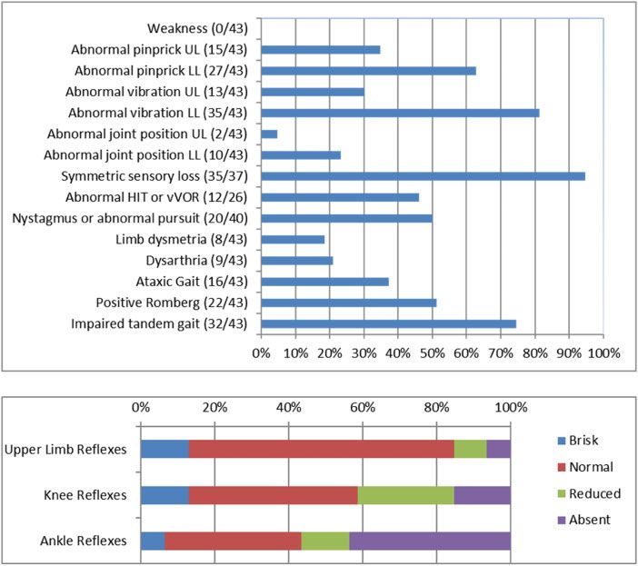
RFC1 expansions in idiopathic sensory neuropathy                                               BRAIN 2021: 144; 1542–1550          | 1543

 5 Department of Neurosciences, Rehabilitation, Ophthalmology, Genetics, Maternal and Child Health (DINOGMI),
   University of Genoa, Genoa, Italy
 6 Neurology Unit, IRCCS San Martino Hospital, Genoa, Italy
 7 Department of Neuromuscular Diseases, UCL Queen Square Institute of Neurology, London, UK
 8 Department of Clinical and Movement Neurosciences, UCL Queen Square Institute of Neurology, London, UK
 9 Department of Neurosciences, Biomedicine and Movement Sciences, University of Verona, Verona, Italy
10 Department of Molecular Medicine, Unit of Genetics, Università degli studi di Pavia, Pavia, Italy
11 Medical Genetics Unit, IRCCS San Martino Hospital, Genoa, Italy
12 Sheffield Diagnostic Genetics Service, Sheffield Children’s NHS Foundation Trust, Western Bank, Sheffield, UK
13 Academic Department of Neurosciences, Sheffield Teaching Hospitals NHS Trust and University of Sheffield,
   Sheffield, UK
14 Department of Brain Sciences, Neuro-otology Unit, Imperial College London, London, UK
15 Department of Clinical and Motor Neurosciences, University College London, London, UK

                                                                                                                                               Downloaded from https://academic.oup.com/brain/article/144/5/1542/6272840 by guest on 30 September 2021
Correspondence to: Andrea Cortese, MD PhD
Department of Neuromuscular Diseases
UCL Queen Square Institute of Neurology
Queen Square, WC1N 3BG
London, UK
E-mail: andrea.cortese@ucl.ac.uk; andrea.cortese@unipv.it

Keywords: sensory neuropathy; chronic idiopathic axonal polyneuropathy; CANVAS; RFC1
Abbreviations: CANVAS = cerebellar ataxia, neuropathy, vestibular areflexia syndrome; CIAP = chronic idiopathic
axonal polyneuropathy

Introduction                                                          general peripheral nerve clinics in multiple centres in Italy and the
                                                                      UK. Patients were classified as having a sensory-motor or sensory
Polyneuropathy is a common health problem leading to neuro-           neuropathy. Importantly, as sporadic CIAP is the focus of the
logical consultation, with an estimated prevalence of 1–2.4% in the   study, patients previously diagnosed with CANVAS or with a fam-
general population and up to 7% in people aged 465 years.1,2          ily history of CANVAS were not included. The following laboratory
    Even after extensive evaluation, 25–32% of patients remain un-    investigations were performed in all cases: fasting blood glucose,
diagnosed and are often labelled as having chronic idiopathic         glycosylated haemoglobin, folate, vitamin B12 and serum protein
axonal polyneuropathy (CIAP).3–6 CIAP refers to a sensory or sen-     immunofixation electrophoresis.12 Depending on the clinical fea-
sory-motor, axonal, slowly progressive neuropathy.7 Clinically, the   tures, additional tests were performed, including those for
disease burden falls predominantly or exclusively on the sensory      markers of systemic autoimmunity (antinuclear antibodies, ex-
fibres, especially at disease onset.8 The slow progression of CIAP    tractable nuclear antigens and antineutrophil cytoplasmic anti-
with an accumulating burden of disability9 has raised the hypoth-     bodies), paraneoplastic disorders (onconeural antibodies), HIV,
esis of an underlying genetic cause.10 However, family history in     hepatitis B and C infection, dosage of vitamin E, genetic testing for
CIAP is usually negative and age at onset is significantly higher     familial amyloid polyneuropathy and/or hereditary sensory neuro-
than in most known inherited neuropathies.9,11 Previous attempts      pathies, fat pad aspiration for amyloid staining, imaging and nerve
at identifying a genetic cause of CIAP were unsuccessful.12,13        biopsy. A detailed algorithm for case enrolment is provided in
    Recently, biallelic intronic AAGGG repeat expansions in the       Fig. 1.
replication factor complex subunit 1 (RFC1) gene were identified          Patients with sensory neuropathy (n = 650) were assessed be-
as the cause of cerebellar ataxia, neuropathy, vestibular areflexia   tween 2010 and 2019 in two Italian University Hospitals (Pavia
syndrome (CANVAS) and a frequent cause of late onset                  and Padova). Of these, 220 could not be contacted or had insuffi-
ataxia.14,15 Notably, a sensory neuropathy/neuronopathy has           cient clinical information and were therefore excluded. Fifty-five
been present in all the patients with biallelic RFC1 expansions to    per cent of 430 patients (n = 238) were identified as having an-
date and can be observed as an isolated complaint in some, pos-       other cause of their sensory neuropathy, including diabetes
sibly reflecting an early stage of this complex neurological          (n = 78), paraneoplastic syndrome (n = 57), B12 and/or folate defi-
disease.16                                                            ciency (n = 47), neuropathy associated with systemic or rheum-
    Therefore, we speculated that biallelic RFC1 expansions could     atological disease (n = 37), genetic (n = 25), acquired or familial
account for a proportion of idiopathic sensory neuropathies also      amyloidosis (n = 6) or hepatitis C, hepatitis B or HIV infection
diagnosed as CIAP.                                                    (n = 28) and were therefore excluded. We identified 192 patients with
                                                                      idiopathic sensory neuropathy and 62 agreed to take part in the
                                                                      study. These patients were clinically reassessed, and a DNA sample
Materials and methods                                                 was obtained for RFC1 testing. A further 163 patients with a diagnosis
                                                                      of CIAP were identified in general peripheral nerve clinics in Italy
Patient selection and clinical evaluation
                                                                      (Genova and Napoli) or the UK (London and Sheffield), including 63
Patients affected by CIAP were identified through the interrogation   patients with sensory neuropathy and 100 with sensory-motor neur-
of clinical and neurophysiological databases from 2010 to 2019 in     opathy. Overall, 100 patients with sensory-motor and 125 with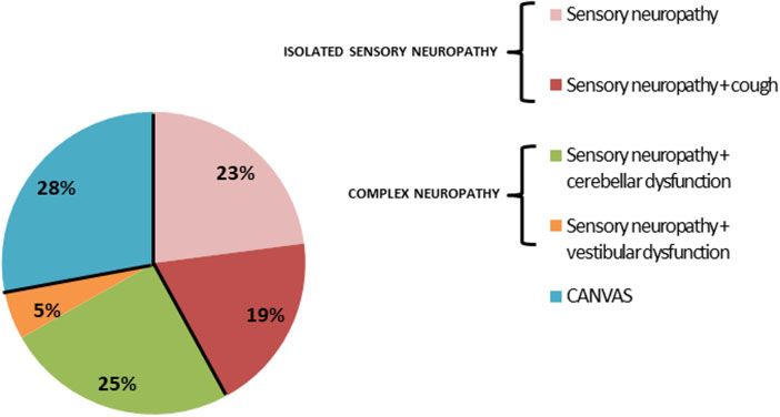
1544     | BRAIN 2021: 144; 1542–1550                                                                                        R. Currò et al.

                                                                                                                                                 Downloaded from https://academic.oup.com/brain/article/144/5/1542/6272840 by guest on 30 September 2021
Figure 1 Flow chart for enrolment of patients with sensory and sensory-motor CIAP. ENMG = electroneuromyography; HSN = hereditary sensory
neuropathy; PN = polyneuropathy.

sensory CIAP were included in the study and tested for biallelic RFC1   Results
expansions.
                                                                        Genetic testing

RFC1 testing                                                            Of 125 patients with sensory neuropathy 43 (34%) carried biallelic
                                                                        AAGGG repeat expansions in RFC1. Conversely, none of 100 axonal
The presence of RFC1 expansions was assessed as previously              sensory-motor neuropathies had biallelic AAGGG expansions.
described.14 Briefly, DNA was tested by flanking PCR and repeat-            In RFC1-positive cases, the expansions size, as measured by
primed PCR for AAGGG repeat expansions in RFC1. Samples with-           Southern blotting, ranged from 249 to 2386 repeat units, with a me-
out amplifiable products on flanking PCR and a positive repeat-         dian size of 661 repeats for the smaller allele and 810 for the larger
primed PCR for the AAGGG repeat were tested by Southern blotting        allele (Supplementary Fig. 1).
in order to confirm the presence and measure the size of the bial-
lelic RFC1 expansions. Non-pathogenic AAAAG or AAAGG expan-
sions were excluded by repeat-primed PCR in all the cases with          Clinical features of RFC1-positive patients
positive repeat-primed PCR for AAGGG expansion.                         Symptoms
                                                                        The demographic data and clinical features are detailed in
Ethics                                                                  Table 1. The median age at disease onset was 56 years (range: 30–
                                                                        75). Twenty-five patients were male and 18 were female. The
The study was approved by the ethics committee of IRCCS
                                                                        family history for neuropathy was unremarkable in all cases
Fondazione Policlinico San Matteo (p-20170028026) and by local in-
                                                                        apart from a patient with a sibling who had a diagnosis of sen-
stitutional review boards. All patients gave informed consent prior     sory neuropathy.
to their inclusion in the study. The study complied with all rele-          At onset, 98% of patients (n = 42) had symptoms of sensory
vant ethical regulations.                                               neuropathy, including distal numbness (n = 21), tingling or pins
                                                                        and needles sensation (n = 14), unsteadiness (n = 19) and pain
                                                                        (n = 10). The distribution of sensory involvement was length-de-
Statistical analysis
                                                                        pendent in 69% of cases (n = 20/29). Symptoms of dysautonomia
Continuous data were expressed as medians (minimum-max-                 (e.g. orthostatic hypotension, erectile dysfunction or altered
imum). Statistical differences between subgroups of patients posi-      sweating) were infrequent at onset (n = 2). Only two patients
tive versus negative for RFC1 expansions were tested using the          reported symptoms suggestive of cerebellar (e.g. slurred speech
Mann-Whitney test for quantitative data and Pearson’s chi-              or difficulties in swallowing) or vestibular dysfunction (e.g. oscil-
squared test for categorical data. All analyses were performed          lopsia) at disease onset. When specifically asked, 70% of patients
using STATA statistical software, version 14.                           (n = 26/37) reported a chronic, paroxysmal dry cough, which was
                                                                        often previously attributed to asthma or gastro-esophageal re-
                                                                        flux. In an asymptomatic 41-year-old male, the sensory neur-
Data availability                                                       opathy was suspected because of reduced or absent reflexes
Anonymized data from this study will be shared by request from          tested during a routine medical visit for workplace health
any qualified investigator.                                             surveillance.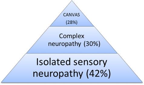
RFC1 expansions in idiopathic sensory neuropathy BRAIN 2021: 144; 1542–1550 | 1545
Previous diagnoses intravenous immunoglobulins or mycophenolate) and three had
B12 replacement therapy.
In 26% (n = 11) of genetically confirmed patients, the neuropathy
Patients were followed up for 4.7 years (range 0–16.7 years).
was previously attributed to a different cause, including inflamma-
Notably, at the most recent examination, 70% of patients (n = 30)
tory (n = 7; one vasculitis, three CIDP, one Sjögren’s syndrome, one
still had symptoms of isolated neuropathy and did not report any
post-infectious, one paraneoplastic), metabolic (n = 3, B12 defi-
vestibular or cerebellar complaint.
ciency) or toxic (n = 1, previous exposure to hexavalent chromium).
Three patients received immunosuppressive therapy (i.e. steroids,
Disability
The disease was slowly progressive in 81% of cases (n = 35) and sta-
Table 1 Clinical features of RFC1 + patients at disease onset ble in 19% (n = 8). Thirty-seven per cent of patients (n = 16) lost in-
and at most recent evaluation dependent walking: 10 required unilateral support, five bilateral
support and one was wheelchair-dependent.
Demographics n = 43
Downloaded from https://academic.oup.com/brain/article/144/5/1542/6272840 by guest on 30 September 2021
Males 25 (58%)
Positive family history 1 (2%) Neurological examination
Age at onset 56 (30–75)
Age at examination 67 (41–86) The data from the neurological examinations are summarized in
Fig. 2. The median disease duration at the most recent examin-
Symptoms Disease onset Most recent evaluation ation was 9.6 years (range: 0.6–31.7 years). Gait was ataxic in 37%
Numbness 21 (49%) 27 (63%) of patients (n = 16), but difficulty in tandem walking was observed
Unsteadiness 19 (44%) 30 (70%) in 74% (n = 32). Romberg was positive in 51% (n = 22), supporting
Tingling/pins and needles 14 (33%) 20 (46%) the presence of a prominent peripheral component of the un-
Pain 10 (23%) 17 (39%) steadiness. Eighty-six per cent of patients (n = 37) had an abnormal
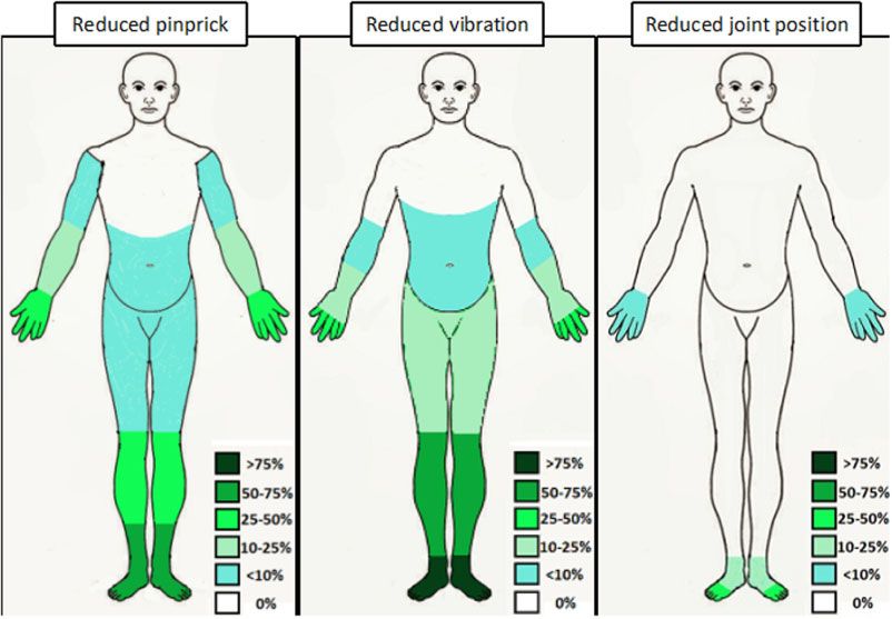
Distribution sensory examination. Pinprick and vibratory sensation were
Length dependent NA 20/29 (69%) impaired in 65% (n = 28) and 81% (n = 35) of patients, respectively.
Non-length-dependent NA 9/29 (31%)
Conversely, joint position was impaired only in 23% of cases
Dysautonomia 2 (5%) 9 (21%)
(n = 10; Fig. 3). Sensory impairment was length-dependent (name-
Dysarthria/dysphagia 1 (2%) 10 (23%)
ly, lower limbs were more affected than upper limbs and distal
Oscillopsia 1 (2%) 4 (9%)
limb segments were more affected than proximal) in 92% (n = 34/
Chronic cough NA 26/37 (70%)
37) and symmetric in 95% (n = 35/37). Nineteen per cent (n = 8) of
NA = not available. patients showed incoordination during cerebellar testing, but the
Figure 2 Detailed neurological examination of RFC1 + patients. HIT = head-impulse test; LL = lower limbs; UL = upper limbs; vVOR = visually-
enhanced vestibulo-ocular reflex.1546 | BRAIN 2021: 144; 1542–1550 R. Currò et al.
Downloaded from https://academic.oup.com/brain/article/144/5/1542/6272840 by guest on 30 September 2021
Figure 3 Graphic representation of the distribution of abnormalities at sensory examination. The different shades of colour correspond to different
percentages of patients with reduced sensation in the specific area.
upper limbs were involved only in half of them. Strength, tone and Additional investigations
plantar reflexes were normal in all patients. Reflexes were usually
Cerebellar atrophy was identified in 26% of patients who under-
decreased or absent, but normal or brisk reflexes were observed in
went brain MRI (n = 7/27). Clinical examination was more sensi-
more than one-third of cases (n = 11 and 5, respectively).
tive in detecting cerebellar abnormalities than brain MRI: only 5
Abnormalities of eye movements were identified in 50% of
of 20 subjects with clinical evidence of cerebellar dysfunction at
cases, as revealed by the presence of nystagmus (n = 19/40) or bro-
examination had definite cerebellar atrophy on brain MRI.
ken pursuit (n = 9/34). Of 26 patients tested, 46% (n = 12) had an
Conversely, subclinical cerebellar atrophy was identified in only
altered bedside head-impulse test.
two cases without cerebellar symptoms or signs at clinical
examination.
Six patients with an abnormal bedside head-impulse
Nerve conduction studies
test underwent vestibular testing, which confirmed a
The detailed nerve conduction study results are reported in bilateral vestibular areflexia in all of them. CSF analysis
Table 2. The median delay from symptom onset to the first nerve was performed in 13 patients to rule out an inflammatory cause of
conduction study was 5 years (range 0–31 years). Axonal loss of the neuropathy and was normal in all. Four patients underwent a
sensory fibres was generally severe, with globally absent sensory sural nerve biopsy, showing in all cases a diffuse and severe reduc-
action potentials in 26% of cases (n = 11), reduced in a length-de- tion of myelinated fibre density with no signs of regeneration
pendent pattern (namely, a greater impairment in the lower than (Supplementary Fig. 2).
in the upper limbs) in 30% of patients (n = 13) and reduced in a
non-length dependent pattern in 44% (n = 19). Sensory conduc-
RFC1 spectrum disorder: clinical syndrome
tion velocities were normal or slightly decreased, consistently
with a loss of fast-conducting large fibres, in all cases (median Based on history, neurological examination and ancillary investi-
value of sural conduction velocity = 41.8 m/s, range: 31.4–51 m/s). gations, after 10 years of disease duration, the clinical syndrome
We observed a clinical-electrodiagnostic dissociation with reduc- could be classified as isolated sensory neuropathy in 42% of
tion in sensory amplitudes being worse than expected based on patients (n = 18), including 19% of cases with sensory neuropathy
clinical findings in 44% (n = 19) of cases: six patients with a nor- and cough (n = 8), in 5% (n = 2) sensory neuropathy with vestibular
mal sensory examination showed diffusely reduced sensory ac- areflexia, in 25% (n = 11) sensory neuropathy with cerebellar dys-
tion potentials, with absent sensory action potentials throughout function and in 28% (n = 12) full CANVAS (Fig. 4).
in two cases; 13 patients with reduced sensation restricted to dis-
tal segments of the lower limbs had reduced or absent sensory
Predictors of positive RFC1 testing in patients with
action potentials in the upper limbs in 38% (n = 5) and 62% (n = 8),
respectively.
sensory neuropathy
Motor nerve conduction studies were normal in all but two To identify the symptoms and signs predictive of positive RFC1
patients with decreased compound muscle action potentials in the testing, we compared 43 cases with sensory neuropathy and bial-
lower limbs. lelic AAGGG expansions with 58 cases also diagnosed withRFC1 expansions in idiopathic sensory neuropathy BRAIN 2021: 144; 1542–1550 | 1547
Table 2 Details of nerve conduction studies in RFC1 + patients
Sensory nerves Reduced SAPs Absent SAPs Amplitude (mV) SCV (m/s)
Reference – – 410 448
Radial 10/21 (48%) 11/21 (52%) 2.6 (0.37–9.4) 50 (39–62)
Ref – – 46 446
Ulnar 11/30 (37%) 18/30 (60%) 2.4 (0.5–6.2) 49.1 (39.7–61)
Reference – – 48 443.5
Median 16/30 (53%) 14/30 (47%) 3.4 (0.3–7.4) 47 (36–56.5)
Reference – – 46 442
Sural 18/39 (46%) 19/39 (49%) 1.8 (0.2–11.6) 41.8 (31.4–51)
Reference – – 46 440
Sup. Peroneal 6/17 (35%) 11/17 (65%) 1.8 (0.19–4) 40 (37–43)
Motor nerves Reduced CMAPs Absent CMAPs Amplitude (mV) MCV (m/s)
Downloaded from https://academic.oup.com/brain/article/144/5/1542/6272840 by guest on 30 September 2021
Reference – – 45 448
Ulnar 1/19 (5%) 0/19 (0%) 9.6 (4.7–16.4) 55 (50–73)
Reference – – 46 446.8
Median 2/25 (8%) 0/25 (0%) 10.4 (4–16.3) 54 (48–62.9)
Reference – – 42 441
Peroneal 2/34 (6%) 0/34 (0%) 5 (0.7–17.1) 45.2 (37.3–52.8)
Reference – – 45 440.6
Tibial 1/21 (4%) 0/21 (0%) 8.1 (2.9–19) 43 (38.8–51.2)
Length-dependent neuropathy = 13 (30%)
Absent LL SAPs/Reduced UL SAPs = 5 LL SAPs 5 UL SAPs = 8
Non length-dependent neuropathy = 30 (70%)
Absent SAPs at four limbs = 11 Absent UL SAPs/ UL SAPs 5 LL SAPs = 9 Globally absent SAPs with at least one SAP preserved = 5
Reduced LL SAPs = 5
Values are expressed as median (range) or n (%). CMAPs = compound muscle action potentials; LL = lower limbs; MCV = motor conduction velocity; SAPs = sensory action
potentials; SCV = sensory conduction velocity; Sup. Peroneal = superficial peroneal; UL = upper limbs.
Figure 4 Distribution of system involvement in RFC1 patients after clinical examination and investigations. Patients with isolated sensory neur-
opathy were further subdivided depending on the presence of cough; patients with complex neuropathy who did not have the full triad of CANVAS
were further classified depending on the presence of vestibular or cerebellar dysfunction.
sensory neuropathy but negative for AAGGG repeat expansions Discussion
(Table 3). The presence of cough, dysarthria and/or dysphagia,
history of falls, clinical evidence of cerebellar or vestibular dys- We identified RFC1 AAGGG biallelic expansions in 34% of patients
function at examination, reduced vibratory sensation above the with sensory CIAP suggesting that RFC1 expansions represent a
knee in the lower limbs and a progressive course were more significant genetic cause of sporadic sensory neuropathy.12,17
frequently observed in cases carrying biallelic RFC1 expansions.
Conversely, the two groups did not significantly differ in gender,
Frequency of RFC1 expansion in the general
age of onset, family history, report of vestibular symptoms, pres-
ence and distribution of sensory symptoms and signs (other than peripheral nerve clinic
proximal reduction of vibratory sensation in the lower limbs), The AAGGG allele frequency in the general population is 0.7–
presence of gait and limb ataxia. 6.8%,14,15,18 resulting in an estimated prevalence at birth of biallelic1548 | BRAIN 2021: 144; 1542–1550 R. Currò et al.
Table 3 Comparison of main clinical features and investigation
results in RFC1-positive and -negative patients
RFC1 + RFC1– P-value
Mean age at onset 56 (30–75) 58 (12–85) 0.17
Male sex 25/43 (58%) 38/60 (63%) 0.59
Cough 26/37 (70%) 5/54 (9%) 0.00
Unsteadiness 30/43 (70%) 29/55 (53%) 0.08
History of falls 15/43 (35%) 6/56 (10%) 0.00
Chronic progression 35/43 (81%) 29/58 (50%) 0.00
Need for walking support 16/43 (37%) 6/58 (10%) 0.00
Sensory symptoms (non- 9/29 (31%) 13/47 (28%) 0.95
length-dependent
distribution)
Downloaded from https://academic.oup.com/brain/article/144/5/1542/6272840 by guest on 30 September 2021
Impaired vibratory sensation 27/43 (63%) 13/57 (22%) 0.00 Figure 5 The ‘iceberg’ hypothesis for RFC1 spectrum disorders.
at knee or above Complex neuropathy was defined by the contemporary presence of
Non length-dependent pat- 30/43 (70%) 15/55 (27%) 0.00 neuropathy and vestibular or cerebellar involvement.
tern at NCS
Absent SAPs at UL 22/35 (63%) 10/40 (25%) 0.00 Cerebellar symptoms were less common and oscillopsia rare.
Cerebellar symptoms 10/43 (23%) 4/56 (7%) 0.02 Although ataxia is a key component of the disease and the leading
Cerebellar signs 20/40 (50%) 12/58 (21%) 0.00 cause of disability during progression, unsteadiness was reported
Vestibular symptoms 4/43 (9%) 1/58 (1%) 0.09
by less than half of cases at onset and observed in one-third at
Vestibular signs 12/26 (46%) 2/37 (5%) 0.00
examination, while complaints of upper limb incoordination were
Values are provided mean (range) or n (%). Values in bold indicate significance. NCS rare (510% of cases).
= nerve conduction studies; SAPs = sensory action potentials; UL= upper limbs. After clinical examination and investigation, the disease was
confirmed as an isolated sensory neuropathy in 42% of RFC1 cases
after a median of 9.6 years of disease duration. Conversely, 58%
carriers at risk of developing the disease ranging from 1 in ’20 000 had a complex neuropathy or CANVAS, usually based on the pres-
to 1 in ’200 individuals. However, full CANVAS is considered rela- ence of subtle clinical signs, including nystagmus, broken pursuits
and abnormal bedside head impulse test, which can be missed if
tively rare, while idiopathic sensory neuropathy is common in the
not specifically looked for. The shorter disease duration in the pre-
ageing population.
sent cohort compared to our previously published CANVAS cohort
We have previously hypothesized that sensory neurons could
(9.6 years, range 0–31 years versus 20 years, range 0–50 years)16
be the first system to be involved in a context of progressive dis-
may explain the lower percentage of complex disease and
ease leading to full CANVAS.16 This was based on the findings
CANVAS and the lower degree of disability of our patients.
from a retrospective cohort of patients affected by late-onset
Interestingly, clinical examination seemed more accurate than
ataxia, including familial cases, enrolled from highly specialized
brain MRI in detecting cerebellar involvement. Indeed, less than
centres and with a specific expertise in neurogenetics and inher-
one-third of patients with signs of cerebellar dysfunction on exam-
ited neuropathies.14,16 Overall, the frequency of carriers of biallelic
ination had evidence of definite cerebellar atrophy on brain MRI,
RFC1 AAGGG expansions in that series was 22%. The frequency in
while vestibular testing was mostly confirmatory of abnormal bed-
sporadic sensory neuropathy has not yet been investigated.
side test. This is in accordance with previous studies, showing that
In the present study, most of the cases were recruited from
investigations seldom show cerebellar atrophy and vestibular are-
general peripheral nerve and electromyography clinics, without a
flexia in patients with a normal neurological examination.16,22
bias towards genetic cases. Also, inclusion criteria were limited to
the presence of sensory neuropathy and not of cerebellar ataxia or
CANVAS. The study showed that in this population the frequency
of RFC1 expansion was as high as 34%, significantly higher than RFC1 sensory neuropathy
the frequency reported in previous ataxic cohorts, ranging from 2 The study also helped to better define the sensory neuropathy in
to 22%.14,18–21 Thus, sensory nerve degeneration seems to play a carriers of biallelic AAGGG expansions in RFC1, including: (i) a
major and early role in RFC1-related ataxic disorders. length dependent distribution of sensory symptoms and signs in
Although larger prospective studies are needed to elucidate the 69% and 92% of cases, respectively, mimicking a distal symmetric-
full spectrum and the natural history of RFC1 spectrum disorders, al polyneuropathy; (ii) the report of loss of sensation and positive
data from our cohort support an ‘iceberg’ hypothesis, where full sensory symptoms (which are unusual in genetic neuropathies) as
CANVAS represents the tip, whereas isolated sensory neuropathy the most common complaint at disease onset, and more frequent
the bulk of the iceberg (Fig. 5). than unsteadiness; (iii) a preferential involvement of pinprick and
vibration sense with relative sparing of position sense; (iv) variable
reflexes, from absent to brisk (in accordance with previous studies
Clinical features and diagnostic clues of the RFC1
showing a preservation of H and T-reflexes, as a result of the spar-
disease spectrum ing of 1A muscle spindle afferent fibres)23,24; (v) a frequent clinical-
RFC1-positive cases typically reported complaints suggestive of neurophysiological dissociation between the length-dependent
distal symmetrical neuropathy including distal sensory loss, par- distribution of sensory symptoms and signs and the widespread
aesthesia, pain. Interestingly, a clinical-neurophysiological dis- impairment of sensory conduction; and (vi) the confirmation that
sociation between relatively mild clinical symptoms and signs, the motor system is typically unaffected in RFC1 disorder, as RFC1
often in a length-dependent distribution, and a more severe and expansions were absent from 100 cases with sensory motor neur-
widespread alteration of sensory action potentials seems typical opathy and RFC1-positive patients showed no or minimal motor
for RFC1 sensory neuropathy and should prompt genetic testing. involvement.RFC1 expansions in idiopathic sensory neuropathy BRAIN 2021: 144; 1542–1550 | 1549
Chronic cough 3. Farhad K, Traub R, Ruzhansky KM, Brannagan TH. Causes of
neuropathy in patients referred as “idiopathic neuropathy”.
The study also shows that a chronic cough is a strong diagnostic
Muscle Nerve. 2016;53:856–861.
clue to suspect RFC1 expansion in patients with a sensory neur-
4. Pasnoor M, Nascimento OJM, Trivedi J, et al. North America and
opathy. Cough has been previously reported in patients affected
South America (NA-SA) neuropathy project. Int J Neurosci. 2013;
by cerebellar ataxia25 and hereditary sensory neuropathy type
123:563–567.
1B,26 but its origin remains unknown. In RFC1 disease, cough often
5. Smith AG, Singleton JR. The diagnostic yield of a standardized
appears years or decades before the onset of sensory disturbances.
approach to idiopathic sensory-predominant neuropathy. Arch
Importantly, patients seldom spontaneously report cough as a
Intern Med. 2004;164:1021–1025.
symptom and need to be directly questioned about it. A history of
6. Visser NA, Notermans NC, Linssen RS, van den Berg LH,
asthma or gastroesophageal reflux should not cause this symptom
Vrancken AF. Incidence of polyneuropathy in Utrecht, the
to be dismissed in the context of a patient with sensory
Netherlands. Neurology. 2015;84:259–264.
neuropathy.
7. Notermans NC, Wokke JH, van der Graaf Y, et al. Chronic idio-
pathic axonal polyneuropathy: A five year follow up. J Neurol
Downloaded from https://academic.oup.com/brain/article/144/5/1542/6272840 by guest on 30 September 2021
Misdiagnoses of RFC1 neuropathy Neurosurg Psychiatry. 1994;57:1525–1527.
8. Notermans NC, Wokke JH, Franssen H, et al. Chronic idiopathic
Positive RFC1 testing led to reclassification of 11 cases who had a polyneuropathy presenting in middle or old age: A clinical and
previous alternative diagnosis, including sensory CIDP, Sjögren’s electrophysiological study of 75 patients. J Neurol Neurosurg
syndrome, paraneoplastic and metabolic neuropathy, and entail- Psychiatry. 1993;56:1066–1071.
ing three cases who had received unnecessary immune treat- 9. Teunissen LL, Notermans NC, Franssen H. Differences between
ments. Therefore, it is of the utmost importance to consider RFC1 hereditary motor and sensory neuropathy type 2 and chronic
in these cases to better inform patients on their condition and idiopathic axonal neuropathy. A clinical and electrophysio-
prognosis, to offer genetic counselling to patients and their fami- logical study. Brain. 1997;120:955–962.
lies and to prevent therapeutic attempts with ineffective and pos- 10. Dyck PJ, Oviatt KF, Lambert EH. Intensive evaluation of referred
sibly detrimental drugs. unclassified neuropathies yields improved diagnosis. Ann
Neurol. 1981;10:222–226.
11. Zis P, Sarrigiannis PG, Rao DG, Hewamadduma C,
Conclusion Hadjivassiliou M. Chronic idiopathic axonal polyneuropathy: A
After the exclusion of the common acquired causes, RFC1 should systematic review. J Neurol. 2016;263:1903–1910.
be considered in the diagnostic algorithm of all patients with spor- 12. England JD, Gronseth GS, Franklin G, et al.; American Academy
adic sensory, but not sensory-motor, CIAP assessed in a general of Physical Medicine and Rehabilitation. Evaluation of distal
peripheral nerve clinic. Early diagnosis of patients carrying biallelic symmetric polyneuropathy: The role of laboratory and genetic
RFC1 expansions, when the sensory neuropathy is still isolated, is testing (an evidence-based review). Muscle Nerve. 2009;39:
crucial both for ensuring optimal patient management and for 116–125.
tracking the full natural history of the disease, including its early 13. Wang W, Wang C, Dawson DB, et al. Target-enrichment
stages, when the neurodegenerative processes will likely be more sequencing and copy number evaluation in inherited polyneur-
amenable to therapeutic interventions.27 opathy. Neurology. 2016;86:1762–1771.
14. Cortese A, Simone R, Sullivan R, et al. Biallelic expansion of an
intronic repeat in RFC1 is a common cause of late-onset ataxia.
Funding Nat Genet. 2019;51:649–658.
15. Rafehi H, Szmulewicz DJ, Bennett MF, et al. Bioinformatics-
A.C. thanks the Medical Research Council (MR/T001712/1), the based identification of expanded repeats: A non-reference in-
Fondazione CARIPLO (2019-1836), the Italian Ministry of Health tronic pentamer expansion in RFC1 causes CANVAS. Am J Hum
Ricerca Corrente 2018–2019, the Inherited Neuropathy Consortium Genet. 2019;105:151–165.
(INC) and Fondazione Regionale per la Ricerca Biomedica for grant 16. Cortese A, Tozza S, Yau WY, et al. Cerebellar ataxia, neur-
support. H.H. and M.M.R. thank the MRC, the Wellcome Trust, the opathy, vestibular areflexia syndrome due to RFC1 repeat ex-
MDA, MD UK, Ataxia UK, The MSA Trust, the Rosetrees Trust and pansion. Brain. 2020;143:480–490.
the NIHR UCLH BRC for grant support. F.M. is supported by the 17. Fridman V, Reilly MM. Inherited neuropathies. Semin Neurol.
European Academy of Neurology (EAN) Research Fellowship 2020. 2015;35:407–423.
18. Aboud Syriani D, Wong D, Andani S, et al. Prevalence of RFC1 -
mediated spinocerebellar ataxia in a North American ataxia co-
Competing interests hort. Neurol Genet. 2020;6:e440.
19. Akçimen F, Ross JP, Bourassa CV, et al. Investigation of the RFC1
The authors report no competing interests.
repeat expansion in a Canadian and a Brazilian Ataxia Cohort:
Identification of novel conformations. Front Genet. 2019;10:1219.
20. Tsuchiya M, Nan H, Koh K, et al. RFC1 repeat expansion in
Supplementary material
Japanese patients with late-onset cerebellar ataxia. J Hum Genet.
Supplementary material is available at Brain online. 2020;65:1143–1147.
21. Wan L, Chen Z, Wan N, et al. Biallelic intronic AAGGG expan-
sion of RFC1 is related to multiple system atrophy. Ann Neurol.
References 2020;88:1132–1143.
1. Hanewinckel R, van Oijen M, Ikram MA, van Doorn PA. The epi- 22. Yip CW, Glaser M, Frenzel C, Bayer O, Strupp M. Comparison of
demiology and risk factors of chronic polyneuropathy. Eur J the bedside head-impulse test with the video head-impulse
Epidemiol. 2016;31:5–20. test in a clinical practice setting: A prospective study of 500 out-
2. Hughes RAC. Peripheral neuropathy. BMJ. 2002;324:466–469. patients. Front Neurol. 2016;7:58.1550 | BRAIN 2021: 144; 1542–1550 R. Currò et al.
23. Burke D, Halmagyi GM. Normal tendon reflexes despite absent 26. Spring PJ, Kok C, Nicholson GA, et al. Autosomal dominant
sensory nerve action potentials in CANVAS: A neurophysio- hereditary sensory neuropathy with chronic cough and
logical study. J Neurol Sci. 2018;387:75–79. gastro-oesophageal reflux: Clinical features in two
24. Infante J, Garcı́a A, Serrano-Cárdenas KM, et al. Cerebellar ataxia, families linked to chromosome 3p22-p24. Brain. 2005;128:
neuropathy, vestibular areflexia syndrome (CANVAS) with chronic 2797–2810.
cough and preserved muscle stretch reflexes: Evidence for select- 27. Coelho T, Maia LF, Martins da Silva A, et al. Tafamidis for trans-
ive sparing of afferent Ia fibres. J Neurol. 2018;265:1454–1462. thyretin familial amyloid polyneuropathy: A randomized, con-
25. Coutinho P, Cruz VT, Tuna A, Silva SE, Guimara ~ es J. Cerebellar trolled trial. Neurology. 2012;79:785–792.
ataxia with spasmodic cough. Arch Neurol. 2006;63:553–555.
Downloaded from https://academic.oup.com/brain/article/144/5/1542/6272840 by guest on 30 September 2021You can also read