Multicenter Epidemiologic Study of Coronavirus Disease-Associated Mucormycosis, India
←
→
Page content transcription
If your browser does not render page correctly, please read the page content below
Multicenter Epidemiologic Study of
  Coronavirus Disease–Associated
        Mucormycosis, India
       Atul Patel,1 Ritesh Agarwal,1,2 Shivaprakash M. Rudramurthy, Manoj Shevkani, Immaculata Xess,
            Ratna Sharma, Jayanthi Savio, Nandini Sethuraman, Surabhi Madan, Prakash Shastri,
            Deepak Thangaraju, Rungmei Marak, Karuna Tadepalli, Pratik Savaj, Ayesha Sunavala,
          Neha Gupta, Tanu Singhal, Valliappan Muthu, Arunaloke Chakrabarti,2 MucoCovi Network3

 During September–December 2020, we conducted a multi-
 center retrospective study across India to evaluate epidemi-
 ology and outcomes among cases of coronavirus disease
                                                                     S   econdary infections are known to complicate the
                                                                         clinical course of coronavirus disease (COVID-19).
                                                                     Bacterial infections are the most common secondary
 (COVID-19)–associated mucormycosis (CAM). Among 287                 infections, but increasing reports of systemic fungal
 mucormycosis patients, 187 (65.2%) had CAM; CAM preva-              infections are causing concern. In the early part of the
 lence was 0.27% among hospitalized COVID-19 patients.               COVID-19 pandemic,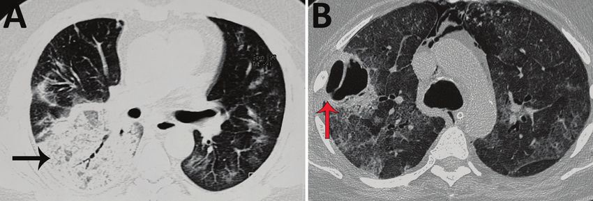
RESEARCH

                                                                                                Figure 1. Locations of 16
                                                                                                healthcare centers participating
                                                                                                in MucoCovi Network study on
                                                                                                coronavirus disease–associated
                                                                                                mucormycosis, India. AIIMS,
                                                                                                All India Institute of Medical
                                                                                                Sciences; CIMS, Care Institute of
                                                                                                Medical Sciences; PD Hinduja,
                                                                                                Parmanand Deepchand Hinduja;
                                                                                                PGIMER, Post Graduate
                                                                                                Institute of Medical Education &
                                                                                                Research; SGPI, Sanjay Gandhi
                                                                                                Postgraduate Institute

We collected data for all confirmed mucormycosis                riod. The prevalence of CAM was calculated as the
cases among patients with and without COVID-19 re-              total number of CAM cases divided by the number of
ported during September 1–December 31, 2020. The                COVID-19 patients treated at the 7 participating cen-
ethics committees of the respective centers approved            ters during the study period. Similarly, the preva-
the study protocol.                                             lence of CAM cases in the intensive care unit (ICU)
                                                                was calculated as the total number of patients devel-
Study Subjects and Definitions                                  oping mucormycosis among COVID-19 patients who
We defined a case of mucormycosis as compatible                 received treatment in the ICU. We classified CAM
clinical and radiologic manifestations and demon-               cases as early when mucormycosis was diagnosed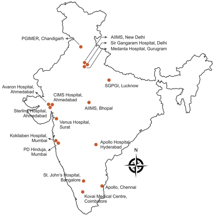
COVID-19–Associated Mucormycosis, India

records: demographic characteristics; underlying              Study Objectives
diseases, such as diabetes mellitus, hematological            Our primary objective was to compare the epidemiol-
malignancy, organ transplantation, and others; days           ogy of mucormycosis between CAM and non-CAM
to the diagnosis of mucormycosis before or after              groups during the study period, including the preva-
COVID-19 diagnosis; anatomic site of mucormycosis             lence, underlying diseases, relationship to COVID-19,
involvement; diagnostic modalities for mucormyco-             site of infection, and outcomes. Our secondary objec-
sis, including microscopy, culture, or histopathology;        tives were to compare CAM versus non-CAM and
treatment details, including antifungal drug therapy,         ascertain whether COVID-19 is a risk factor for mu-
surgical therapy, and other treatments; site of case          cormycosis death.
management, including home, hospital ward, or ICU;
immunosuppressive treatment received, such as glu-            Sample Processing
cocorticoid and other drugs; and outcome at 6 and 12          Tissue biopsies from mucormycosis-affected ana-
weeks. We classified multiple underlying diseases by          tomical sites were used for conventional microscopy,
using a hierarchical model. For instance, if a patient        culture, and histopathology, as appropriate, at the
had hematologic malignancy and then diabetes mel-             respective health centers. Microscopy was performed
litus developed due to the patient’s therapy, we con-         by using potassium hydroxide mount with or with-
sidered hematologic malignancy as the primary risk            out calcofluor stain. The samples were inoculated on
factor. On the other hand, for patients with COVID-19         2 sets of Sabouraud dextrose agar and incubated at
and preexisting uncontrolled diabetes, we regarded            25°C and 37°C. Positive cultures were identified by
diabetes as the primary underlying disease.                   macroscopic and microscopic characteristics. Tissue
                                                              samples submitted for histopathology were exam-
Treatment Details                                             ined by using hematoxylin and eosin, periodic acid
All patients received treatment for COVID-19 and              Schiff, or Gomori methenamine silver stain.
mucormycosis according to protocol at the respec-
tive treating institution. We recorded the information        Statistical Methods
regarding the type, dose, and duration of glucocor-           We performed data analysis using SPSS Statistics 21.0
ticoid drugs used for managing COVID-19, where                (IBM, Inc, https://www.ibm.com). We provide de-
available, by using dexamethasone-equivalent dose;            scriptive statistics as frequencies, mean (SD), or me-
0.75 mg dexamethasone is equivalent to 4 mg meth-             dian (interquartile range [IQR]), as appropriate. We
ylprednisolone or 5 mg prednisolone. We classified            compared categorical variables by using χ2 or Fischer
glucocorticoid use as not indicated when any steroid          exact test and analyzed differences between continu-
was used for managing nonhypoxemic COVID-19,                  ous data by using Mann-Whitney U tests. We per-
appropriate when dexamethasone-equivalent doses               formed multivariate logistic regression analyses to
of 6 mg/day were used for 10 days, or indicated but           identify factors predicting development of late CAM
inappropriate when dexamethasone-equivalent doses             and mucormycosis mortality rates. We considered
>6 mg/day were used for >10 days. To treat mucor-             p
RESEARCH

Table 1. Baseline characteristics among patients with mucormycosis, with and without COVID-19, India*
Variables                                                            CAM, n = 187            Non-CAM, n = 100                        p value
Mean age, y (SD)                                                       56.9 (12.5)                46.9 (16.4)                        0.0001
Sex                                                                                                                                   0.003
   M                                                                   150 (80.2)                  64 (64.0)
   F                                                                    37 (19.8)                  36 (36.0)
Underlying disease                                                                                                                   0.0001
   None                                                                     0                      19 (19.0)
   COVID-19 only                                                        61 (32.6)                       0
      Glucocorticoids for COVID-19                                    48/61 (78.7)                     NA
   Diabetes mellitus                                                   113 (60.4)                  67 (67.0)
   Traumatic inoculation (dental surgery, trauma, and burns)             3 (1.6)                    9 (9.0)
   Hematological malignancy                                              2 (1.1)                      2 (2)
   Renal transplantation                                                 3 (1.6)                        0
  Other†                                                                 5 (2.7)                      3 (3)
Glucocorticoids                                                        146 (78.1)                   6 (6.0)                          0.0001
Site of involvement
   Rhino-orbital                                                       117 (62.6)                  50 (50.0)                          0.07
   Rhino-orbito-cerebral                                                44 (23.5)                  34 (34.0)                          0.07
   Pulmonary                                                            16 (8.6)                    6 (6.0)                           0.42
   Renal                                                                 1 (0.5)                    1 (1.0)                           0.66
   Other (e.g., cutaneous, stomach)                                      5 (2.7)                    9 (9.0)                           0.03
   Disseminated                                                          4 (2.1)                        0                             0.41
Microscopy                                                                                                                            0.10
   Negative smear                                                       30 (16.0)                  10 (10.0)
   Aseptate hyphae                                                     153 (81.8)                  84 (84.0)
   Septate hyphae                                                        1 (0.5)                        0
   Septate and aseptate hyphae                                           3 (1.6)                    6 (6.0)
Culture                                                                                                                               0.04
   No growth                                                            87 (46.5)                  61 (61.0)
   Mucorales                                                            99 (52.9)                  37 (37.0)
   Mucorales and Aspergillus species                                     1 (0.5)                    1 (1.0)
   Aspergillus species                                                      0                       1 (1.0)
Histopathology diagnostic of mucormycosis‡                          143/155 (92.3)               37/44 (84.1)                         0.10
Management and outcome
   Hypoxemia during hospitalization                                     74 (39.6)                  12 (12.0)                         0.0001
   Admission to the intensive care unit                                 58 (31.0)                   9 (9.0)                          0.0001
   Treatment
      Liposomal amphotericin B                                         136 (72.7)                   84 (84)                          0.002
      Amphotericin D deoxycholate                                       31 (16.6)                   5 (5.0)                          0.005
      Posaconazole                                                      73 (39.0)                  14 (14.0)                         0.0001
      Isavuconazole                                                     19 (10.2)                   2 (2.0)                           0.01
   Combined antifungal therapy                                                                                                       0.0001
      Single antifungal drug                                            95 (50.8)                  88 (88.0)
      Concurrent                                                        13 (7.0)                    1 (1.0)
      Sequential                                                        79 (42.5)                  11 (11.0)
   Combined medical and surgical therapy                               131 (70.1)                  73 (73.0)                          0.60
   Outcome
      Death
COVID-19–Associated Mucormycosis, India
whom 48 (78.7%) received glucocorticoid treatment
for COVID-19 management. Other risk factors, in-
cluding hematologic malignancy and solid organ
transplantation, were noted in few among the study
population (Table 1).
Clinical Manifestations and Site of Involvement
A greater percentage of patients with CAM had
hypoxemia requiring ICU admission during hos-
pitalization than the non-CAM group (Table 1).
The rhino-orbital region was the most common
mucormycosis site (58.2%), followed by rhino-or-
bital-cerebral, pulmonary, and other sites (Table 1).
However, site of involvement was similar in both
the CAM and the non-CAM groups. Toothache, Figure 2. Cumulative number of mucormycosis cases during
loosening of teeth, and radiologic involvement of September–December 2019 and September–December
2020 in 10 health centers, India. White bar section indicates
the jaw were noted in many CAM patients (Figure coronavirus disease–associated mucormycosis (CAM); black
3) but were not seen in non-CAM patients. One bar sections indicate non-CAM cases. During 2019, 112 cases of
participating center reported jaw involvement in mucormycosis were detected, but a total of 231 cases, 92 non-
10/47 (21.3%) contributed CAM cases (Figure 3). CAM and 139 CAM, were detected in 2020.
The common form of pulmonary involvement was
cavitary lung disease (Figure 4). Treatment
Liposomal amphotericin B was the most used anti-
Diagnosis fungal agent in both groups. However, the use of li-
Mucormycosis diagnosis was made by direct micros- posomal amphotericin B was much lower in the CAM
copy in 237/287 (82.6%) patients. Histopathology group (72.7%) compared with the non-CAM group
demonstrated aseptate hyphae in 180/199 (90.5%) (84%). Posaconazole and isavuconazole were more
patients. Culture identified the etiologic agent in frequently used in CAM patients than in the non-CAM
138/287 (48.1%) cases (Table 1). The isolated Muco- group. A combination of antifungal therapy, such as
rales included Rhizopus arrhizus, Rhizomucor pusillus, amphotericin B plus triazoles, either concurrent or se-
Apophysomyces variabilis, Lichtheimia corymbifera, and quential, was used much more often in CAM patients
others. We did not note association of any species (49.5%) than in non-CAM (12%) patients. Combined
with any anatomic infection site. medical and surgical management was performed in
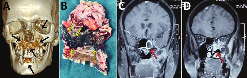
Figure 3. Radiographic images and surgical specimens demonstrating rhino-orbital-cerebral coronavirus disease–associated
mucormycosis in patients from India, 2020. A) Three-dimensional reconstruction of computed tomography scan of 54-year-old male
patient. Black arrows indicate patchy osteonecrosis involving the upper jaw, right orbital wall, and paranasal sinuses. B) Surgical
specimen from the maxilla of 54-year-old male patient showing black necrotic paranasal sinus with palatal involvement indicated by
yellow arrows. C, D) Magnetic resonance imaging (MRI) of coronal section of paranasal sinus and brain of 51-year-old female patient.
Red arrow in panel C indicates enhancing cavernous sinus lesion; D) red arrow in panel D indicates right ethmoid and maxillary
sinusitis. Scale bar indicates 7 cm.
Emerging Infectious Diseases • www.cdc.gov/eid • Vol. 27, No. 9, September 2021 2353RESEARCH
Figure 4. Noncontrast
computed tomography scan
of the thorax of a patient with
coronavirus disease–associated
mucormycosis, India, 2020.
A) Pulmonary mucormycosis
demonstrated as a large area
of consolidations with patchy air
trapping (black arrow), patchy
ground-glass opacities, and septal
thickening; B) large thick-walled
cavity (red arrow) with surrounding
ground-glass opacities.
71.1% (204/287) of patients and was similar in the 2 associated with better survival at 6 and 12 weeks (Ta-
groups. Major resection of the affected site was per- ble 2; Appendix Table 2).
formed in 59/284 patients; the remaining patients un-
derwent partial resection or debridement. Subgroup Analysis of CAM
The median time to CAM diagnosis was 18 (IQR
Outcomes 11–27) days (Figure 5). Among 187 CAM patients,
Mortality rates were similar between CAM and non- 158 (84.2%) were classified as late CAM (Table 3).
CAM groups; the combined 6-week mortality rate Some (33/187; 17.6%) patients were managed for
was 38.3% (110/287 patients) and the 12-week mor- COVID-19 at home before developing CAM. Among
tality rate was 45.7% (117/256 patients) (Table 1). 187 CAM patients, 74 (55.6%) were hypoxemic. Glu-
Univariate analysis showed that combined medical cocorticoid drugs were administered in various dos-
and surgical management improved survival in the es; the median cumulative dexamethasone-equivalent
rhino-orbital-cerebral group but did not improve dose was 84 mg (range 18–1,343 mg). Of note, only
outcomes for patients with infections at other sites 49/146 (33.6%) patients received steroids at appropri-
(Appendix Table 1, https://wwwnc.cdc.gov/EID/ ate levels (Table 3). Tocilizumab was administered to
article/27/9/21-0934-App1.pdf). On multivariate 5 (2.7%) patients for COVID-19 management.
logistic regression analysis, we found age, site of The demographic characteristics, underly-
involvement (rhino-orbital-cerebral or pulmonary), ing diseases, and site of involvement were similar
and ICU admission were associated with increased among patients with early and late CAM. However,
mortality rates. In contrast, sequential treatment with we saw diabetic ketoacidosis more often in patients
a combination of antifungal drugs was independently with early CAM (28%) than late CAM (5%). A higher
Table 2. Multivariate analysis of factors predicting death at 6 weeks among patients with mucormycosis, India*
Variables Survivors, n = 177 Non-survivors, n = 110 Odds ratio (95% CI) p value
Mean age, y (SD) 52.6 (15.1) 54.7 (14.0) 1.02 (1.00–1.04) 0.03
Underlying disease
None 10 (5.6) 9 (8.2) Referent Referent
Isolated COVID-19 42 (23.7) 19 (17.3) 0.56 (0.17–1.83) 0.34
Diabetes mellitus 109 (61.6) 71 (64.5) 0.92 (0.32–2.64) 0.88
Traumatic inoculation 8 (4.5) 4 (3.6) 1.30 (0.25–6.80) 0.76
Others 5 (2.8) 3 (2.7) 1.20 (0.18–7.81) 0.85
Renal transplantation 1 (0.6) 2 (1.8) 6.87 (0.42–113.19) 0.18
Hematological malignancy 2 (1.1) 2 (1.8) 1.60 (0.14–18.72) 0.71
Site of involvement
Rhino-orbital 117 (66.1) 50 (45.5) Referent Referent
Rhino-orbito-cerebral 39 (22) 39 (35.5) 2.39 (1.30–4.40) 0.005
Pulmonary 8 (4.5) 14 (12.7) 3.26 (1.05–10.11) 0.04
Other† 13 (7.3) 7 (6.4) 1.29 (0.43–3.86) 0.64
Admission to the intensive care unit 32 (18.1) 35 (31.8) 2.87 (1.43–5.75) 0.003
Combined medical surgical therapy 135 (76.3) 69 (62.7) 0.77 (0.41–1.45) 0.41
Combination of antifungals
Single antifungal drug 95 (53.7) 88 (80) Referent Referent
Concurrent 9 (5.1) 5 (4.5) 0.37 (0.09–1.44) 0.15
Sequential 73 (41.2) 17 (15.5) 0.17 (0.87–0.35) 0.0001
*Values are no. (%) except as indicated. Bold text indicates statistical significance. COVID-19, coronavirus disease.
†Includes cutaneous, stomach, disseminated, or other.
2354 Emerging Infectious Diseases • www.cdc.gov/eid • Vol. 27, No. 9,, September 2021COVID-19–Associated Mucormycosis, India
proportion of patients with late CAM received glu-
cocorticoid treatment (Table 3). Whereas amphoteri-
cin B remained the most common antifungal drugs
used in both groups, posaconazole, isavuconazole,
or a sequential use of antifungal agents (i.e., ampho-
tericin B followed by posaconazole or isavuconazole)
was more often seen in patients with late CAM. We
saw no statistically significant difference in 6- and 12-
week mortality rates between the early and late CAM
groups (Table 3).
We also explored factors associated with late CAM
development (Table 4). After adjusting for age, sex, and
underlying risk factors, we found hypoxemia due to
COVID-19 and inappropriate glucocorticoid adminis- Figure 5. Waterfall plot showing the number of days between the
tration were associated with development of late CAM. diagnosis of coronavirus disease (COVID-19) and COVID-19–
associated mucormycosis (CAM). Each vertical line represents a
case-patient. Red indicates late CAM (mucormycosis developing
Discussion
>8 days after COVID-19 diagnosis); black indicates early CAM
In our study, the prevalence of CAM was 0.27% in (mucormycosis developing 8 days after COVID-19 diagnoses. Hy- 187 CAM cases, 61 (32.6%) had COVID-19 as the
poxemia due to COVID-19 and inappropriate use of only underlying disease; 13 of those cases were not
glucocorticoid drugs were independently associated treated with glucocorticoid or other immunomodu-
with development of late CAM. The mortality rate for latory therapies. Whether COVID-19 itself causes
CAM patients was high (44%) but was comparable immune dysregulation and predisposes patients to
to rates for non-CAM (49%) patients. Older age (>54 invasive mucormycosis remains an unproven possi-
years), admission to an ICU, and pulmonary or brain bility (16–18). We did not find that COVID-19 was an
involvement by Mucorales were independently asso- independent predictor of late CAM, possibly because
ciated with a higher risk for death. The sequential use of the lower numbers of patients in our cohort with
of antifungal drugs at any site was associated with COVID-19 as the only underlying disease without
improved survival at 6 and 12 weeks, irrespective of any other risk factor. Lymphopenia is common in
anatomical site of mucormycosis. COVID-19, and progressive lymphopenia has been
In our study, 74.6% of patients affected by mu- shown to correlate with COVID-19 severity (19). The
cormycosis were men, as observed in previous stud- persisting immune dysregulation during the recov-
ies (11–13). We found diabetes mellitus was the most ery phase of COVID-19 infection also confers ad-
common underlying disease for both CAM and non- ditional risk. Unfortunately, we have not evaluated
CAM patients. SARS-CoV-2 has been shown to affect the effect of lymphopenia on the development or
the beta cells of the pancreas, resulting in metabolic outcome of CAM. Tocilizumab use in COVID-19 has
derangement, possibly causing diabetes mellitus been reported as a risk factor for invasive candidiasis
(14,15). Whether more frequent diagnosis (20%) of di- (20). However, only 2.7% of the CAM patients in this
abetes mellitus during the evaluation for CAM com- study received tocilizumab.
pared with non-CAM (10%) is related to SARS-CoV-2 The high mortality rate for CAM is a major concern
infection, glucocorticoid therapy, or a chance occur- (7). Patients with CAM were older (56.9 years) than
rence remains unclear. Unfortunately, we do not have non-CAM patients (46.9 years). Evidence suggests that
glycated hemoglobin values taken at admission for older age imparts increased risk for hospitalization,
all newly detected diabetes cases in our study, so we respiratory failure, ICU admission, and attendant glu-
cannot determine if these patients had diabetes mel- cocorticoid therapy in COVID-19 (21,22). Further, age
litus before CAM developed. >54 years also was associated with an increased risk
We found inappropriate glucocorticoid use was for death among our cohort. The site of mucormycosis
independently associated with late CAM. Among involvement and the survival at 6 and 12 weeks was
Emerging Infectious Diseases • www.cdc.gov/eid • Vol. 27, No. 9, September 2021 2355RESEARCH
Table 3. Characteristics of early and late CAM among patients with COVID-19, India*
Variables Early CAM, n = 29† Late CAM, n = 158‡ p value
Mean age, y (SD) 51.8 (14.2) 57.8 (11.9) 0.015
Sex 0.10
F 9 (31.0) 28 (17.7)
M 20 (69.0) 130 (82.3)
Glucocorticoids 8 (27.6) 138 (87.3) 0.0001
Tocilizumab 0 5 (3.2) 0.33
Underlying diseases 0.52
COVID-19 only 11 (37.9) 50 (31.6)
Diabetes mellitus 16 (55.2) 97 (61.4)
Diagnosed during current illness 6 33
Diabetic ketoacidosis§ 8 8
Traumatic inoculation: dental surgery, trauma, and burns 0 3 (1.9)
Hematological malignancy 0 2 (1.3)
Renal transplantation 0 3 (1.9)
Other: liver cirrhosis, immunosuppression, and others 2 (6.9) 3 (1.9)
Site of involvement 0.88
Rhino-orbital 17 (58.7) 100 (63.3)
Rhino-orbito-cerebral 8 (27.6) 36 (22.8)
Pulmonary 3 (10.3) 13 (8.2)
Renal 0 1 (0.6)
Other: e.g., cutaneous, stomach 0 5 (3.2)
Disseminated 1 (3.4) 3 (1.9)
Hypoxemia during hospitalization 9 (31.0) 65 (41.1) 0.19
ICU admission 12 (41.4) 46 (29.1) 0.31
Glucocorticoid treatment for COVID-19 N = 17 N = 133
Appropriate 11 (64.7) 44 (33.1)
Not indicated 4 (23.5) 46 (34.6)
Indicated, but inappropriately high dose 2 (11.8) 43 (32.3)
Treatment
Liposomal amphotericin B 26 (89.7) 110 (71.9) 0.06
Amphotericin D deoxycholate 3 (10.3) 28 (17.7) 0.33
Posaconazole 4 (13.8) 69 (43.7) 0.02
Isavuconazole 0 19 (12.0) 0.049
Combined antifungal therapy 0.004
Single antifungal drug 23 (79.4) 72 (45.6)
Concurrent 1 (3.4) 12 (7.6)
Sequential 5 (17.2) 74 (46.8)
Combined medical and surgical therapy 18 (62.1) 113 (71.5) 0.31
Outcomes
Death at 6 weeks 12 (41.4) 58 (36.7) 0.63
Death at 12 weeks, n = 170 13/22 (59.1) 62/148 (41.9) 0.17
*Values are no. (%) except as indicated. CAM, COVID-19–associated mucormycosis; COVID-19, coronavirus disease; ICU, intensive care unit.
†Early CAM is considered mucormycosis diagnosed 8 days of COVID-19 diagnosis.
§Diabetic ketoacidosis was more frequent among patients with early CAM (p = 0.0001).
similar in CAM and non-CAM groups. We expected a sential in mucormycosis management. Liposomal
higher proportion of pulmonary mycosis because re- amphotericin B is the drug of choice, but isavucon-
spiratory viral infections, such as influenza, often are azole also is recommended in primary therapy. Tri-
associated with secondary invasive aspergillosis (8). azoles, including posaconazole and isavuconazole,
However, we did not observe an increased occurrence commonly are used in the consolidation phase or as
of pulmonary mucormycosis compared with infec- salvage therapy (23). The role of combination antifun-
tions in other sites among the CAM group. Consider- gal treatment in mucormycosis is not clearly support-
ing the low rate of pulmonary involvement, we believe ed by evidence (24). The combination of surgery and
that CAM can be attributed to the systemic effects of antifungal therapy was associated with better sur-
COVID-19 or its treatment, rather than a sole alteration vival in the rhino-orbital-cerebral group in this study,
in the lungs. Several pulmonary mucormycosis cases conforming with previous experiences (6,11,25).
also might have remained undiagnosed because of However, the same was not true for mucormycosis in
challenges in obtaining diagnostic respiratory samples other anatomic sites. Early diagnosis of mucormyco-
among critically ill COVID-19 patients. sis and the more frequent use of consolidation thera-
Appropriate and timely antifungal therapy and py or combination of antifungals in this study could
surgical resection, when feasible, are considered es- be one explanation; another could be fewer surgeries
2356 Emerging Infectious Diseases • www.cdc.gov/eid • Vol. 27, No. 9,, September 2021COVID-19–Associated Mucormycosis, India
performed in patients with other than rhino-orbital Bangladesh, China, Iran, Mexico, and Pakistan, from
mucormycosis. which data on mucormycosis are still limited (26).
We found the sequential use of antifungal drugs, Further studies should compare data from coun-
amphotericin B then posaconazole or isavuconazole, tries with high rates of diabetes and mucormycosis
was independently associated with improved sur- with that of data from the United States and Europe,
vival among mucormycosis patients. However, the where mucormycosis predominantly is encountered
lack of randomization, possibility of case selection, in hematological malignancies and organ transplan-
and chance survival are potential biases. In addi- tation. Given the large number of late CAM cases,
tion, the optimal duration and dose of amphotericin healthcare-associated mucormycosis remains a dis-
B and posaconazole are not clear. The usefulness of tinct possibility (27,28). Contaminated ventilation
antifungal combination administered simultaneous- systems, air conditioners, and ongoing construction
ly could not be ascertained due to the small num- in hospitals have been reported to cause outbreaks
ber of patients receiving concurrent therapy in our of mucormycosis in the past (28). However, we did
study. A randomized controlled trial could affirm not estimate the burden of Mucormycetes spores in
the role of a combination of antifungals or mainte- the hospital environment (29). We also do not have
nance therapy in mucormycosis. data on the timing of amphotericin B use, timing of
We expected better survival for the CAM patients surgery, or duration of sequential antifungal thera-
in this study. Contrary to the prevailing practices py, which are critical factors that have a bearing on
(11,24), a combination of antifungal agents was more mucormycosis outcomes; hence, we could not ana-
frequently used (50%) in CAM patients than in non- lyze these factors. Other unexplored factors, includ-
CAM patients (12%). Also, hospitalized CAM patients ing genetic predisposition, might explain the high
were closely monitored. The treatment practices used prevalence of CAM and non-CAM in India. Thus,
for the CAM group, especially those with late CAM, prospective studies from the rest of the world, es-
were distinct from those for the non-CAM group and pecially those severely affected by the COVID-19
those for patients with early CAM. The occurrence pandemic, would be needed to ascertain the epide-
of a mold infection and the apprehension associated miology of CAM. The strength of our study is the
with the COVID-19 pandemic could have resulted in large number of patients, which lends credibility to
more frequent use of combination therapy in CAM. our observations.
However, we saw no difference in mortality rates In conclusion, mucormycosis is a rare but criti-
between CAM and non-CAM patients. Of course, in- cal problem complicating the later part of the clini-
creased risk for death due to COVID-19 itself cannot cal course of COVID-19 in India, possibly due to im-
be ruled out for these CAM patients. proper glucocorticoid usage. We found no difference
Our study’s first limitation is that we collected in the risk factors, site of involvement, and outcome
data from a single country. The predominant risk of mucormycosis complicating COVID-19 cases com-
factor for mucormycosis in our study was diabetes, pared with non–COVID-19 cases. Nevertheless, the
which is also the case in some countries, including prevalence of mucormycosis has increased greatly in
Table 4. Multivariate analysis of factors predicting the development of late CAM among COVID-19 patients, India*
Variables Early CAM, n = 29† Late CAM, n = 158‡ Odds ratio (95% CI) p value
Mean age, y (SD) 51.8 (14.2) 57.8 (11.9) 1.02 (0.96–1.07) 0.62
Sex
M 20 (69.0) 130 (82.3) 0.25 (0.06–1.10) 0.07
F 9 (31.0) 28 (17.7) Referent
Underlying disease
Isolated COVID-19 11 (23.7) 50 (17.3) 1.71 (0.25–11.96) 0.59
Diabetes mellitus 16 (61.6) 97 (64.5) 5.84 (0.70–48.89) 0.10
Others§ 2 (4.5) 11 (3.6) Referent
Hypoxemia due to COVID-19 9 (31.0) 65 (41.1) 11.84 (1.43–98.06) 0.02
Glucocorticoid usage N = 17 N = 133
Appropriate 11 (64.7) 44 (33.1) Referent
Not indicated 4 (23.5) 46 (34.6) 66.93 (7.05–635.19) 0.0001
Indicated, but inappropriately high dose 2 (11.8) 43 (32.3) 9.91 (1.39–70.77) 0.02
*Values are no. (%) except as indicated. Bold text indicates statistical significance. CAM, COVID-19–associated mucormycosis; COVID-19, coronavirus
disease.
†Early CAM is considered mucormycosis diagnosed 8 days of COVID-19 diagnosis.
§Includes traumatic inoculation, cirrhosis, immunosuppression, renal transplantation, and hematological malignancy.
Emerging Infectious Diseases • www.cdc.gov/eid • Vol. 27, No. 9, September 2021 2357RESEARCH
India, coinciding with the country’s COVID-19 epi- patients hospitalized with COVID-19: incidence and
demic. Clinicians should be vigilant for mucormyco- predictive factors. Clin Microbiol Infect. 2021;27:451–7.
https://doi.org/10.1016/j.cmi.2020.10.021
sis in the patients recovering from COVID-19 illness, 3. Seaton RA, Gibbons CL, Cooper L, Malcolm W, McKinney R,
especially among patients with new or previously di- Dundas S, et al. Survey of antibiotic and antifungal
agnosed diabetes mellitus and clinical manifestations prescribing in patients with suspected and confirmed
of facial or orbital pain or black or blood-stained nasal COVID-19 in Scottish hospitals. J Infect. 2020;81:952–60.
https://doi.org/10.1016/j.jinf.2020.09.024
discharge. In addition, we found improper glucocor- 4. Nucci M, Barreiros G, Guimarães LF, Deriquehem VAS,
ticoid use for the COVID-19 treatment to be an ad- Castiñeiras AC, Nouér SA. Increased incidence of candidemia
ditional risk factor in CAM. Therefore, treating physi- in a tertiary care hospital with the COVID-19 pandemic.
cians should ensure they use appropriate drugs and Mycoses. 2021;64:152–6. https://doi.org/10.1111/myc.13225
5. van Arkel ALE, Rijpstra TA, Belderbos HNA,
doses in treating COVID-19 patients. van Wijngaarden P, Verweij PE, Bentvelsen RG. COVID-19–
associated pulmonary aspergillosis. Am J Respir Crit Care
Members of the MucoCovi Network in India: Kamalesh Med. 2020;202:132–5. https://doi.org/10.1164/rccm.202004-
Patel (Sterling Hospital, Ahmedabad); Inderpaul Singh 1038LE
Sehgal, Ashish Bhalla, and G.D. Puri (Postgraduate 6. Chong WH, Saha BK, Ananthakrishnan Ramani, Chopra A.
State-of-the-art review of secondary pulmonary infections in
Institute of Medical Education and Research, Chandigarh);
patients with COVID-19 pneumonia. Infection. 2021 Mar 11
Gagandeep Singh and Manish Soneja (All India Institute of [Epub ahead of print]. https://doi.org/10.1007/
Medical Sciences, New Delhi); Sunil Kumar (Apollo s15010-021-01602-z
Hospital, Chennai); Priyadarshini A. Padaki (St. John’s 7. Garg D, Muthu V, Sehgal IS, Ramachandran R, Kaur H,
Bhalla A, et al. Coronavirus disease (Covid-19) associated
Medical College, Bengaluru); Mahathi Kandala and
mucormycosis (CAM): case report and systematic review
J. Prathiba (Apollo Hospital, Hyderabad); Gayathri Devi of literature. Mycopathologia. 2021;186:289–98.
Rajagopal (Kovai Medical Center and Hospital, Coimbatore); https://doi.org/10.1007/s11046-021-00528-2
Hemal Shah and Reedham Mehta (CIMS Hospital, 8. Ahmadikia K, Hashemi SJ, Khodavaisy S, Getso MI,
Alijani N, Badali H, et al. The double-edged sword of
Ahmedabad); Amir Keshri and Prabhakar Mishra (Sanjay
systemic corticosteroid therapy in viral pneumonia: a case
Gandhi Postgraduate Institute of Medical Sciences, report and comparative review of influenza-associated
Lucknow); Vikas Gupta and Ganakalyan Behera (All India mucormycosis versus COVID-19 associated mucormycosis.
Institute of Medical Sciences, Bhopal); and Umang Agarwal Mycoses. 2021 Feb 16 [Epub ahead of print].
https://doi.org/10.1111/myc.13256
and Irfana Mohammed (Hinduja Hospital, Mumbai).
9. Moorthy A, Gaikwad R, Krishna S, Hegde R, Tripathi KK,
Kale PG, et al. SARS-CoV-2, uncontrolled diabetes and
corticosteroids—an unholy trinity in invasive fungal infections
Acknowledgments of the maxillofacial region? A retrospective, multi-centric
We thank Prashant Sood for critical evaluation of the analysis. J Maxillofac Oral Surg. 2021 Mar 6 [Epub ahead of
manuscript and the following for their assistance in print]. https://doi.org/10.1007/s12663-021-01532-1
conducting the study: Ketan Patel, Renu Yadav, Camilla 10. Joshi SR, Das AK, Vijay VJ, Mohan V. Challenges in diabetes
care in India: sheer numbers, lack of awareness and inadequate
Rodrigues, Anjali Shetty, Shaoli Basu, Sangeeta Varty, Savari control. J Assoc Physicians India. 2008;56:443–50.
Desai, Arprit Sharma, Ashish Hegde, and Farah Jiijina. 11. Patel A, Kaur H, Xess I, Michael JS, Savio J, Rudramurthy S,
et al. A multicentre observational study on the epidemiology,
risk factors, management and outcomes of mucormycosis in
About the Authors India. Clin Microbiol Infect. 2020;26:944.e9–15.
https://doi.org/10.1016/j.cmi.2019.11.021
Dr. Patel is an infectious disease specialist at the Sterling 12. Jeong W, Keighley C, Wolfe R, Lee WL, Slavin MA,
Hospital, Ahmedabad, India, and a fellow of the Kong DCM, et al. The epidemiology and clinical
Infectious Disease Society for America. His research manifestations of mucormycosis: a systematic review
interests include fungal infections. Dr. Agarwal is a and meta-analysis of case reports. Clin Microbiol Infect.
2019;25:26–34. https://doi.org/10.1016/j.cmi.2018.07.011
pulmonologist at Postgraduate Institute of Medical 13. Prakash H, Ghosh AK, Rudramurthy SM, Singh P, Xess I,
Education & Research, Chandigarh, India. His research Savio J, et al. A prospective multicenter study on mucormycosis
interests include allergic and chronic lung aspergillosis. in India: epidemiology, diagnosis, and treatment. Med Mycol.
2019;57:395–402. https://doi.org/10.1093/mmy/myy060
14. Müller JA, Groß R, Conzelmann C, Krüger J, Merle U,
References Steinhart J, et al. SARS-CoV-2 infects and replicates in cells
1. Baiou A, Elbuzidi AA, Bakdach D, Zaqout A, Alarbi KM, of the human endocrine and exocrine pancreas. Nat Metab.
Bintaher AA, et al. Clinical characteristics and risk factors for 2021;3:149–65. https://doi.org/10.1038/s42255-021-00347-1
the isolation of multi-drug-resistant Gram-negative bacteria 15. Accili D. Can COVID-19 cause diabetes? Nat Metab.
from critically ill patients with COVID-19. J Hosp Infect. 2021;3:123–5. https://doi.org/10.1038/s42255-020-00339-7
2021;110:165–71. https://doi.org/10.1016/j.jhin.2021.01.027 16. Files JK, Boppana S, Perez MD, Sarkar S, Lowman KE, Qin K,
2. Ripa M, Galli L, Poli A, Oltolini C, Spagnuolo V, Mastrangelo et al. Sustained cellular immune dysregulation in
A, et al.; COVID-BioB study group. Secondary infections in individuals recovering from SARS-CoV-2 infection. J Clin
2358 Emerging Infectious Diseases • www.cdc.gov/eid • Vol. 27, No. 9,, September 2021COVID-19–Associated Mucormycosis, India
Invest. 2021;131:e140491. https://doi.org/10.1172/
EID Podcast
JCI140491
17. Potenza L, Vallerini D, Barozzi P, Riva G, Forghieri F,
Zanetti E, et al. Mucorales-specific T cells emerge in the course
of invasive mucormycosis and may be used as a surrogate
diagnostic marker in high-risk patients. Blood. 2011;118:5416–
9. https://doi.org/10.1182/blood-2011-07-366526 Livestock, Phages,
MRSA, and
18. Ghuman H, Voelz K. Innate and adaptive immunity to
mucorales. J Fungi (Basel). 2017;3:48. https://doi.org/
10.3390/jof3030048
19. Zhang X, Tan Y, Ling Y, Lu G, Liu F, Yi Z, et al. Viral and
host factors related to the clinical outcome of COVID-19.
Nature. 2020;583:437–40. https://doi.org/10.1038/
People in Denmark
s41586-020-2355-0
20. Antinori S, Bonazzetti C, Gubertini G, Capetti A, Pagani C,
Morena V, et al. Tocilizumab for cytokine storm syndrome
in COVID-19 pneumonia: an increased risk for candidemia?
Autoimmun Rev. 2020;19:102564. https://doi.org/10.1016/
j.autrev.2020.102564
21. Levin AT, Hanage WP, Owusu-Boaitey N, Cochran KB,
Walsh SP, Meyerowitz-Katz G. Assessing the age specificity
of infection fatality rates for COVID-19: systematic review,
meta-analysis, and public policy implications. Eur J Epidemiol.
2020;35:1123–38. https://doi.org/10.1007/s10654-020-00698-1
22. Pijls BG, Jolani S, Atherley A, Derckx RT, Dijkstra JIR,
Franssen GHL, et al. Demographic risk factors for COVID-19
infection, severity, ICU admission and death: a meta-analysis
of 59 studies. BMJ Open. 2021;11:e044640. https://doi.org/
10.1136/bmjopen-2020-044640
23. Cornely OA, Alastruey-Izquierdo A, Arenz D, Chen SCA,
Dannaoui E, Hochhegger B, et al.; Mucormycosis ECMM MSG
Global Guideline Writing Group. Global guideline for the
diagnosis and management of mucormycosis: an initiative of
the European Confederation of Medical Mycology in
cooperation with the Mycoses Study Group Education and Methicillin-resistant Staphylococcus aureus, bet-
Research Consortium. Lancet Infect Dis. 2019;19:e405–21.
https://doi.org/10.1016/S1473-3099(19)30312-3 ter known as MRSA, is often found on human skin.
24. Jeong W, Keighley C, Wolfe R, Lee WL, Slavin MA, Chen SC, But MRSA can also cause dangerous infections that
et al. Contemporary management and clinical outcomes of
mucormycosis: a systematic review and meta-analysis
are resistant to common antimicrobial drugs. Epide-
of case reports. Int J Antimicrob Agents. 2019;53:589–97. miologists carefully monitor any new mutations or
https://doi.org/10.1016/j.ijantimicag.2019.01.002
25. Muthu V, Agarwal R, Dhooria S, Sehgal IS, Prasad KT,
transmission modes that might lead to the spread of
Aggarwal AN, et al. Has the mortality from pulmonary this infection.
mucormycosis changed over time? A systematic review and Approximately 15 years ago, MRSA emerged in
meta-analysis. Clin Microbiol Infect. 2021;27:538–49.
https://doi.org/10.1016/j.cmi.2020.12.035 livestock. From 2008 to 2018, the proportion of in-
26. Prakash H, Chakrabarti A. Global Epidemiology of fected pigs in Denmark rocketed from 3.5% to 90%.
mucormycosis. J Fungi (Basel). 2019;5:26. https://doi.org/
10.3390/jof5010026 What happened, and what does this mean for hu-
27. Rammaert B, Lanternier F, Zahar JR, Dannaoui E, man health?
Bougnoux ME, Lecuit M, et al. Healthcare-associated mucor-
mycosis. Clin Infect Dis. 2012;54:S44–54. In this EID podcast, Dr. Jesper Larsen, a senior re-
https://doi.org/10.1093/cid/cir867 searcher at the Statens Serum Institut, describes the
28. Walther G, Wagner L, Kurzai O. Outbreaks of mucorales and
the species involved. Mycopathologia. 2020;185:765–81. spread of MRSA from livestock to humans.
29. Prakash H, Singh S, Rudramurthy SM, Singh P, Mehta N,
Shaw D, et al. An aero mycological analysis of Mucormycetes Visit our website to listen:
in indoor and outdoor environments of northern India. Med https://go.usa.gov/x74Jh
Mycol. 2020;58:118–23. https://doi.org/10.1093/mmy/myz031
Address for correspondence: Arunaloke Chakrabarti, Department
of Medical Microbiology, Postgraduate Institute of Medical
Education & Research, Sector-12, Chandigarh 160012, India; email:
arunaloke@hotmail.com
Emerging Infectious Diseases • www.cdc.gov/eid • Vol. 27, No. 9, September 2021 2359You can also read