Novel R225C variant identified in the HGD gene in Jordanian patients with alkaptonuria
←
→
Page content transcription
If your browser does not render page correctly, please read the page content below
AIMS Molecular Science, 8(1): 60–75.
                                                                 DOI: 10.3934/molsci.2021005
                                                                 Received: 18 December 2020
                                                                 Accepted: 18 February 2021
                                                                 Published: 25 February 2021
http://www.aimspress.com/journal/Molecular

Research article

Novel R225C variant identified in the HGD gene in Jordanian patients

with alkaptonuria

Nesrin R. Mwafi1,*, Dema A. Ali2, Raida W. Khalil3, Ibrahim N. Alsbou'4, Ahmad M. Saraireh5

1
    Department of biochemistry and molecular biology, Faculty of Medicine, Mutah University,
    Al-karak 61710, Jordan
2
    Cell Therapy Center, The University of Jordan, Amman 11942, Jordan
3
    Department of Biotechnology and Genetics Engineering, University of Philadelphia, Amman
    19392, Jordan
4
    Medical Laboratory Science, Faculty of Science, Mutah University, Al-karak 61710, Jordan
5
    Faculty of Medicine, Mutah University, Al-karak 61710, Jordan

* Correspondence: Email: drnesrin@mutah.edu.jo; Tel: 00962796777785.

Abstract: Alkaptonuria (AKU) is a rare metabolic disease which is inherited as an autosomal
recessive trait. It is characterized by the accumulation of homogentisic acid over time in various
tissues of the body particularly connective tissues. This genetic disease is caused by mutation of the
Homogentisate 1,2-dioxygenase (HGD) gene which encodes for enzyme essential for the catabolism
of phenylalanine and tyrosine. The aim of the present study is to investigate variant types in
Jordanian patients with alkaptonuria. Genomic DNA was extracted from whole blood samples of the
participated AKU family members (n = 23). The 14 exons of HGD gene for the proband were
amplified using specific PCR primers. The sequenced data were analysed and the pathogenicity of
the identified variants were predicted using the online bioinformatics programs: PolyPhen2, SIFT
and Mutation taster. The analysis showed that the proband was compound heterozygous for the
missense mutations A122V and R225C found within E6 and E10, respectively. R225C variant is
novel and the genotyping of the family members indicated that HGDA122V and HGDR225C alleles were
fully segregated. Moreover, the cousins of the proband who are AKU patients inherited the
homozygous pattern of the novel mutation. This study extends the pathogenic mutations spectrum of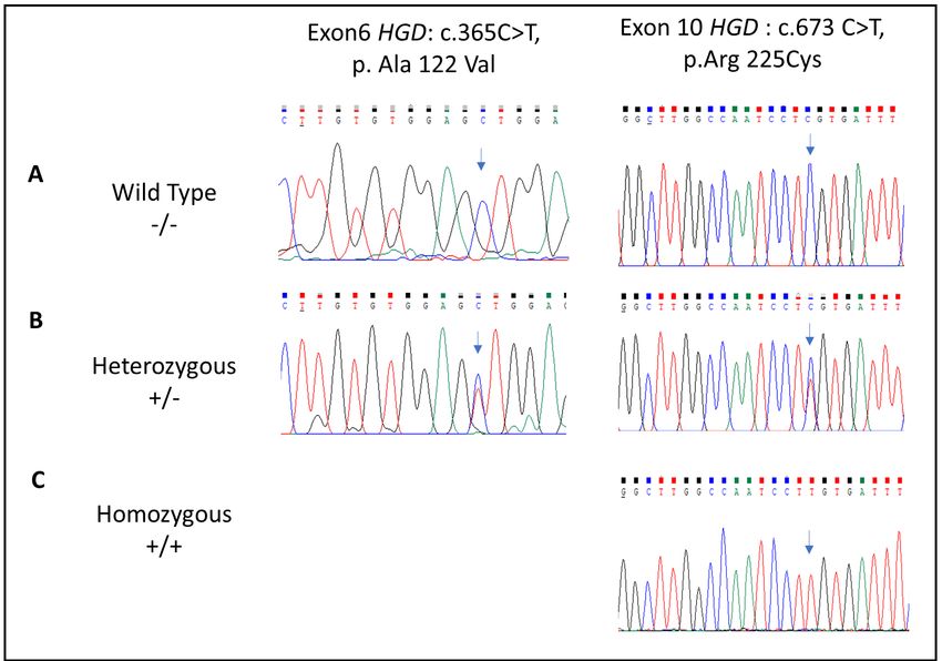
61

the HGD gene. It identified the novel mutation R225C and at the same time confirmed the high
prevalence of the founder mutation A122V in Jordanian AKU patients.

Keywords: alkaptonuria; autosomal recessive; compound heterozygous; Homogentisate 1,2-
dioxygenase gene; missense mutation

Abbreviations: AKU: Alkaptonuria; GC-MS: Gas Chromatography-Mass Spectrometry analysis;
HGD: Homogentisate 1,2-dioxygenase; MLPA: multiplex ligation-dependent probe amplification

1.   Introduction

      Alkaptonuria (AKU, OMIM 203500 ) is a rare metabolic disease that is inherited as Mendelian
autosomal recessive trait and was firstly described by Garrod [1,2]. The global AKU prevalence rate
is extremely low and was estimated as 1:250,000 live births [3]. However, the incidence rate is
higher in certain regions such as Jordan, India, Slovakia and the Dominican Republic [4]. AKU is
resulting from a defect in homogentisate 1,2-dioxygenase (HGD) enzyme (EC 1.13.11.5) which is
involved in the catabolic pathway of phenylalanine and tyrosine amino acids [5]. Consequently,
homogentisic acid is generated as an intermediary product and released to the circulation. A portion
of the circulating homogentisic acid is excreted in the urine which turns black in colour on standing
or upon alkalinisation [6]. The residual portion of homogentisic acid in bloodstream will be
distributed to various connective tissues and deposited mainly in cartilages, tendons and ligaments as
ochronotic pigment after oxidation and polymerization reactions [7,8]. The gene encoding for HGD
enzyme is located on the human chromosome 3 at q21–q23 and it is expressed particularly in hepatic
and renal cells [9,10]. HGD gene consists of 14 exons with transcript length of 1674 nucleotides
(NCBI Reference Sequence: NM_000187.4) that encode for 445-amino acids protein.
      The initial clinical manifestations of AKU are homogentisic aciduria, ochronosis and
arthropathy of large joints such as knees and hips [11]. At advanced stage of the disease, AKU
patients presented with additional clinical features including Achilles tendon rupture, arthroplasty,
aortic valve disease and stones formation commonly renal and prostatic ones [12]. However, the
severity of the disease varies between AKU patients but it increases with age due to the ongoing
accumulation of the homogentisic acid in various body tissues and fluids [13]. Therefore, individual
AKU patients may not have similar signs and symptoms even between the same family siblings [14].
Taylor et al. (2011) showed that joint tissues are prone to pigmentation only after the incidence of
focal cellular and extracellular matrix changes [15]. Moreover, the authors revealed that there is a
spectrum of pigmentation in articular cartilages in which HGA-derived pigment is initially deposited
at the boundaries of subchondral bone and calcified cartilages and ended with complete pigmentation
of cartilage matrix at more advanced ochronosis [15]. Various radiographic and imaging techniques
such as MRI and CT-scan are widely used to determine the extent of and severity of joints and spinal
damage [3]. Moreover, the quality of life rather than the life span is strictly affected, mainly as a

AIMS Molecular Science                                                         Volume 8, Issue 1, 60–75.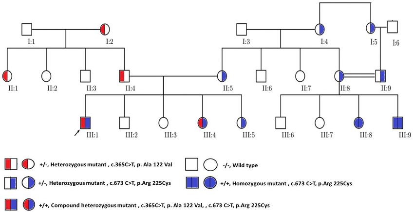
62

result of alteration of mechanical properties of large joints as well as degenerative changes and
calcifications of spinal intervertebral discs [16].
      The disease is primarily diagnosed by measuring the level of homogentisic acid in blood and
urine samples using gas chromatography-mass spectrometry analysis (GC-MS) [17]. Differential
diagnosis is mandatory because the spinal and joints symptoms of different disorders such as
osteoarthritis, rheumatoid arthritis and ankylosing spondylitis are similar to those associated with
AKU [18,19]. The clinical trials SONIA 1 and SONIA 2 approved the efficacy and safety of
nitisinone as a therapy for AKU [20,21]. Nitisinone has a role in delaying the disease progression
through blocking of homogentisic acid formation and subsequent accumulation in tissues [22,23].
Corneal keratopathy secondary to hypertyrosinemia is an eye pathology reported in some AKU
patients who are receiving nitisinone therapy, a major adverse effect which can be reversed by dose
reduction and diet restriction [24]. However, the lifestyle of AKU patients must be adjusted to slow
and manage the symptoms of the disease [14]. In addition, regulating nutritional habits to lower
protein intake significantly reduces the level of circulating tyrosine and a subsequent reduction in
homogentisic acid was also observed [25].
      AKU patients are either homozygous or compound heterozygous for the various identified
variants of the HGD gene [26,27]. In addition to DNA sequencing technique, multiplex ligation-
dependent probe amplification (MLPA) analysis can also be used for detection of AKU mutations
because of the recent discovery of numerous large-scale deletions in AKU [13]. The HGD mutation
database was constructed in 2010 and so far a list of 213 unique DNA variants has been reported
among AKU patients worldwide [28]. Remarkably, HGD mutations in the Arab populations are
rarely reported [29,30]. In Jordanian society, only two reports were published despite the high
prevalence of the disease and the identification of more than 40 cases in south Jordan [31,32]. The
aim of the present study is to investigate pathogenic variants among members of single Jordanian
family diagnosed with AKU. As a consequence, our results identified a novel HGD mutation in
addition to the common A122V variant. The novel mutation occurring in exon 10 has not been listed
yet in the HGD mutation database and other databases including: AKU database, ClinVar, HGMD
and ApreciseKUre. To the best of our knowledge this is the first study to publish this unique
mutation globally.

2.   Materials and methods

2.1. Sample collection and genomic DNA extraction

     The study was conducted during the period of September 2019 to June 2020. Peripheral blood
and urine samples were collected from Jordanian AKU family members (n = 23). There was no
consanguinity relationship between the parents enrolled in the current study except the mother II:8
and the father II:9 who are first degree cousins as depicted in the family pedigree (Figure 1). To
investigate HGD variants among participants, genomic DNA was extracted from whole blood using
Quick-gDNA Miniprep Kit (Zymo Research, USA) according to the manufacturer’s instructions.
Briefly, the procedure started with the mixing of 100 µl of whole blood with 400 µl of Genomic
Lysis Buffer (4:1) in a 1.5 ml micro centrifuge tube. Then, the sample was incubated for 10 minutes

AIMS Molecular Science                                                        Volume 8, Issue 1, 60–75.
63

at room temperature. The lysate was transferred to Zymo-Spin column in a collection tube and
centrifuged at 10,000× g for 1 minute. After centrifugation, 200 µl DNA Pre-Wash Buffer was added
to the spin column and then re-centrifuged at 10,000× g for 1 minute. Finally, 500 µl of g-DNA
Wash Buffer was added to the spin column and centrifuged at 10,000× g for 1 minute. The DNA was
eluted from the column using 100 μl of elution buffer.
     The quantity and quality (260/280 and 260/230) of the extracted DNA were measured by
NanoDrop 2000 spectrophotometer (Thermo Fisher Scientific, USA). The extracted DNA was stored
at −20°C until analysis.

      Figure 1. Pedigree chart showing the relationship between all participated family
      members. The black arrow located at the down left corner of AKU patient III:1 indicates
      the proband in this study. The pedigree also illustrates the segregation of AKU alleles
      through the family members. WT indicates wild-type.

2.2. Diagnosis of AKU patients

      AKU patients were diagnosed based on family history, clinical examination and routine analysis
for homogentisic aciduria using ferric chloride solution or by overnight standing of urine as
illustrated in Figure 2. Presence of homogentisic acid was additionally confirmed by measuring
urinary homogentisic acid level using GC-MS analysis. Written informed consents were obtained
from all participants prior to inclusion in the study. The study protocol was approved by the
Committee of Research Ethics in Mutah University, Jordan (Certificate of approval No. 201957;
approval date September 18, 2019). All followed procedures were in accordance with the
Declaration of Helsinki and its contemporary amendments.

AIMS Molecular Science                                                        Volume 8, Issue 1, 60–75.
64
Figure 2. Urine sample collected from AKU patient. (A) is a fresh urine sample; (B)
after 24 hours standing, the colour of the urine was converted to brownish-black due to
oxidation of urinary homogentisic acid.
2.3. PCR amplifications of HGD exons and sanger sequencing
The 14 exons of the human HGD gene for the proband III:1 were amplified from the extracted
genomic DNA using the specific primers listed in Table 1. This was followed by a segregation
analysis for exon 6 and exon 10 for all participated members (Figure 1). The primers pair of each
exon was designed to include the flanking intronic regions containing the splice sites sequences
using Primer3 (v. 0.4.0) online tool [33,34]. The primers were purchased from Macrogen, South
Korea. PCR reaction was prepared according to the standard protocol using Phusion High-Fidelity
PCR Master Mix (Thermo Fisher, USA), 300 nM of each of the forward and reverse primers and 100
ng of genomic DNA. PCR reactions were executed under the standard programme with specific
annealing temperature for each primer pair (the range was between 52–60°C).
An initial denaturation for 30 sec at 98ºC, followed by 35 cycles of 10 sec denaturation at 98ºC,
annealing for 30 sec at 52–60ºC, extension for 30 sec at 72ºC followed by the final extension step at
72ºC for 7 min. PCR products were separated on 2% agarose gel electrophoresis and then purified
with ExoSAP-IT PCR Product Cleanup Reagent (Applied biosystems, USA) according to the
manufacturer’s instructions. Briefly, 5 µL of a post-PCR reaction product was mixed with 2 µL of
ExoSAP-IT reagent. The mix were then incubated at 37°C for 15 minutes to degrade remaining
primers and nucleotides followed by another incubation at 80°C for 15 minutes to inactivate
ExoSAP-IT reagent. PCR purified fragments were sequenced from both directions using the BigDye
terminator V3.1 Cycle Sequencing kit (Applied Biosystems, USA) and the ABI 3730xl gene analyser
at Macrogen Inc, South Korea. Segregation analysis for the candidate variants was then performed
for all participated members from the enrolled family.
AIMS Molecular Science Volume 8, Issue 1, 60–75.65
Table 1. PCR primers for genomic amplification of the human HGD exons (5’ → 3’).
PCR product
Exon Forward Reverse
size (bp)
E1 GAGTTAGACAATTCTTTCAGC ATGAACAAAGGCAAGGGATG 418
E2 GCAATATCCAGCACTCTTCTGA CCCCTATGACTTGGGAAACC 437
E3 GGGGCAAGTCACATCAAAAG GCTGGCAGGAAGTTCATTCT 416
E4 TTGGCAGCATGGAAATAACC TTTGAGCAGAAAACAGACACACT 517
E5 AGCATGAAAAGCAGCATCAG ACGCAGGTGGTTTTGTCTCT 560
E6 GTCAGTAAATTCAGGCTCCTTAGA TCCATCCTCCCTTTTCTGTTT 521
E7 CGCTATTCTTTCATTCCCTCA GTCCAGAAGAGATGGGCAAA 530
E8 ACAAGTTCCTTGCCTGGTGA CTCAGATTCCCTCCTCGTTG 439
E9 CCAAGCAGCTCAACAAACAA AGTGAGACAGCGAAGGGAGA 319
E10 CTCTCTTCCCTTCCCCTCAC TTTGTAGTGCCGTAGTGGTATGA 551
E11 TCTCCCAAAGGACGGTAAAA CTCCCTCACCAAAGGACAAA 392
E12 CAGATCCCTACCCCAAACCT CACGAGCCAAATGAACCTCT 600
E13 TGCCAAGAATGCCAATATGA CCCTCTTTTGACTCTTCCTCTG 478
E14 ACCAGAGCCACAACTCAGG CTGCCAGGTTTGTCTCATCA 576
2.4. Analysis of sequenced data
The sequence data were analysed with Chromas Pro software (Technolysium LTD, South
Brisbane, Australia). The effects of missense variants identified in the coding regions were predicted
using in silico tools: PolyPhen2, SIFT and Mutation taster. Variants occurring with a frequency of
≥1% were classified as benign. The identified variants were queried in the HGD mutation database,
AKU database, ClinVar, Human Gene Mutation Database (HGMD) and ApreciseKUre database.
3. Results
Jordanian family with total number of 23 individuals (10 males and 13 females) participated in
the current study. The sociodemographic characteristics of the participants are shown in Table 2. The
proband III:1 was diagnosed with AKU when he was 11 years old. The proband’s mother confirmed
the existence of the black urine phenomenon since childhood and the brownish-black colour of the
nappies drove her attention to seek medical advice. The proband III:1 was not diagnosed as AKU
patient until 2010 after a visit to a local paediatrician. Subsequently, his sister III:4 who also showed
the black urine sign was diagnosed with AKU. Additionally, two AKU patients (III:8 and III:9) who
are cousins of the proband were diagnosed with the disease during the screening of the proband
family members and relatives. Genomic DNA was extracted from whole blood samples of all
participants to identify healthy from carriers since carriers are asymptomatic. The relationship
between the family members is illustrated in the pedigree shown in Figure 1.
The PCR amplification of the 14 exons was performed for genotyping of the proband III:1. The
analysis of exons sequences revealed the existence of two different pathogenic mutations in exons 6
and 10 and subsequent identification of different HGD alleles as shown in Figure 3.
AIMS Molecular Science Volume 8, Issue 1, 60–75.66
Table 2. Sociodemographic characteristics of the study participants.
Sociodemographic Variable Frequency Family member code in pedigree
Gender
• Male 10 See pedigree (square indicates
• Female 13 male and circle indicates female)
Age
• 1–20 6 III: 2,3,4,5,7,8
• 21–40 4 II: 7 and III: 1,6,9
• 41–60 8 II: 1,2,3,4,5,6,8,9
• 60–80 5 I: 1,2,3,4,5
Marital status
• Single 9 III: 1,2,3,4,5,6,7,8,9
• Married (non-consanguineous) 12 I: 1,2,3,4,5 and II: 1,2,3,4,5,6,7
• Consanguineous marriage (first- 2 II: 8,9
cousins)
Nationality
• Jordanian 23 All participants
• Others 0
Diagnosed with AKU (urinary level of
homogentisic acid)/date
• Healthy (ND*) 10 I: 1,3 / II: 2,3,6,7 and III:2,3,6,7
• AKU carrier (ND*)/genotyped in 9 I: 2,4,5 / II: 1,4,5,8,9 and III: 5
this study
• AKU patient (2g/24h & 1.5 2 III: 1,4
g/24h, respectively) /diagnosed in
2010
• AKU patient (2.2g/24h & 1.9 2 III: 8,9
g/24h, respectively) / diagnosed
in 2020
Occupation status
• Not working 5 I: 1,2,3,4,5
• Student 8 III: 1,2,3,4,5,6,7,8
• Working 10 II: 1,2,3,4,5,6,7,8,9 and III: 9
Educational level
• Illiterate 4 I: 2,3,4,5
• Attending school 6 III: 2,3,4,5,7,8
• Attending university 3 III: 1,6,9
• Qualified/ academic level 10 I: 1 and II: 1,2,3,4,5,6,7,8,9
Note: ND*: not detected.
AIMS Molecular Science Volume 8, Issue 1, 60–75.67
Figure 3. DNA sequence analysis of exon 6 and exon 10 of the HGD gene in the family
members enrolled in our study. (A) chromatograms show the wild-type sequence in
healthy participants; (B) chromatograms show heterozygous variants in AKU carriers; (C)
chromatogram shows the homozygous pattern of the novel variant R225C identified in
AKU patients.
The fathers I:1 and I:3 have two normal alleles whereas the mother I:2 was heterozygous with
one normal allele assigned as HGDNr and a mutated allele, carrying the conservative missense
variant A122V, denoted as HGDA122V within exon 6, c.365C>T transition mutation. This mutation is
recurrent and has been reported before in the HGD mutation database and was classified as
pathogenic mutation with hexamer disruption effect on HGD protein [3,27]. On the other hand, the
mutated allele found in the carrier members I:4 and I:5 from the first generation showed different
pathogenic missense mutation located in exon 10 (c.673 C>T, p.R225C). Subsequently, this allele
was assigned as HGDR225C. Segregation analysis of participated members from the second generation
(II: 1–9) revealed that II:2, II:3, II:6 and II:7 were wild-type (WT) and healthy, II:1 & II:4 were
carrier for the HGDA122V mutation and II:5, II:8 and II:9 were carriers for the HGDR225C mutation as
they inherited the corresponding maternal mutated allele and the paternal normal allele. To
investigate for segregation of the previous two mutated alleles (HGDA122V and HGDR225C), we
genotyped all participated siblings from the third generation. Mutational analysis revealed that III:1
and III:4 were compound heterozygous as they inherited the mutated alleles from their corresponding
AIMS Molecular Science Volume 8, Issue 1, 60–75.68
parent and denoted as HGDA122V/HGDR225C. The daughter III:5 did not show the phenotype of AKU
but genotyping analysis demonstrated that she was AKU carrier with heterozygous pattern
(HGDNr/HGDR225C) as she inherited the mutated variant from her mother and the normal allele from
her father. The distribution of different alleles in progeny is shown in Figure 1. On the other hand,
the sibling III:8 and III:9 were HGDR225C homozygous. The chromatograms of the alleles are
illustrated in Figure 3. Sanger sequencing determined that variants are fully segregated with the
disease in affected family members. This segregation mode is in agreement with the autosomal
recessive pattern of AKU inheritance which is strongly suggesting that these missense mutations are
pathogenic. Moreover, the two missense variants (c.365C>T, p.Ala122Val and c.673 C>T, p.Arg
225Cys) were detected with allele frequency of 0.005569% and 0.001196% respectively in the
gnomAD database. Both variants were identified to be deleterious, probably damaging and disease
causing in SIFT, PolyPhen2 and Mutation taster, respectively as shown in Table 3.
4. Discussion
AKU is a degenerative disease and several studies showed that it is resulted due to structural
rather than regulatory gene mutation [27,35]. The signs and symptoms of the diseases are mainly
caused by the accumulation of homogentisic acid in tissues instead of its conversion into
maleylacetoacetic acid leading to the deposition of ochronotic dark pigment in connective tissues
particularly the skin, sclera of the eye, cardiac valves, spine and large joints cartilages [27]. In the
study performed on large cohort of AKU patients, Ascher et al. reported that there is no phenotype-
genotype correlation between the severity of the disease and the type of HGD variant [27].
Numerous variants of the HGD gene have been reported and listed in the HGD mutation database
and AKU database. Therefore, this disorder displays an outstanding allelic heterogeneity [7,28]. The
genetic analysis of DNA extracted from diagnosed AKU patients showed that the disease is
presented either as homozygous or compound heterozygous pattern of the different pathogenic
variants of the HGD gene [27,28].
Middle East and Arab countries including Jordan are regions known with high rate of
consanguinity and endogamy due to cultural, ethnical, socioeconomic and historical reasons [36]. A
study conducted in Jordan demonstrated that there is a strong association between consanguinity and
autosomal recessive diseases due to increased level of homozygosity between offspring of
consanguineous matings [37]. Our study was conducted in a small village in south Jordan known
with high rate of marriages between relatives or within one’s own tribe or community. Consequently,
families are genetically isolated and share at least one common ancestor. This will help in
highlighting specific conditions related to AKU disease at the molecular level and identifying allelic
variants directly implicated in the lack of HGD enzyme functionality.
AIMS Molecular Science Volume 8, Issue 1, 60–75.69
Table 3. The effects of the missense mutations found in Jordanian family members as predicted by PolyPhen2, SIFT and Mutation taster.
Variant Mutation Database Protein Prediction
Exon Chromosome Nucleotide Protein effect Variant
MAF
number location change Effect Family HGD Refer
dbSNP ID PolyPhen- Mutation gnomAD
(GRCh37) ENST00000 ENST000002 member ClinVar mutation SIFT # ences
2* Taster ^ (%)
283871.10 83871.10 database
(HGD-201) (HGD-201)
0.01/ 0.996/ 0.999/
I:1, II:1,II:4, Likely [3,32,
E6 3:120369690 c.365C>T p.Ala122Val Missense Pathogenic rs544956641 Deleteri Probably Disease 0.005569
III:1, III:4 Pathogenic 41]
ous damaging causing
I:4, I:5, II:5,
0/ 1/ 0.999/
II:8, II:9, III:1,
E10 3:120363267 c.673C>T p.Arg225Cys Missense N/A N/A rs756789146 Deleteri Probably Disease 0.001196 N/A
III:4, III:5,
ous damaging causing
III:8, III:9
Notes: #: 0.0 to 0.05, Variants with scores in this range are considered deleterious, Variants with scores closer to 0.0 are more confidently; *: 0.85 to 1.0, Variants with scores
in this range are more confidently predicted to be damaging; ^: Score range from 0 to 1 and variants with higher scores are predicted to be more likely to be pathogenic.
AIMS Molecular Science Volume 8, Issue 1, 60–75.70
Titus et al. (2000) showed that the HGD protomer consists of a 280 amino acids N-terminal
domain and a 140 amino acids C-terminal domain [38]. Additionally, the authors revealed that the
HGD protomer associates as a hexamer arranged as a dimer of trimers to form the functional HGD
enzyme [38]. Homogentisic acid is a substrate of this enzyme and it binds the catalytic site at Glu
341, His335 and His371 amino acids in presence of the cofactor Fe2+ [10]. The activity of this
enzyme is highly affected by mutations in the HGD sequence. Actually, the complex structure of the
protein can be disrupted in numerous and different ways depending on the type of mutation. Some of
the mutations specifically affect the stability of the protomer itself (protomer destabilization) such as
G205V and A267V variants while others interfere with the catalytic side (active site disruption) such
as R330S and P332R variants or can affect the hexamer assembly (hexamer disruption) such as
G152R and G185R variants [27,39]. In the study of Bernini et al., the authors suggested a new
strategy for the treatment of AKU through the targeting of the defective HGD enzyme by
pharmacological chaperones which can restore the structural stability of the native HGD enzyme
disrupted by the various missense mutations [40]. These chaperones are able to rescue the activity
and functionality of the HGD enzyme partially or completely so considered as promising AKU
treatment [40].
In our study we identified a novel missense mutation R225C in addition to the founder missense
mutation A122V which was previously reported in Jordanian AKU patients [32]. The proband III:1
in this study and his sister III:4 were found compound heterozygous carrying the novel mutation
from their mother and the recurrent mutation from their father. Further analysis of the variants in
their relatives revealed that their cousins were homozygous for the novel mutation. The proband’s
cousins are generated from first degree consanguineous mating and the parents who are AKU
carriers inherited the novel mutation from their maternal grandmother. Our results clearly
demonstrate that the mother and the father of the proband do not share common ancestor because
they inherited two different alleles from their corresponding maternal grandmothers. Indeed, the
presence of two variants indicates that there are two independent founders implicated in the
prevalence of AKU in Jordan.
A122V is relatively common AKU mutation which is present in AKU chromosomes from
different geographical regions [3,41]. Phornphutkul et al. (2002) reported the mutation of A122V in
addition to 23 novel variants found in the study conducted on 58 AKU patients [3]. Molecular
dynamics simulation and functional analysis showed that the common A122V variant exerts a
negative impact on the HGD enzyme function through a disruption effect on the hexamer [27]. We
analysed the effect of the novel R225C mutation on the structure of the HGD protein complex using
the computational tool mSCM-PPI2. We found that the reported mutation causes destabilization of
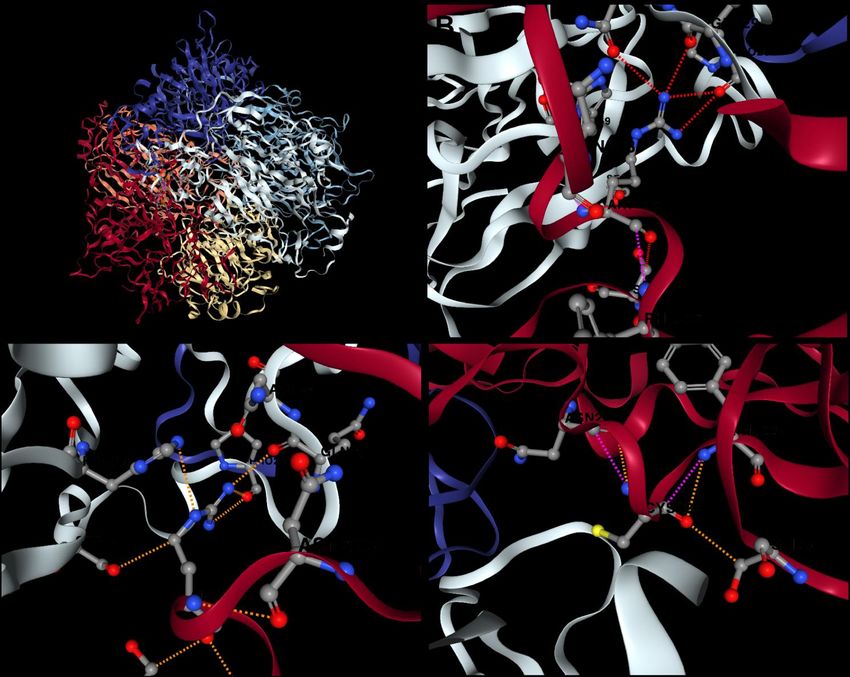
the hexamer due to disruption of protomer-protomer interactions as illustrated in Figure 4. The
analysis showed that there was a decrease in binding affinity by a factor close to 1 kcal/mol
(ΔΔGAffinity = −0.98 kcal/mol) highlighting the vital role of the wild-type residue.
AIMS Molecular Science Volume 8, Issue 1, 60–75.71
Figure 4. The predicted effect of the missense novel AKU mutation on the structure of
HGD protein complex using mSCM-PPI2 tool. (A) The hexameric structure of the active
HGD protein which consists of six chains A, B, C, D, E and F (red, orange, yellow, grey,
light blue and purple respectively); (B) Nitrogen atoms (dark blue ball) in the side chain
of arginine (position 225) found in WT chain A (red) make four hydrogen bonds (dashed
red line) with residues of adjacent chain D (grey); (C) Arginine at position 225 in WT
chain A (red) makes five polar bonds (dashed orange line) with residues of adjacent
chain D (grey); (D) Cysteine (yellow ball refers to sulphur atom) at position 225 in
mutated chain A (red) has low affinity to bind residues in chain D because it lost all polar
and H-bonds present in WT chain resulting in loose intermolecular interactions between
chains and a subsequent reduction in hexamer stability.
Interestingly, the amino acid arginine at position 225 (Figure 5) appears remarkable and
different studies reported the substitution of arginine (R) with other amino acids such as histidine (c.
674G>A) [42], leucine (c. 674G>T) [3], proline (c. 674G>C) [39] or cysteine (c.673 C>T) as
detected in our study.
AIMS Molecular Science Volume 8, Issue 1, 60–75.72
R225C
*R225H
*R225L
A122V *R225P
5’ UTR 3’ UTR
Exon 6 Exon 10
Length: 92 bases Length: 125 bases
Figure 5. Distribution of the HGD variants identified in the present study. The common
A122V was identified in exon 6 and the novel R225C at exon 10. Previously reported
missense variants which are occurring at the same position of exon 10 but with different
substituted amino acids were marked with red asterisk (R225H [42], R225L [3] & R225P [39]).
Additionally, the novel mutation was not found in coincidence in the same haplotype with the
second HGD mutation A122V which strongly suggests that the novel variant does not represent a
frequent polymorphism. However, Usher et al. (2015) showed that the mutation at position 225 in
the protein reduces the hexamer stability because arginine at 225 in wild-type protein is located at
the interface between protomers and it is involved in intermolecular interactions [39]. Consequently,
any alteration at this position is expected to disrupt theses intermolecular interactions and the
subsequent destabilisation of the hexamer [39]. Furthermore, the investigation of the effect of the
novel AKU-associated mutation on the enzyme structure and function and the evaluation of its
potential pathogenicity using different prediction tools specifically designed for interpretation of
missense variants confirmed that the previously unreported AKU variant at exon 10 is pathogenic
rather than benign polymorphism. This novel mutation is going to be submitted to HGD mutation
database.
5. Conclusions
The current study provides insight on AKU alleles present in the members of the third
generation of an AKU family from south of Jordan. In summary, our study bares a novel missense
pathogenic variant R225C in addition to the recurrent mutation A122V in HGD gene among
Jordanian AKU family members. Since Jordan is a country with high rate of consanguineous
marriages, there is a need to start a national screening project including different areas of Jordan to
diagnose more AKU patients. Our plan in the future is to do genetic analysis study on larger cohort
as other pathogenic variants of the HGD may be detected among Jordanian AKU patients. Moreover,
and in term of prevention of recurrent occurrence of the identified pathogenic variants, testing for at-
risk relatives and prenatal molecular diagnosis for pregnancies at increased risk are recommended.
AIMS Molecular Science Volume 8, Issue 1, 60–75.73
Acknowledgments
This study was funded by the Deanship of Scientific research and graduate studies at
Philadelphia University in Jordan.
Conflict of interest
All authors declare no conflicts of interest in this paper.
References
1. Garrod AE (1902) The Incidence of Alkaptonuria : A Study in Chemical Individuality. Lancet 2:
1616–1620.
2. Garrod AE (1908) The Croonian Lectures on inborn errors of metabolism, lecture II
Alkaptonuria. Lancet 2: 73–79.
3. Phornphutkul C, Introne WJ, Perry MB, et al. (2002) Natural history of alkaptonuria. N Engl J
Med 347: 2111–2121.
4. Mistry JB, Bukhari M, Taylor AM (2013) Alkaptonuria. Rare Dis 1: 1–7.
5. La Du BN, Zannoni VG, Laster L, et al. (1958) The nature of the defect in tyrosine metabolism
in alcaptonuria. J Biol Chem 230: 251–260.
6. Al-Shagahin HM, Mwafi N, Khasawneh M, et al. (2019) Ear, nose, and throat manifestations of
alkaptonuria patients from Jordan. Indian J Otol 25: 109–113.
7. Sakthivel S, Zatkova A, Nemethova M, et al. (2014) Mutation screening of the HGD gene
identifies a novel alkaptonuria mutation with significant founder effect and high prevalence.
Ann Hum Genet 78: 155–164.
8. Helliwell TR, Gallagher JA, Ranganath L (2008) Alkaptonuria--a review of surgical and autopsy
pathology. Histopathology 53: 503–512.
9. Fernandez-Canon JM, Granadino B, Beltran-Valero de Bernabe D, et al. (1996) The molecular
basis of alkaptonuria. Nat Genet 14: 19–24.
10. Zatkova A (2011) An update on molecular genetics of Alkaptonuria (AKU). J Inherit Metab Dis
34: 1127–1136.
11. Alsbou M, Mwafi N (2013) A previously undiagnosed case of alkaptonuria: A case report. Turk
J Rheumatol 28: 132–135.
12. Ranganath LR, Milan AM, Hughes AT, et al. (2020) Reversal of ochronotic pigmentation in
alkaptonuria following nitisinone therapy: Analysis of data from the United Kingdom National
Alkaptonuria Centre. JIMD Rep 1–13.
13. Zatkova A, Ranganath L, Kadasi L (2020) Alkaptonuria: Current Perspectives. Appl Clin Genet
13: 37–47.
14. Ranganath LR, Jarvis JC, Gallagher JA (2013) Recent advances in management of alkaptonuria
(invited review; best practice article). J Clin Pathol 66: 367–373.
AIMS Molecular Science Volume 8, Issue 1, 60–75.74
15. Taylor AM, Boyde A, Wilson PJ, et al. (2011) The role of calcified cartilage and subchondral
bone in the initiation and progression of ochronotic arthropathy in alkaptonuria. Arthritis Rheum
63: 3887–3896.
16. Wu K, Bauer E, Myung G, et al. (2018) Musculoskeletal manifestations of alkaptonuria: A case
report and literature review. Eur J Rheumatol 6: 98–101.
17. Thapa M, Yu J, Lee W, et al. (2015) Determination of homogentisic acid in human plasma by
GC-MS for diagnosis of alkaptonuria. Anal Sci Technol 28: 323–330.
18. Groseanu L, Marinescu R, Laptoiun D, et al. (2010) A late and difficult diagnosis of ochronosis.
J Med Life 3: 437–443.
19. Balaban B, Taskaynatan M, Yasar E, et al. (2006) Ochronotic spondyloarthropathy: Spinal
involvement resembling ankylosing spondylitis. Clin Rheumatol 25: 598–601.
20. Ranganath LR, Milan AM, Hughes AT, et al. (2016) Suitability Of Nitisinone In Alkaptonuria 1
(SONIA 1): an international, multicentre, randomised, open-label, no-treatment controlled,
parallel-group, dose-response study to investigate the effect of once daily nitisinone on 24-h
urinary homogentisic acid excretion in patients with alkaptonuria after 4 weeks of treatment.
Ann Rheum Dis 75: 362–367.
21. Ranganath LR, Psarelli EE, Arnoux J, et al. (2020) Efficacy and safety of once-daily nitisinone
for patients with alkaptonuria (SONIA 2): an international, multicentre, open-label, randomised
controlled trial. Lancet Diabetes Endocrinol 8: 762–772.
22. Ranganath LR, Khedr M, Milan AM, et al. (2018) Nitisinone arrests ochronosis and decreases
rate of progression of Alkaptonuria: Evaluation of the effect of nitisinone in the United
Kingdom National Alkaptonuria Centre. Mol Genet Metab 125: 127–134.
23. Keenan CM, Preston AJ, Sutherland H, et al. (2015) Nitisinone Arrests but Does Not Reverse
Ochronosis in Alkaptonuric Mice. JIMD Rep 24: 45–50.
24. Khedr M, Judd S, Briggs MC, et al. (2018) Asymptomatic Corneal Keratopathy Secondary to
Hypertyrosinaemia Following Low Dose Nitisinone and a Literature Review of Tyrosine
Keratopathy in Alkaptonuria. JIMD Rep 40: 31–37.
25. Hughes JH, Wilson P, Sutherland H, et al. (2020) Dietary restriction of tyrosine and
phenylalanine lowers tyrosinemia associated with nitisinone therapy of alkaptonuria. J Inherit
Metab Dis 43: 259–268.
26. Kılavuz S, Derya Bulut F, Kör D, et al. (2018) Demographic, Phenotypic and Genotypic
Features of Alkaptonuria Patients: A Single Centre Experience. J Pediatr Res 3: 7–11.
27. Ascher DB, Spiga O, Sekelska M, et al. (2019) Homogentisate 1,2-dioxygenase (HGD) gene
variants, their analysis and genotype–phenotype correlations in the largest cohort of patients
with AKU. Eur J Hum Genet 27: 888–902.
28. Zatkova A, Sedlackova T, Radvansky J, et al. (2012) Identification of 11 Novel Homogentisate
1, 2 Dioxygenase Variants in Alkaptonuria Patients and Establishment of a Novel LOVD-Based
HGD Mutation Database. JIMD Rep 4: 55–65.
29. Abdulrazzaq YM, Ibrahim A, Al-khayat AI, et al. (2009) R58fs mutation in the HGD gene in a
family with alkaptonuria in the UAE. Ann Hum Genet 73: 125–130.
AIMS Molecular Science Volume 8, Issue 1, 60–75.75
30. Ladjouze-Rezig A, Rodriguez de Cordoba S, Aquaron R (2006) Ochronotic rheumatism in
Algeria: clinical, radiological, biological and molecular studies- a case study of 14 patients in 11
families. Joint Bone Spine 73: 284–292.
31. Al-Sbou M, Mwafi N, Lubad MA (2012) Identification of forty cases with alkaptonuria in one
village in Jordan. Rheumatol Int 32: 3737–3740.
32. Nemethova M, Radvanszky J, Kadasi L, et al. (2016) Twelve novel HGD gene variants
identified in 99 alkaptonuria patients: Focus on “black bone disease” in Italy. Eur J Hum Genet
24: 66–72.
33. Koressaar T, Remm M (2007) Enhancements and modifications of primer design program
Primer3. Bioinformatics 23: 1289–1291.
34. Untergasser A, Cutcutache I, Koressaar T, et al. (2012) Primer3-new capabilities and interfaces.
Nucleic Acids Res 40: e115.
35. Zatkova A, Nemethova M (2015) Genetics of alkaptonuria – an overview. Eur Pharm J 62: 27–32.
36. Ben Halim N, Ben Alaya Bouafif N, Romdhane L, et al. (2013) Consanguinity, endogamy, and
genetic disorders in Tunisia. J Community Genet 4: 273–284.
37. Hamamy HA, Masri AT, Al-Hadidy AM, et al. (2007) Consanguinity and genetic disorders.
Profile from Jordan. Saudi Med J 28: 1015–1017.
38. Titus GP, Mueller HA, Burgner J, et al. (2000) Crystal structure of human homogentisate
dioxygenase. Nat Struct Biol 7: 542–546.
39. Usher J, Ascher D, Pires D, et al. (2015) Analysis of HGD Gene Mutations in Patients with
Alkaptonuria from the United Kingdom: Identification of Novel Mutations. JIMD Rep 24: 3–11.
40. Bernini A, Galderisi S, Spiga O, et al. (2017) Toward a generalized computational workflow for
exploiting transient pockets as new targets for small molecule stabilizers: application to the
homogentisate 1,2-dioxygenase mutants at the base of rare disease Alkaptonuria. Comput Biol
Chem 70: 133–141.
41. Vilboux T, Kayser M, Introne W, et al. (2009) Mutation spectrum of homogentisic acid oxidase
(HGD) in alkaptonuria. Hum Mutat 30: 1611–1619.
42. Beltrán-Valero De Bernabé D, Granadino B, Chiarelli I, et al. (1998) Mutation and
polymorphism analysis of the human homogentisate 1,2- dioxygenase gene in alkaptonuria
patients. Am J Hum Genet 62: 776–784.
©2021 the Author(s), licensee AIMS Press. This is an open access
article distributed under the terms of the Creative Commons
Attribution License (http://creativecommons.org/licenses/by/4.0)
AIMS Molecular Science Volume 8, Issue 1, 60–75.You can also read