The Diagnosis and Management of Extranodal NK/T-Cell Lymphoma, Nasal-Type and Aggressive NK-Cell Leukemia
←
→
Page content transcription
If your browser does not render page correctly, please read the page content below
J Clin Exp Hematopathol
                                                                                                                                  Vol. 51, No. 1, May 2011

     Review Article

     The Diagnosis and Management of Extranodal NK/T-Cell
     Lymphoma, Nasal-Type and Aggressive NK-Cell Leukemia

                                                                      Yok-Lam Kwong

         Natural killer (NK) cell lymphomas are rare malignancies. They are classified as extranodal NK/T-cell lymphoma, nasal
     type, and aggressive NK cell leukemia. NK cell neoplasms are prevalent in Asian and South American populations, but are
     extremely rare in the West. They can be classified clinically into nasal, non-nasal, and aggressive lymphoma/leukemia subtypes.
     For nasal NK cell lymphomas, combined chemotherapy and radiotherapy are indicated for stage I/II disease. Chemotherapy is
     the main treatment for stage III/IV nasal NK cell lymphomas, as well as the non-nasal and aggressive subtypes. Regimens
     containing drugs not affected by the P-glycoprotein, particularly in combination with L-asparaginase, have resulted in much
     improvement in treatment outcome for high-risk, refractory or relapsed patients. Autologous or allogeneic hematopoietic stem
     cell transplantation should be considered for selected patients. Epstein-Barr virus DNA load as a surrogate marker for
     prognostication, and clinical stratification of patients should be incorporated in clinical management algorithms. 〔J Clin Exp
     Hematopathol 51(1) : 21-28, 2011〕

     Keywords: natural killer cell lymphoma, natural killer cell leukemia, chemotherapy, radiotherapy, hematopoietic stem cell
               transplantation

                                                                                          sues, and occasionally skeletal muscles.3
                              INTRODUCTION
         Natural killer (NK) cells are cytotoxic cells, which are
                                                                                          HISTOLOGICAL PERSECTIVES OF NK-CELL
     capable of lysing tumor cells, and cells infected by bacteria
                                                                                          MALIGNANCIES
     and virus.1-3 Morphologically, NK cells appear as large gran-
     ular lymphocytes with pale cytoplasm and abundant azuro-                                 Destructive midline facial lesions have traditionally been
     philic granules. The bone marrow is the main site of develop-                        referred to as lethal midline granuloma.2 In Caucasian pa-
     ment of NK cells, which represent a distinct lineage of                              tients, Wegener’ s granulomatosis, sarcoidosis, carcinomas
     lymphocytes different from T cells. However, the two line-                           and conventional sinonasal diffuse large B cell lymphomas
     ages are developmentally related, with a bipotential T/NK cell                       are the common causative lesions.4 However, in Asian and
     progenitor that can develop into NK cells (without rearrange-                        South American patients, these lesions often show atypical
     ment of the T cell receptor, TCR, genes), or alternatively into                      lymphoid cells in a polymorphic inflammatory infiltrate of
     T cells (with rearrangement of the TCR genes).1 Because of a                         polymorphs, eosinophils and plasma cells, leading to its de-
     common ontogeny, NK cells express T cell antigens, includ-                           scription as “polymorphic reticulosis”.4 Immunohistochemically,
     ing CD2, CD7 and CD8. They are negative for surface CD3,                             the abnormal lymphoid cells expressed the T lineage antigen
     but express cytoplasmic CD3 epsilon (e) chain. NK cells also                         CD3. With the recognition of angiocentricity and angiode-
     express “NK-lineage associated” markers, including CD16,                             struction in these lesions, they were categorized as angiocen-
     CD56 and CD57.2 Of these antigens, CD56 is generally                                 tric T cell lymphomas in the Revised European American
     regarded as an NK cell marker, although it can also be ex-                           Lymphoma (REAL) classification.5
     pressed on NK-like T cells, neural and neuroendocrine tis-
                                                                                          NK CELL LYMPHOMAS
     Received : November 11, 2010
     Accepted : November 14, 2010                                                             With the availability of anti-CD56 antibodies, most angio-
     Department of Medicine, Queen Mary Hospital, Hong Kong                               centric T cell lymphomas of the nose and upper aerodigestive
     Address correspondence and reprint request to Kwong Y.L., M.D., Department of        tract were found to express CD56.6 The diagnosis was further
     Medicine, Professorial Block, Queen Mary Hospital, Pokfulam Road, Hong Kong,
     China                                                                                improved by the utilization of monoclonal anti-CD3 antibod-
     Email : ylkwong@hkucc.hku.hk                                                         ies that stained only T cells (which expressed surface CD3)

                                                                                     21

④10-011.mcd   Page 1                                                                                                                      11/05/25 18:05     v4.21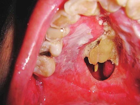
Kwong Y
but not NK cells (which only expressed cytoplasmic CD3e
but not surface CD3).7 Hence, NK cells are surface CD3- ,
cytoplasmic CD3e+, and CD56+, whereas T cells are surface
CD3+ and CD56+ . Once NK cells became distinguishable
from T cells, most nasal angiocentric T cell lymphomas were
found to be actually NK cell lymphomas. In the World
Health Organization (WHO) classification system, these lym-
phomas are now classified as extranodal NK/T cell lympho-
mas, nasal type.8 The notation of NK/T cell is to cater for the
very rare finding of true surface CD3+ and CD56+ cytotoxic T
cell lymphomas occurring in the nose, which may be indistin-
guishable clinically from NK cell lymphomas. However, in
clinical practice nearly all NK/T cell lymphomas are actually Fig. 1. Mosquito bite hypersensitivity developing into a cutane-
NK cell lymphomas. ous NK cell lymphoma. (1A) Cutaneous NK cell lymphoma. (1B)
Previous scars from mosquito bites on the other leg (arrow).
INFECTION OF NK LYMPHOMA CELLS BY
EPSTEIN-BARR VIRUS CLINICAL FEATURES OF NK CELL
LYMPHOMAS
NK lymphoma cells are almost invariably infected with
Epstein Barr virus (EBV).2,3 Analysis of the EBV terminal The WHO classification divides NK cell lymphomas into
repeat region reveals a clonal episomal pattern, suggesting two subtypes, extranodal NK/T cell lymphoma, nasal type,8
that the virus may be of pathogenetic significance. The con- and aggressive NK cell leukemia.12 Clinically, NK cell lym-
sistent presence of EBV has important implications. In situ phomas can be divided into three categories : nasal, non-
hybridization (ISH) for the EBV encoded early small RNA nasal, and aggressive lymphoma/leukemia subtypes (Table
(EBER) is an accurate localization method for NK lymphoma 1).2,3
cells,9 particularly in sites where EBV is usually absent, such
as the liver and bone marrow.2 In fact, the presence of EBV
Nasal NK cell lymphoma
in the neoplastic cells is a pre-requisite in the WHO classifica-
tion criteria for NK/ T cell lymphomas.10 Nasal NK cell lymphomas refer generally to tumors aris-
ing in the nose and the upper airway.13 The male to female
ratio is approximately 3 : 1, with disease peaking in the fifth
Chronic active EBV infection and related conditions
decade of life. NK cell lymphoma is the commonest histo-
NK cell lymphomas may be preceded by uncommon dis- logic subtype in nasal lymphomas in Asian patients.13 Nasal
eases related to EBV infection. Chronic active EBV infection NK cell lymphomas present as destructive mass lesions in-
(CAEBV) is a rare condition involving predominantly chil- volving the nasal cavity, nasopharynx, paranasal sinuses, ton-
dren and young adults of Japanese, Korean and Chinese sils, hypopharynx, and larynx. Destruction of the hard palate
decent.11 Patients present with chronic fever, lymphadenop- leads to a characteristic midline perforation, from which the
athy, hepatosplenomegaly and peripheral blood cytopenias, term “lethal midline granuloma” was originally derived (Fig. 2).
lasting six months or longer. Blood EBV antibody titers and
EBV DNA loads are very high. Biopsies of involved tissues
Non-nasal NK cell lymphomas
show EBV-positive lymphoid infiltrates of either NK cell or T
cell lineage. The disease is progressive, culminating in an Non-nasal NK cell lymphomas may involve any anatomic
NK cell leukemia/lymphoma or a T cell lymphoma. site.14 Male predominance and age of presentation are similar
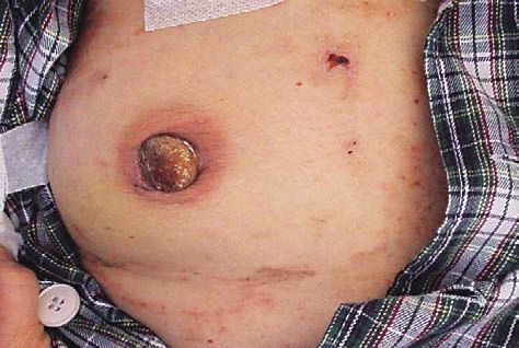
Mosquito bite hypersensitivity is a dermatologic condition, to nasal NK cell lymphomas. Common primary sites include
with affected individuals developing severe blistering reac- the skin, gastrointestinal tract, salivary glands, spleen and
tions to mosquito or insect bites, often associated with fever testis (Fig. 3). Interestingly, primary sites of non-nasal NK
and hepatosplenomegaly.11 These lesions heal spontaneously cell lymphomas are also sites where nasal NK cell lympho-
with scarring, but with time a lesion may continue to deterio- mas disseminate to. Hence, it is important for a primary nasal
rate and develop into a lymphoma of T cell and occasionally NK cell lymphoma to be excluded before a non-nasal NK cell
NK cell lineage (Fig. 1). lymphoma is diagnosed. In practice, patients with NK cell
lymphomas involving sites other than the nasal areas should
undergo imaging studies and a nasal panendoscopy to exclude
an occult nasal primary.2
22
④10-011.mcd Page 2 11/05/11 17:05 v4.21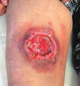
Diagnosis and management of NK cell malignancies

                               Table 1. Clinicopathological features of different subtypes of natural killer cell
                                        neoplasms

                                                           Nasal NK cell           Non-nasal NK cell     Aggressive NK cell
                                  Clinical features
                                                            lymphoma                  lymphoma           lymphoma/leukemia
                                Sex                     Male > Female          Male > Female             Male = Female
                                Median age              50-60 years            50-60 years               30-40 years
                                Anatomical sites        Nose, para-nasal       Skin, gastrointestinal    Blood, bone marrow,
                                involved                sinuses, orbits        tract, salivary glands,   liver, spleen, lymph
                                                                               testis, other organs      nodes
                                                                               and tissues
                                Clinical presentation   Nasal bleeding,        Ulceration and mass       Fever, jaundice,
                                                        obstruction, hard      lesion                    enlargement of liver
                                                        palatal perforation                              and/or spleen,
                                                                                                         lymphadenopathy
                                                                               Usually advanced in
                                                        Stage I/II : good
                                Prognosis                                      stage, aggressive,        Highly fatal
                                                        Stage III/LV : poor
                                                                               poor outcome

                                                                                      Aggressive NK cell leukemia/lymphoma
                                                                                       Aggressive NK cell leukemia/lymphoma is a catastrophic
                                                                                   disorder.15 Men and women are equally affected, with dis-
                                                                                   ease presenting usually at the third decade. Clinical features
                                                                                   include high fever, significant weight loss, jaundice, skin
                                                                                   infiltration, lymphadenopathy and hepatosplenomegaly.
                                                                                   Marrow hemophagocytosis leading to severe anemia and
                                                                                   thrombocytopenia may be present. Circulating lymphoma
                                                                                   cells vary morphologically from large granular lymphocytes
                                                                                   to frank blast. Liver function derangement and disseminated
                                                                                   intravascular coagulopathy appear progressively. The clinical
                                                                                   course is often lethal, with survival measured just in weeks.

       Fig. 2. Palatal perforation due to a nasal NK cell lympho-                     Patterns of dissemination of NK cell lymphomas
       ma                                                                              Nasal NK cell lymphomas are locally malignant, with
                                                                                   distant organ involvement occurring in only about 10% of
                                                                                   patients at presentation. Fewer than 10% of cases show mar-
                                                                                   row infiltration.16 On the contrary, non-nasal NK cell lym-
                                                                                   phomas are generally disseminated. With modern imaging
                                                                                   techniques, most apparently non-nasal cases can be found to
                                                                                   have nasal involvement, implying that they are in fact dis-
                                                                                   seminated nasal NK cell lymphomas. Aggressive NK cell
                                                                                   leukemia/lymphoma is disseminated at presentation. It can be
                                                                                   differentiated from a rare terminal leukemic form of
                                                                                   nasal/non-nasal NK cell lymphoma by the absence of a pre-
                                                                                   vious history, a shorter illness, a younger age of presentation,
                                                                                   and an extremely aggressive course.

                                                                                      Peripheral blood and marrow involvement in nasal and
       Fig. 3. Non-nasal NK cell lymphoma infiltrating the right                      non-nasal NK cell lymphomas
       nipple
                                                                                      Peripheral blood cytopenias may be found in about 10-
                                                                                   15% of cases of nasal and non-nasal NK cell lymphomas, and

                                                                              23

④10-011.mcd   Page 3                                                                                                                11/05/11 17:05    v4.21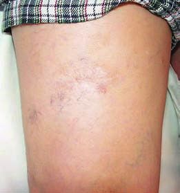
Kwong Y

     are predominantly due to active hemophagocytosis in the                 nodal lymphomas may not always be accurate. To improve
     marrow. The hemophagocytic cells represent activated retic-             patient stratification for treatment, several prognostic models
     uloendothelial cells and on their own do not indicate marrow            have been applied. The international prognostic index (IPI),
     infiltration. To detect possible lymphomatous infiltration,             taking into account the stage, age, performance status, number
     EBER ISH is a more specific and sensitive test. Staging of              of extranodal sites and the lactate dehydrogenase (LDH) lev-
     nasal and non-nasal NK cell lymphomas should always in-                 el, has been shown to be relevant in NK cell lymphomas.20
     clude EBER ISH in the marrow.2 Positive results indicated               Two other prognostic models based on the IPI concept has
     by EBER positive cells in the marrow heralds a grave                    also been formulated. When B symptoms, stage, LDH level
     prognosis.17                                                            and regional lymph node involvement are considered, stage
                                                                             I/II nasal NK cell lymphomas can be better stratified into
                                                                             different risk groups.21 In another prognostic model, non-
       Initial assessment of NK cell lymphomas
                                                                             nasal type, stage, performance status and number of extrano-
         A complete history and physical examination is needed.              dal involvement are found to predict outcome.22
     For biopsy of the involved organ, the specimen should be as
     sizeable as possible, because zonal necrosis is characteristi-
                                                                             MANAGEMENT OF NK CELL MALIGNANCIES
     cally found, and a small biopsy may only contain necrotic
     tissue. The biopsies should be sent fresh unfixed to the path-              The optimal treatment strategy of NK cell lymphoma has
     ology laboratory for cryostat sectioning or flow cytometry.             until recently not been well defined. With increased under-
     This will enable surface CD3 to be detected, which distin-              standing of these malignancies, several important principles
     guishes between T and NK cell lymphomas. If fresh tumor                 have emerged.23 For nasal NK cell lymphomas, the best
     biopsies are not available, NK and T cell lymphomas can be              treatment results are obtained with a combination of chemo-
     differentiated by TCR gene rearrangement studies, with the              therapy and radiotherapy.24,25 Chemotherapy is the mainstay
     TCR genes germline in NK cell lymphomas but clonally rear-              of treatment for non-nasal NK cell lymphoma and aggressive
     ranged in T cell lymphomas.2,3                                          NK cell leukemia/lymphoma. Different from conventional
         Radiologic imaging is an essential initial evaluation of all        lymphomas, anthracyclines have not been shown to be neces-
     clinical subtypes of NK cell lymphomas. Computerized to-                sary for effective treatment.20 Frontline high dose chemother-
     mographic (CT) scan is better for detection of bony involve-            apy and hematopoietic stem cell transplantation (HSCT) is not
     ment, whereas magnetic resonance imaging (MRI) is superior              indicated.26 Even for cases with relapsed lymphoma, the use
     in defining soft tissue infiltration. Positron emission tomog-          of HSCT will still have to be evaluated on an individual
     raphy (PET) is very useful for detection of involvement of              basis.26 Novel treatment approaches are needed to improve
     other systemic sites. NK cell lymphomas are 18-                         the outcome of patients with advanced diseases.
     fluorodeoxyglucose (FDG) avid, with standardized uptake
     value maximum of about 5-10.18 Detailed initial imaging is
                                                                               Nasal NK cell lymphoma
     essential for assessment of response. For nasal NK cell lym-
     phoma, radiologic assessment is particularly critical for accu-             For localized stage I/II nasal NK cell lymphoma, radio-
     rate planning of subsequent radiotherapy.                               therapy used to be the primary treatment. Systemic failure
                                                                             occurred in at least 30% of patients, suggesting that subclini-
                                                                             cal dissemination of lymphoma had occurred in these appa-
       Quantification of circulating plasma EBV DNA
                                                                             rently early-stage patients.2 The use of primary chemother-
         In EBV-associated lymphoid malignancies, increases in               apy was also associated with treatment failure in about 40%
     circulating EBV DNA are found, due to viral DNA release                 of patients, necessitating the use of salvage radiotherapy.2
     from apoptosis of proliferating tumor cells.19 Serial EBV               Therefore, combined chemotherapy and radiotherapy appears
     DNA quantifications by quantitative polymerase chain reac-              to be the treatment of choice. Chemotherapy and radiother-
     tion in NK cell lymphoma have been found to correlate with              apy can be given sequentially or concomitantly,24,25 both
     disease control.19 EBV DNA quantification can be performed              methods giving similar treatment results.
     in plasma or whole blood. However, peripheral blood mono-                   Several points are worthy of note. Radiation dosage is
     nuclear cells are not a suitable source, because circulating            typically about 50 Gy, and smaller dosages are associated
     lymphoma cells are absent.                                              with inferior outcome when used alone.2 Whether the dose of
                                                                             radiotherapy can be decreased when concomitant chemother-
                                                                             apy or radio-sensitizer is used remains to be defined.25 Early
       Staging and prognostication of NK cell lymphomas
                                                                             use of radiotherapy is important, whether concomitantly or
        As NK cell lymphomas are almost exclusively extranodal,              sequentially with chemotherapy.20 Non-anthracycline based
     conventional lymphoma staging procedures designed for                   chemotherapy is effective and may be preferable to

                                                                        24

④10-011.mcd   Page 4                                                                                                         11/05/11 17:05    v4.21
Diagnosis and management of NK cell malignancies
anthracycline-containing regimens, particularly in elderly pa- refractory NK cell lymphomas is difficult. The therapeutic
tients. Combined chemotherapy and radiotherapy can be ex- use of L-asparaginase, either singly or in combination chemo-
pected to be curative in at least 70-80% of patients with stage therapy, has led to favorable responses. Different prepara-
I/II nasal NK cell lymphomas.20,24,25 tions, including E. coli derived, Erwinia-derived, and pegy-
Interestingly, very late relapses of early-stage nasal NK lated forms, appear to have similar treatment results.31,32
cell lymphoma as local or systemic recrudescence have been Prospective studies examining L-asparaginase in combination
described to happen after more than ten years to up to thirty chemotherapy, such as the SMILE regimen, have also shown
years.27,28 It is unknown if these relapses were derived from very good efficacies.29,30 The main side effects of
the original tumors or represented new lymphomas. Lasparaginase include hyperbilirubinemia, liver dysfunction,
Therefore, life-long follow up is recommended even for pa- leucopenia, infections, hyperglycemia, and hypersensitivity
tients who are in prolonged remission. reactions. Other drug combinations, including ifosfamide,
methotrexate, etoposide, and predonisolone, have also been
reported to result in an OR rate of 43.8% and 5-year overall
Advanced-stage nasal and non-nasal NK cell lymphoma
survival (OS) of 24.8%.33
Chemotherapy is the mainstay of treatment for advanced-
stage NK cell lymphomas. Conventional CHOP or CHOP-
Aggressive NK-cell leukemia/lymphoma
like regimens give poor outcome, with CR achieved in < 20%
of patients.2 The unsatisfactory result of CHOP may be due Aggressive NK-cell leukemia/lymphoma is a devastating
to expression of the multi-drug resistance 1 (MDR-1) gene, illness, with few treatment successes reported.2,3 Treatment
leading to high levels of P-glycoprotein and therefore active results of anthracycline-based regimens were dismal, and in
export of many chemotherapeutic drugs including one series only three of 13 patients achieved CR, which
anthracyclines.3 Hence, non-anthracycline containing regi- merely extended the survival for several weeks.15 Treatment
mens may actually be more effective in these patients. with L-asparaginse-containing regimens followed by alloge-
A novel regimen SMILE, comprising dexamethasone, neic HSCT had resulted in prolonged survivals in a few
methotrexate , ifosfamide, L-asparaginase, and etoposide cases.34 This approach will need to be validated.
(Table 2) has been shown in phase I and phase II studies to be
promising.29,30 The regimen is based on the use of drugs not
Hematopoietic stem cell transplantation (HSCT)
exported by P-glycoprotein together with L-asparaginase,
which has been shown to have considerable activities in NK Because of unsatisfactory treatment outcome of advanced-
cell lymphomas as a single agent.31,32 The treatment results of staged, relapsed or refractory diseases, the role of autologous
SMILE are remarkable. In patients with relapsed or refrac- and allogeneic HSCT has been explored as consolidation or
tory NK cell lymphoma, SMILE treatment resulted in an salvage therapy.26 However, owing to the relative rarity of
overall response (OR) rate of 74%, and complete remission NK cell lymphoma, prospective studies of HSCT have not
(CR) rate of 35-50%.29,30 Important side effects include neu- been performed.
tropenia and infections, so that aggressive granulocyte colony
stimulating factor support is needed. However, the long-term
Autologous HSCT
results still remain to be defined.
In a recent retrospective multi-centre analysis, NK cell
lymphoma patients who received autologous HSCT were
Relapsed and refractory NK cell lymphomas
compared with matched patients who were treated with che-
The management of patients with relapsed or therapy- motherapy or radiotherapy only.35 The disease status before
Table 2. SMILE protocol for advanced stage and relapse natural killer cell
malignancies
Drugs Dosage Administration Days
Methotrexate with leucovorin 2 g/m2 Intravenous 1
Ifosfamide with mesna 1.5 g/m
2
Intravenous 2, 3, 4
Dexamethasone 40 mg Intravenous or oral 2, 3, 4
Etoposide 100 mg/m
2
Intravenous 2, 3, 4
L-asparaginase Intravenous 8, 10, 12, 14, 16, 18, 20
2
6, 000 U/ m
Granulocyte colony stimulating factor started on day 6. Cycles to be repeated every 28 days.
25
④10-011.mcd Page 5 11/05/11 17:05 v4.21Kwong Y
HSCT was the most important factor correlating with out-
Response evaluation
come. Patients with early-stage disease had better outcome
than those with advanced or refractory disease. Although NK cell lymphomas are predominantly extranodal, so that
patients undergoing HSCT appeared to have a slightly lower conventional criteria of response evaluation for nodal lym-
relapse rate, a treatment-related-mortality of 8.5% was also phoma may not be easily applicable. As NK cell lymphomas
observed, so that the OS was not different in the two groups.35 are FDG-avid, PET/CT scan is a useful modality.
Several issues in autologous HSCT remain controversial. Quantification of circulating EBV DNA is another way of
Patients with early stage disease limited to the nasal areas are measuring tumor load. The use of PET/CT and EBV DNA
potentially curable with combined chemotherapy and radio- quantification in documenting clinical and molecular remis-
therapy, so that it is doubtful if frontline autologous HSCT sion will have to be validated prospectively.
may be beneficial.26 In fact, retrospective analyses appear to
show that HSCT in these patients did not confer survival
CONCLUSIONS
advantage. In patients with advanced-stage or relapsed dis-
ease, the results of HSCT remain poor. Whether the applica- NK cell malignancies are divided clinically into nasal,
tion of prognostic models to identify patients who may benefit non-nasal and aggressive subtypes. There is an almost invar-
from early use of HSCT will have to be defined.35 Finally, iable association with clonal episomal EBV infection. The
conditioning regimens for HSCT in NK cell lymphoma have diagnosis of NK cell lymphoma should be based on morpho-
most often been those used in B-cell lymphomas, including logic, immunophenotypic, and molecular approaches.
CBV (etoposide, carmustine, and cyclophosphamide) and Combination chemotherapy and radiotherapy is currently the
BEAM (carmustine, etoposide, cytarabine, and melphalan). standard treatment for nasal NK cell lymphoma. For other
However, whether these regimens are optimal, or other more subtypes of NK cell lymphoma, chemotherapy is the mainstay
effective drugs have to be used, will need to be defined. of treatment. Non P-glycoprotein dependent drugs appear to
be efficacious. The optimal timing and indications of autolo-
gous and allogeneic HSCT need to be evaluated.
Allogeneic HSCT
The potential benefits of allogeneic over autologous
REFERENCES
HSCT are related a putative graft-versus-lymphoma effect.2,3
This possibility is attractive, since NK lymphoma cells ex- 1 Spits H, Lanier LL, Phillips JH: Development of human T and
press EBV viral antigens, which should theoretically be tar- natural killer cells. Blood 85:2654-2670, 1995
geted by donor derived cytotoxic T cells reactive to EBV. 2 Kwong YL: Natural killer-cell malignancies: diagnosis and treat-
However, available studies of allogeneic HSCT in NK cell ment. Leukemia 19:2186-2194, 2005
lymphomas are very limited. Problems affecting the interpre- 3 Oshimi K: Progress in understanding and managing natural killer-
tation of these studies include differences in donor source cell malignancies. Br J Haematol 139:532-544, 2007
(including HLA-matched siblings, unmatched donors, or cord 4 Batsakis JG, Luna MA: Midfacial necrotizing lesions. Semin
blood), heterogeneity of conditioning regimens (presence or Diagn Pathol 4:90-116, 1987
absence of total-body irradiation), and timing of HSCT (at 5 Harris NL, Jaffe ES, Stein H, Banks PM, Chan JK, et al : A
remission, during relapse or refractory disease). In a review revised European-American classification of lymphoid neo-
of cases reported in the literature, the majority of patients who plasms: a proposal from the International Lymphoma Study
received allogeneic HSCT had nasal NK cell lymphoma and, Group. Blood 84:1361-1392, 1994
at the time of transplantation, 69% had recognizable or refrac- 6 Wong KF, Chan JK, Ng CS, Lee KC, Tsang WY, et al : CD56
tory diseases. Half of the patients were alive after HSCT, (NKH1)-positive hematolymphoid malignancies: an aggressive
with 25% dying from transplantation-related complications, neoplasm featuring frequent cutaneous/mucosal involvement, cy-
and the rest from progressive lymphoma.26 In the largest toplasmic azurophilic granules, and angiocentricity. Hum Pathol
retrospective series to date, the 2-year OS was 40%.36 23:798-804, 1992
Interestingly, a patient with NK cell lymphoma relapse after 7 Chan JK, Tsang WY, Ng CS: Clarification of CD3 immunoreac-
allogeneic HSCT achieved a durable remission with discon- tivity in nasal T/natural killer cell lymphomas: the neoplastic cells
tinuation of immunosuppression, implying the presence of a are often CD3e+. Blood 87:839-841, 1996
graft-versus-lymphoma effect.37 Hence, reduced-intensity- 8 Chan JKC, Quintanilla-Martinez L, Ferry JA, Peh S-C:
conditioning allogeneic HSCT may also have a role in de- Extranodal NK/T-cell lymphoma, nasal type. In: Swerdlow SH,
creasing treatment-related mortality while preserving the ben- Campo E, Harris NL, Jaffe ES, Pileri SA, et al. (eds): World
efits of alloreactivity against lymphoma cells.38 International Health Organization Classification of Tumours, WHO
collaborative trials are needed to define the optimal use of Classification of Tumours of Haematopoietic and Lymphoid
allogeneic HSCT in NK cell malignancies. Tissues. 4th ed, International Agency for Research on Cancer
26
④10-011.mcd Page 6 11/05/11 17:05 v4.21Diagnosis and management of NK cell malignancies
(IARC), Lyon, pp. 285-288, 2008 and extranodal NK cell lymphoma, nasal type. Ann Oncol
9 Chan JK, Yip TT, Tsang WY, Ng CS, Lau WH, et al. : Detection 21:1032-1040, 2010
of Epstein-Barr viral RNA in malignant lymphomas of the upper 23 Kwong YL, Anderson BO, Advani R, Kim WS, Levine AM, et
aerodigestive tract. Am J Surg Pathol 18:938-946, 1994 al. : Management of T-cell and natural-killer-cell neoplasms in
11 Cohen JI, Kimura H, Nakamura S, Ko YH, Jaffe ES: Epstein- Asia: Consensus statement from the Asian Oncology Summit
Barr virus-associated lymphoproliferative disease in non- 2009. Lancet Oncol 10:1093-1101, 2009
immunocompromised hosts: a status report and summary of an 24 Yamaguchi M, Tobinai K, Oguchi M, Ishizuka N, Kobayashi Y, et
international meeting, 8-9 September 2008. Ann Oncol 20:1472- al. : Phase I/II study of concurrent chemoradiotherapy for local-
1482, 2009 ized nasal natural killer/T-cell lymphoma: Japan Clinical
12 Chan JK, Jaffe ES, Ralfkiaer E, Ko YH: Aggressive NK-cell Oncology Group Study JCOG0211. J Clin Oncol 27:5594-5600,
leukaemia. In: Swerdlow SH, Campo E, Harris NL, Jaffe ES, 2009
Pileri SA, et al. (eds): World Health Organization Classification 25 Kim SJ, Kim K, Kim BS, Kim CY, Suh C, et al. : Phase II trial of
of Tumours, WHO Classification of Tumours of Haematopoietic concurrent radiation and weekly cisplatin followed by VIPD che-
and Lymphoid Tissues. 4th ed, International Agency for Research motherapy in newly diagnosed, stage IE to IIE, nasal, extranodal
on Cancer (IARC), Lyon, pp. 276-277, 2008 NK/T-cell lymphoma: Consortium for improving survival of lym-
13 Cheung MM, Chan JK, Lau WH, Foo W, Chan PT, et al. : phoma study. J Clin Oncol 27:6027-6032, 2009
Primary non-Hodgkin’ s lymphoma of the nose and nasophar- 26 Kwong YL: High-dose chemotherapy and hematopoietic SCT in
ynx: clinical features, tumor immunophenotype, and treatment the management of natural killer-cell malignancies. Bone Marrow
outcome in 113 patients. J Clin Oncol 16:70-77, 1998 Transplant 44:709-714, 2009
14 Chan JK, Sin VC, Wong KF, Ng CS, Tsang WY, et al. : Nonnasal 27 Ishida F, Nishina S, Asano N, Sasaki S, Sekiguchi N, et al. : Late
lymphoma expressing the natural killer cell marker CD56: a clini- relapse of extranodal natural killer/T cell lymphoma, nasal type,
copathologic study of 49 cases of an uncommon aggressive neo- after more than ten years. Leuk Lymphoma 51:171-173, 2010
plasm. Blood 89:4501-4513, 1997 28 Au WY, Kim SJ, Yiu HH, Ngan RK, Loong F, et al. :
15 Suzuki R, Suzumiya J, Nakamura S, Aoki S, Notoya A, et al. : Clinicopathological features and outcome of late relapses of natu-
NK-cell Tumor Study Group. Aggressive natural killer-cell leuke- ral killer cell lymphomas 10-29 years after initial remission. Am J
mia revisited: large granular lymphocyte leukemia of cytotoxic Hematol 85:362-363, 2010
NK cells. Leukemia 18:763-770, 2004 29 Yamaguchi M, Suzuki R, Kwong YL, Kim WS, Hasegawa Y, et
16 Wong KF, Chan JK, Cheung MM, So JC: Bone marrow involve- al. : Phase I study of dexamethasone, methotrexate, ifosfamide, L-
ment by nasal NK cell lymphoma at diagnosis is uncommon. Am J asparaginase, and etoposide (SMILE) chemotherapy for advanced-
Clin Pathol 115:266-270, 2001 stage, relapsed or refractory extranodal natural killer (NK)/T-cell
17 Lee J, Suh C, Huh J, Jun HJ, Kim K, et al. : Effect of positive lymphoma and leukemia. Cancer Sci 99:1016-1020, 2008
bone marrow EBV in situ hybridization in staging and survival of 30 Kwong YL, Yamaguchi M, Maeda Y, Hashimoto C, Kim WS, et
localized extranodal natural killer/T-cell lymphoma, nasal-type. al. : NK-cell Tumor Study Group. Phase II study of SMILE che-
Clin Cancer Res 13:3250-3254, 2007 mothearpy for newly-diagnosed stage IV, relapsed or refractory
18 Khong PL, Pang CB, Liang R, Kwong YL, Au WY: Fluorine-18 extranodal NK/T-cell lymphoma, nasal type: NKTSG study.
fluorodeoxyglucose positron emission tomography in mature T- Haematologica 95:0299, 2010 (Abstract)
cell and natural killer cell malignancies. Ann Hematol 87:613- 31 Jaccard A, Petit B, Girault S, Suarez F, Gressin R, et al. : L-
621, 2008 asparaginase-based treatment of 15 western patients with extra-
19 Au WY, Pang A, Choy C, Chim CS, Kwong YL: Quantification nodal NK/T-cell lymphoma and leukemia and a review of the
of circulating Epstein-Barr virus (EBV) DNA in the diagnosis and literature. Ann Oncol 20:110-116, 2009
monitoring of natural killer cell and EBV positive lymphomas in 32 Yong W, Zheng W, Zhu J, Zhang Y, Wang X, et al. : L-
immunocompetent patients. Blood 104:243-249, 2004 asparaginase in the treatment of refractory and relapsed extranodal
20 Chim CS, Ma SY, Au WY, Choy C, Lie AK, et al. : Primary NK/T-cell lymphoma, nasal type. Ann Hematol 88:647-652, 2009
nasal natural killer cell lymphoma: long-term treatment outcome 33 Kim BS, Kim DW, Im SA, Kim CW, Kim TY, et al. : Effective
and relationship with the International Prognostic Index. Blood second-line chemotherapy for extranodal NK/T-cell lymphoma
103:216-221, 2004 consisting of etoposide, ifosfamide, methotrexate, and predniso-
21 Lee J, Suh C, Park YH, Ko YH, Bang SM, et al. : Extranodal lone. Ann Oncol 20:121-128, 2009
natural killer T-cell lymphoma, nasal-type: a prognostic model 34 Ito T, Makishima H, Nakazawa H, Kobayashi H, Shimodaira S, et
from a retrospective multicenter study. J Clin Oncol 24:612-618, al. : Promising approach for aggressive NK cell leukaemia with
2006 allogeneic haematopoietic cell transplantation. Eur J Haematol
22 Suzuki R, Suzumiya J, Yamaguchi M, Nakamura S, Kameoka J, et 81:107-111, 2008
al. : NK-cell Tumor Study Group. Prognostic factors for mature 35 Lee J, Au WY, Park MJ, Suzumiya J, Nakamura S, et al. :
natural killer (NK) cell neoplasms: aggressive NK cell leukemia Autologous hematopoietic stem cell transplantation in extranodal
27
④10-011.mcd Page 7 11/05/11 17:05 v4.21Kwong Y
natural killer/T cell lymphoma: A multinational, multicenter, of the tumor after withdrawal of cyclosporine in relapsed extra-
matched controlled study. Biol Blood Marrow Transplant nodal natural killer/T cell lymphoma following allogeneic hema-
14:1356-1364, 2008 topoietic stem cell transplantation. Am J Hematol 82:937-939,
36 Murashige N, Kami M, Kishi Y, Kim SW, Takeuchi M, et al. : 2007
Allogeneic haematopoietic stem cell transplantation as a promis- 38 Sato E, Ohga S, Kuroda H, Yoshiba F, Nishimura M, et al. :
ing treatment for natural killer-cell neoplasms. Br J Haematol Allogeneic hematopoietic stem cell transplantation for Epstein-
130:561-567, 2005 Barr virus-associated T/natural killer-cell lymphoproliferative dis-
37 Kako S, Izutsu K, Oshima K, Sato H, Kanda Y, et al. : Regression ease in Japan. Am J Hematol 83:721-727, 2008
28
④10-011.mcd Page 8 11/05/11 17:05 v4.21You can also read