COVID-19 pandemic as a risk factor for the reactivation of herpes viruses
←
→
Page content transcription
If your browser does not render page correctly, please read the page content below
Epidemiology and Infection                            COVID-19 pandemic as a risk factor for the
        cambridge.org/hyg
                                                              reactivation of herpes viruses
                                                              M. D. Maldonado1               , J. Romero-Aibar2 and M. A. Pérez-San-Gregorio3
                                                              1
                                                                Department of Medical Biochemistry, Molecular Biology, and Immunology, University of Seville Medical School,
        Review                                                Seville, Spain; 2Superior Laboratory Technician, Department of External Analysis Service, IRNAS (CSIC) Seville,
        Cite this article: Maldonado MD, Romero-              Seville, Spain and 3Department of Personality, Evaluation and Psychological Treatment, University of Seville,
        Aibar J, Pérez-San-Gregorio MA (2021).                Seville, Spain
        COVID-19 pandemic as a risk factor for the
        reactivation of herpes viruses. Epidemiology            Abstract
        and Infection 149, e145, 1–5. https://doi.org/
        10.1017/S0950268821001333                               The appearance on the skin of herpes virus lesions, concomitantly with the coronavirus dis-
                                                                ease 2019 (COVID-19) pandemic, leads us to suspect an underlying infection with severe
        Received: 23 March 2021
                                                                acute respiratory syndrome coronavirus-2 (SARS-CoV-2). Diagnostic reverse transcriptase
        Revised: 29 April 2021
        Accepted: 9 June 2021                                   polymerase chain reaction tests and immunoglobulin M (IgM) and IgG seroconversion stud-
                                                                ies have therefore been carried out. We present three cases of herpes virus infections in
        Keywords:                                               immunocompetent patients: one of the infections was herpes simplex 1 in a 40-year-old
        COVID-19; herpes infections; immune system;             woman, and the other two were herpes varicella-zoster infections in a 62-year-old man and
        psychoneuroimmunology; SARS-CoV-2
                                                                a 25-year-old woman. The patients were in the care of the southern health district of
        Author for correspondence:                              Seville of the SAS (Andalusian Health Service) during the Spanish state of alarm over the
        M. D. Maldonado,                                        COVID-19 pandemic. The SARS-CoV-2 infection was confirmed in only one of the three
        Email: aibar@us.es                                      cases. In this study, we briefly review the etiopathogenic role of the COVID-19 pandemic situ-
                                                                ation, whereby immunodeficiencies are generated that favour the appearance of other viral
                                                                infections, such as herpes virus infections.

                                                              Introduction
                                                              Severe acute respiratory syndrome coronavirus-2 (SARS-CoV-2) was first discovered and iso-
                                                              lated in Wuhan, China, in December 2019. This is a new virus of zoonotic origin that has
                                                              caused one of the largest known global pandemics [1]. SARS-CoV-2 is a virus with a great
                                                              capacity for infecting and transmitting. Nevertheless, not everyone responds in the same
                                                              way to this virus. Most patients show symptoms as if they were suffering from a simple
                                                              cold. However, other sufferers develop pneumonia and even toxic shock that leads to death
                                                              [2, 3]. There have been patients who have debuted with mucosal cutaneous manifestations,
                                                              including enanthemas secondary to vasculitis, hives, varicella-like lesions, livedo reticularis,
                                                              Covid toe, erythema multiforme and pityriasis rosea [4–6].
                                                                  Certain pathogens use latency as an escape or evasion mechanism for the host’s immune sys-
                                                              tem. This is the case of the Herpesviridae family, such as herpes simplex type 1 and 2 (HSV-1
                                                              and HSV-2), varicella zoster virus (VZV), cytomegalovirus (CMV), human herpes virus 6, 7
                                                              (HHV 6, 7) and Epstein–Barr virus (EBV), although the cells in which they establish latency
                                                              do vary. The most common entry point for human herpes viruses is the pharynx, although
                                                              they can also enter through the genital or parenteral route. Once inside, they will use various
                                                              mechanisms to colonise human cells and spread, including the use of receptors and co-receptors
                                                              on the surface of human cells. After contact, the virus fuses its lipid envelope with the host cell
                                                              membrane and releases its nucleocapsid together with the integument proteins into the cytosol.
                                                              From here, it will introduce its viral-DNA to the nucleus of the host cell, replicating rapidly and
        © The Author(s), 2021. Published by                   generating infection. After the initial infection, all herpes viruses remain in a state of latency, in
        Cambridge University Press. This is an Open           different host cells, from where they can be later reactivated [7]. Here, we evaluate three cases of
        Access article, distributed under the terms of
        the Creative Commons Attribution-
                                                              herpes infection, in which HSV-1 and VZV were held responsible. Although the host has a
        NonCommercial-NoDerivatives licence (http://          strong and effective immune system, the virus remains quartered in Gasser’s ganglion
        creativecommons.org/licenses/by-nc-nd/4.0/),          (HSV-1) or in the ganglia of the nerve roots (VZV), without generating symptoms or pathology.
        which permits non-commercial re-use,                  In a latently infected neuron, virus-specific proteins are not produced, and as a result, the host’s
        distribution, and reproduction in any medium,
                                                              immune system does not identify the presence of the virus and does not target the latently
        provided the original work is unaltered and is
        properly cited. The written permission of             infected neuron for destruction. The host may suffer from an immunodeficiency, triggered by
        Cambridge University Press must be obtained           any of a large number of reasons, including stress, insomnia, insolation, malnutrition, taking
        for commercial re-use or in order to create a         immunosuppressive drugs, pregnancy, ageing, infections by other viruses and debilitating dis-
        derivative work.                                      eases. When this occurs, the herpes virus appears on the skin, either: in the facial area
                                                              (HSV-1), such as lips, nose, cheekbones and even in the eyes, which can cause herpetic keratitis
                                                              with the risk of blindness, or it appears in the cervical, dorsal or lumbar areas (VZV) causing a
                                                              herpetic neuropathy [8, 9]. The lesions appear as raised erythematous plaques upon which
                                                              maculo-papulous vesicles settle, spreading through the affected dermatome, and usually

Downloaded from https://www.cambridge.org/core. IP address: 46.4.80.155, on 13 Oct 2021 at 07:38:24, subject to the Cambridge Core terms of use, available at https://www.cambridge.org/core/terms.
https://doi.org/10.1017/S0950268821001333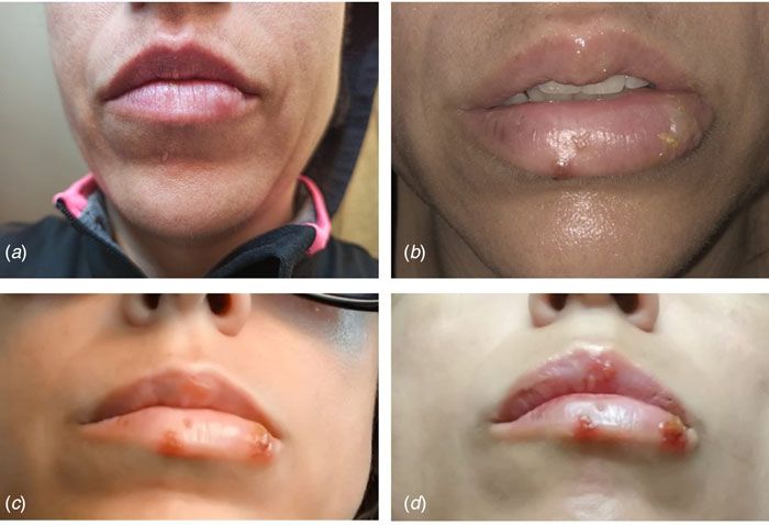
2                                                                                                                                                  M. D. Maldonado et al.

       accompanied by pain, itching and burning in the area. Herpes virus                         history revealed no data of interest, except for the situation of
       infections can occur as primary or nosocomial pathogens, but clin-                         social isolation due to the pandemic. Nasopharyngeal and blood
       ical manifestations are most commonly a re-activation of a latent                          samples obtained from the patient revealed a negative RT-PCR
       viral infection [10].                                                                      and negative antibody (IgM and IgG) test for SARS-CoV-2.
           It is common knowledge that lifestyles with physical activity,
       and social, family and cultural contacts constitute healthy habits
                                                                                                  Presentation of case 3: herpes varicella zoster infection with
       that improve immune defences and the mental and emotional
                                                                                                  subsequent positive diagnosis of SARS-CoV-2
       state [11]. The confinement of families in their homes, towns, cit-
       ies and even countries, isolated from other relatives, friends and                         A 25-year-old woman, suffering from a fever of 38 °C and the
       neighbours due to the coronavirus disease 2019 (COVID-19) pan-                             appearance of very itchy vesicular lesions on her right-hand
       demic has, for many people, presented a very stressful life event,                         side lumbar area, came to the emergency room of her Primary
       triggering never-before-lived experiences that have threatened the                         Care Centre, during the month of February 2021, which coincided
       subject’s health and well-being. The wide-ranging consequences                             with the third wave of COVID-19 in Andalusia (Spain). She pre-
       of these restrictive measures include a sedentary lifestyle, obesity,                      sented vesicular injuries that later spread to her arms and legs
       hypertension, increases in cortisol and immunosuppression [12].                            (Fig. 3). She was initially diagnosed with varicella zoster infection.
           The three cases presented herein show that there is a close rela-                      The patient was discharged with a treatment based on paraceta-
       tionship between the appearance of herpes infections and the                               mol and topical calamine powders for the itchy vesicles. After a
       COVID-19 pandemic situation, and that a secondary immuno-                                  week of evolution, she presented a worsening of her general con-
       deficiency state is generated, either due to the SARS-CoV-2 infec-                         dition, with a fever of up to 40 °C with chills, an unproductive dry
       tion or due to the stress generated by the pandemic itself.                                cough, dyspnoea, fatigue and leg pain. She was treated again, this
                                                                                                  time as an in-patient in the hospital, and an RT-PCR was per-
                                                                                                  formed against the SARS-CoV-2 virus, through which she was
       Presentation of case 1: herpes varicella zoster infection
                                                                                                  diagnosed as being positive.
       We report the case of a 62-year-old Spanish man, subjected to the
       first Spanish total lockdown from 14th March 2020 to 21st June
                                                                                                  Discussion
       2020. On 11th May 2020, the patient presented elevated ery-
       thematous lesions with vesicles on his left side compatible with                           The appearance of the herpes virus infection in times of the
       a varicella zoster infection (Fig. 1), having been diagnosed and                           COVID-19 pandemic in patients, with or without respiratory
       evaluated by his Primary Care Centre. General malaise with                                 symptoms, should make us aware of the possibility of having an
       fever and diarrhoea accompanied the dermatological lesions.                                underlying SARS-CoV-2 infection [4]. The SARS-CoV-2 virus
       The patient was quickly treated, first 24–48 h, with acyclovir for                         is a new contagious beta-coronavirus that is transmitted person
       7 days, oral and topically, as well as antipyretics for the fever.                         to person mainly via the air. Initially, it affects the respiratory sys-
       On questioning, he emphasised that he had previously been in                               tem by entering the host’s respiratory epithelial cells, through the
       good health, had received all necessary vaccinations and that he                           S protein (transmembrane spike glycoprotein) of its outer capsule,
       had not previously suffered from any fever, that he was not a                              using the ACE2 receptor [13]. The infection is established in the
       drug user, and that he drank no alcoholic beverages, and had                               pneumocytes, causing the activation of innate immunity with the
       not travelled. He also stated that he had been grieving the loss                           release of type 1 interferons (INF-alpha and INF-beta) from the
       of a family member to COVID-19. In short, a patient with no                                infected cells. Furthermore, molecular patterns associated with
       medical history of interest except for (a) benign prostate hyper-                          the pathogen or damage, known as PAMP and DAMP respect-
       trophy, compatible with his age, under treatment with silodyx, a                           ively are generated and recognised, which can lead to the activa-
       specific alpha-adrenergic antagonist, and (b) his low mood. The                            tion of alveolar macrophages (phenotype M1) responsible for the
       evolution of the patient was good. Nasopharyngeal and blood                                release of pro-inflammatory cytokines such as interleukin (IL)-1
       samples obtained from the patient revealed a negative reverse                              beta, tumour necrosis factor-alpha, IL-6, IL-8 and IL-12 [14].
       transcriptase polymerase chain reaction (RT-PCR) and negative                              IL-12, in turn, activates natural killer (NK) cells and the specific
       antibody [immunoglobulin M (IgM) and IgG] test for                                         or adaptive immune response with the activation of T and B lym-
       SARS-CoV-2.                                                                                phocytes. Up to this point, we would be facing a standard viral
                                                                                                  defence [15]. However, SARS-CoV-2 has a series of evasion
                                                                                                  mechanisms that allow it to circumvent our immune system,
       Presentation of case 2: herpes simplex 1 infection
                                                                                                  thereby leaving the host vulnerable and thus facilitating replica-
       We report the case of a 40-year-old Spanish woman, subjected to                            tion of the virus and the increase in viral load. From among the
       the extension of the third state of alarm in Spanish from 9th                              evasion mechanisms used by SARS-CoV-2 we highlight (a) alter-
       November 2020 until 9th May 2021. On 13th December 2020,                                   ation of the synthesis and functionality of INF type 1 (INF-alpha
       the patient reported a prodromal phase of 2 days, with a burning                           and beta) and 2 (INF-gamma). This allows the virus to replicate in
       sensation in her lower lip and hyperesthesia in the area around                            host cells without opposition or without an effective antiviral state
       the mouth. A large vesicle subsequently appeared on her lower                              [2, 16]; (b) cytokine storm or excessive activation of M1 macro-
       lip together with other smaller vesicles that broke at different                           phages with an inordinate amount of pro-inflammatory cytokines
       times, which suppurated and left ulcerations and scabs that remit-                         released into the serum, whose synergistic effects would be
       ted after 10 days of evolution (Fig. 2). This clinical state was                           responsible for the severity of patients infected by SARS-CoV-2
       accompanied by fever and general malaise. She was diagnosed                                [14]. Cytokines released in large quantities cause fever, increased
       with herpes simplex 1 infection and treated symptomatically by                             acute-phase proteins, increased adhesion molecules in the vascu-
       her Primary Care Centre with paracetamol, antihistamines and                               lar endothelium, edema, activation of the coagulation system with
       topical aloe-vera, with a good evolution. The patient’s personal                           disseminated intravascular coagulation, increased cell catabolism

Downloaded from https://www.cambridge.org/core. IP address: 46.4.80.155, on 13 Oct 2021 at 07:38:24, subject to the Cambridge Core terms of use, available at https://www.cambridge.org/core/terms.
https://doi.org/10.1017/S0950268821001333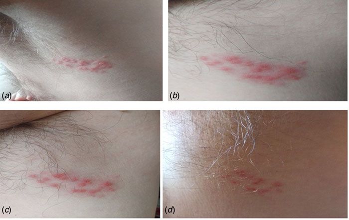
Epidemiology and Infection                                                                                                                                                     3

                                                                                                                                Fig. 1. Case 1. (A) Onset of lesions with raised vesicles
                                                                                                                                over erythematous areas; (B and C) evolutionary periods
                                                                                                                                with favourable remission of the herpetic infection and
                                                                                                                                (D) advanced period of remission with vesicular drying
                                                                                                                                and peeling of the skin. Negative RT-PCR for
                                                                                                                                SARS-CoV-2.

                                                                                                                                Fig. 2. Case 2. (A) Prodormal period with burning area
                                                                                                                                and itching on the lower lip; (B and C) florid period of
                                                                                                                                infection with increased number of vesicles on the lips
                                                                                                                                and (D) referral period. Negative RT-PCR for SARS-CoV-2.

        and decreased cardiac output with multi-organ damage [15, 17].                             in social disconnection, but also in the levels of anxiety, stress
        This altered immune response facilitates the replication of the                            and depression in the population [21]. This has provoked an
        virus and the increase in the viral load, by means of causing the                          unprecedented situation in the Spanish population that has gen-
        NK cells and CD8+ lymph T cells to become exhausted and                                    erated increases in the levels of cortisol, catecholamines and cer-
        hence the coronavirus cannot be eliminated [18]. In the same                               tain opiates, substances which are generally immunosuppressive
        way as occurs in other states of hyperactivation of the immune                             and involve several pathways, including lymphocytopenia and
        system, such as burn patients, polytraumatised patients and                                hypogammaglobulinaemia [22].
        head trauma patients, the increase in adhesion molecules could                                Over recent decades, the interaction between the neuroendo-
        generate a state of immunodeficiency of T and B lymphocytes                                crine and immune systems has frequently been suspected. The
        which would remain attached to the endothelium of the blood                                integration between the two systems is based on a bidirectional
        vessels and therefore unable to enter the site where they should                           communication, which in immunobiochemical and molecular
        perform their function [19, 20]. In addition, the SARS-CoV-2                               terms, implies having intercellular signals and common recogni-
        infection produces a reduction in the percentages of monocytes,                            tion systems. In this respect, it is currently recognised and
        eosinophils and basophils [2].                                                             accepted that many neuroendocrine signals are produced by
           On the other hand, the different confinements of people, in                             immunocompetent cells, and that these same cells also express
        their homes, towns, provinces and even countries, carried out                              specific receptors for these neuroendocrine signals [23–26]. On
        during the COVID-19 pandemic has led not only to an increase                               the contrary, typical cytokines produced by immunocompetent

Downloaded from https://www.cambridge.org/core. IP address: 46.4.80.155, on 13 Oct 2021 at 07:38:24, subject to the Cambridge Core terms of use, available at https://www.cambridge.org/core/terms.
https://doi.org/10.1017/S0950268821001333
4                                                                                                                                                   M. D. Maldonado et al.

       Fig. 3. Case 3. (A) Close-up of herpes varicella zoster
       lesion; (B and C) initiation of vesicles in the right lumbar
       dorsum and (D) posterior extension of the vesicles to the
       right leg. Positive RT-PCR for SARS-CoV-2.

       cells, as well as the receptor molecules for these cytokines, are pro-                     CD8+ T lymphocytes prior to the onset of herpes varicella zoster.
       duced and expressed by neuroendocrine cells [27]. This neuroen-                            Furthermore, Balc’h et al. [33] found 18 SARS-CoV-2-positive
       docrine–immune interaction is currently of recognised and                                  patients with lymphopenia and reactivation of herpes simplex
       growing importance and is considered to play a fundamental                                 virus and cytomegalovirus; these authors consider that the
       role in the generation of diseases [28]. It has been observed how                          SARS-CoV-2 infection may actually constitute the risk factor
       disturbances in the interactions among the nervous, immune and                             for reactivation.
       endocrine systems are implicated in various diseases such as an                               In summary, we show three clinical cases of the herpes virus
       increased number of infections, decreased responses to vaccines,                           infection, two of which yield negative results on RT-PCR for
       the delayed healing of wounds and a higher incidence of onco-                              SARS-CoV-2 and one testing positive. All these cases occurred
       logical diseases and autoimmune diseases [29]. Both, the stress                            during the COVID-19 pandemic period on Spanish territory.
       generated during the pandemic period and the SARS-CoV-2 inva-                              The lesions presented by our patients and those in other afore-
       sion itself are generative elements of immunodeficiency in                                 mentioned cases, support the hypothesis that a herpes infection
       humans, a situation that could be exploited by the herpes virus                            can manifest itself prior to suffering from a SARS-CoV-2 infec-
       to reactivate and infect the host [27]. Hence, it is no surprise                           tion and, although it needs to be tested in larger cohorts of
       that the herpes virus infection may be the first manifestation, as                         patients, performing a PCR test on subjects with active herpes
       if it were a prodromal period, prior to the appearance of symp-                            virus infections could increase the number of cases of SARS-
       toms of the SARS-CoV-2 infection. This is the series of events                             CoV-2 detected early, precisely at the stage when this deadly
       in our third clinical case, that of the 25-year-old woman, whose                           virus is most infectious.
       SARS CoV-2 infection initially debuted with disseminated herpes
       varicella zoster (Fig. 3).                                                                 Acknowledgements. This study was supported by Seville University
           A previous case report by Elsaie et al. [4] suggested that herpes                      (Immunology area), Department of Medical Biochemistry, Molecular
                                                                                                  Biology and Immunology; also, by the Ministry of Economy, Industry and
       varicella zoster might be an indicator for latent COVID-19 infec-
                                                                                                  Competitiveness (MINECO 2017), reference: BFU2017-85832-R.
       tion; the authors provide two clinical cases of patients with
       SARS-CoV-2 infection who initially presented herpes varicella                              Author contributions.
       zoster lesions. Jimenez-Cauhe et al. [30] have found erythema                              M. D. Maldonado conducted the literature review, interpreted the immunological and
       multiforme-like lesions in the skin of four hospitalised patients                          clinical data of the patients and she was the major contributor to the
       with the COVID-19 infection. It remains unclear, however,                                  manuscript. J. Romero-Aibar participated in the care and analytical follow-up of the
                                                                                                  patients as a senior laboratory technician and helped in revising the manuscript. M. A.
       whether they were specific lesions of SARS-CoV-2, or of other                              Perez-San-Gregorio performed the analysis of the mental state of the patients and direc-
       virus infections, or the result of the drugs administered during                           ted the review of the psychological literature.
       the admission of the patients in hospital. Ferreira et al. [31]
       describe a case of a 39-year-old man, who was immunocompetent                              Conflict of interest. The authors declare that they have no conflict of interest.
       with SARS-CoV-2 and co-infected with herpes varicella zoster.
       The authors argue that SARS-CoV-2 probably induced a retro-                                Ethical standards. Written informed consent was obtained from the
                                                                                                  patients for publication, of this cases report included in this article, according
       active reactivation of the herpes virus. Tartari F et al. [32]
                                                                                                  to the specifications established by the Ethics Committee of the University of
       reported four more cases of herpes virus infections in patients                            Seville for the publication of clinical cases.
       with positive SARS-CoV-2. The authors explain that all their
       patients showed a decrease in peripheral blood of the T lympho-                            Data availability statement. Data supporting the findings of this study are
       cyte subpopulations, and specifically a decrease in CD3+ and                               available at SAS (Andalusian Health Service Spain). Restrictions apply to the

Downloaded from https://www.cambridge.org/core. IP address: 46.4.80.155, on 13 Oct 2021 at 07:38:24, subject to the Cambridge Core terms of use, available at https://www.cambridge.org/core/terms.
https://doi.org/10.1017/S0950268821001333
Epidemiology and Infection 5
availability of these data, which were used under license for this study. The 15. Abbas AK, Lichtman AH and Pillais S (2020) Basic Immunology, 6th
data have been made available, to the authors, with the permission of affected Edn. Philadelphia EEUU: Elsevier.
patients. 16. Acharya D, Liu G and Gack MU (2020) Dysregulation of type I interferon
responses in COVID-19. Nature Reviews Immunology 20, 397–398.
17. Tay MZ et al. (2020) The trinity of COVID-19: immunity, inflammation
References
and intervention. Nature Reviews Immunology 20, 363–374.
1. Chih-Cheng L et al. (2020) Severe acute respiratory syndrome coronavirus 2 18. Zheng M et al. (2020) Functional exhaustion of antiviral lymphocytes in
(SARS-CoV-2) and coronavirus disease-2019 (COVID-19): the epidemic and COVID-19 patients. Cellular & Molecular Immunology 17, 533–535.
the challenges. International Journal of Antimicrobial Agents 55, 105924. 19. Maldonado MD et al. (2007) Melatonin as pharmacologic support in
2. Huang C et al. (2020) Clinical features of patients infected with 2019 burn patients: a proposed solution to thermal injury-related lymphocyto-
novel coronavirus in Wuhan, China. Lancet (London, England) 395, penia and oxidative damage. Critical Care Medicine 35, 1–9.
497–506. 20. Cao X (2020) COVID-19: immunopathology and its implications for ther-
3. Singh AK et al. (2020) Prevalence of co-morbidities and their association apy. Nature Reviews Immunology 20, 269–270.
with mortality in patients with COVID-19: a systematic review and 21. Ozamiz-Etxebarria N et al. (2020) Stress, anxiety, and depression levels in
meta-analysis. Diabetes, Obesity and Metabolism 22, 1915–1924. the initial stage of the COVID-19 outbreak in a population sample in the
4. Elsaie ML, Youssef EA and Nada HA (2020) Herpes zoster might be an northern Spain. Reports in Public Health 36, e00054020.
indicator for latent COVID 19 infections. Dermatologic Therapy 33, 22. Eisenbergera NI et al. (2010) Inflammation and social experience: an
e13666. inflammatory challenge induces feelings of social disconnection in add-
5. Estebanez A et al. (2020) Cutaneous manifestations in COVID- 19: a new ition to depressed mood. Brain, Behavior, and Immunity 24, 558–563.
contribution. Journal of the European Academy of Dermatology and 23. Irwin MR (2008) Human psychoneuroimmunology: 20 years of discovery.
Venereology: JEADV 34, e250–e251. Brain Behavior and Immunity 22, 129–139.
6. Wollina U et al. (2020) Cutaneous signs in COVID-19 patients: a review. 24. Ray A, Gulati K and Stress RN (2017) Anxiety, and immunomodulation:
Dermatologic Therapy 33, e13549. a pharmacological analysis. Vitamins & Hormones 103, 1–25.
7. Madavaraju K et al. (2021) Herpes simplex virus cell entry mechanisms: 25. Bekhbat M et al. (2021) Adolescent stress sensitizes the adult neuroim-
an update. Frontiers in Cellular and Infection Microbiology 10, 617578. mune transcriptome and leads to sex-specific microglial and behavioural
8. Inbaraj LR et al. (2021) High susceptibility to varicella among urban and phenotypes. Neuropsychopharmacology 0, 1–10.
rural pregnant women in South India: a brief report. Epidemiology and 26. Maldonado MD (2010) Evidence of melatonin synthesis and release in
Infection 149, 1–14. doi: 10.1017/S0950268821000492. mast cells. Possible modulatory role on inflammation. Pharmacological
9. Kubota Y et al. (2019) Disseminated zoster in an adult patient with exten- Research 62, 282–287.
sive burns: a case report. Virology Journal 16, 68. 27. Raony I et al. (2020) Psycho-neuroendocrine-immune interactions in
10. Kushawaha A, Mobarakai N and Tolia J (2009) A 46-year-old female COVID-19: potential impacts on mental health. Frontiers in
presenting with worsening headache, nuchal rigidity and a skin rash in Immunology 11, 1170.
varicella zoster virus meningitis: a case report. Cases Journal 2, 6299. 28. Ziemssen T and Kern S (2007) Psychoneuroimmunology – crosstalk between
11. Filgueira T et al. (2021) The relevance of a physical active lifestyle and the immune and nervous systems. Journal of Neurology 254, II8–II11.
physical fitness on immune defense: mitigating disease burden, with 29. Ziemssen T (2012) Psychoneuroimmunology – psyche and autoimmun-
focus on COVID-19 consequences. Frontiers in Immunology 12, 587146. ity. Current Pharmaceutical Design 18, 4485–4488.
12. Wollenstein-Betech S et al. (2020) Physiological and socioeconomic char- 30. Jimenez-Cauhe J et al. (2020) Erythema multiforme-like eruption in
acteristics predict COVID-19 mortality and resource utilization in Brazil. patients with COVID-19 infection: clinical and histological findings.
PLoS One 15, e0240346. Clinical and Experimental Dermatology 45, 892–895.
13. Hoffmann M et al. (2020) The novel coronavirus 2019 (2019-nCoV) uses 31. Ferreira F et al. (2020) COVID-19 and herpes zoster co-infection present-
the SARS-coronavirus receptor ACE2 and the cellular protease TMPRSS2 ing with trigeminal neuropathy. European Journal of Neurology 24(9),
for entry into target cells. bioRxiv. 01.31.929042. doi: 10.1101/ 1748–1750.
2020.01.31.929042. 32. Tartari F et al. (2020) Herpes zoster in COVID-19-positive patients.
14. Li S et al. (2020) SARS-CoV-2 triggers inflammatory responses and cell International Journal of Dermatology 59, 1028–1029.
death through caspase-8 activation. Signal Transduction and Targeted 33. Balc’h L et al. (2020) Herpes simplex virus and cytomegalovirus reactiva-
Therapy 5, 235. tions among severe COVID-19 patients. Critical Care 24, 530.
Downloaded from https://www.cambridge.org/core. IP address: 46.4.80.155, on 13 Oct 2021 at 07:38:24, subject to the Cambridge Core terms of use, available at https://www.cambridge.org/core/terms.
https://doi.org/10.1017/S0950268821001333You can also read