Cellular Growth and Form - Course 4: Cellular scaling Thomas Lecuit chaire: Dynamiques du vivant - Collège de France
←
→
Page content transcription
If your browser does not render page correctly, please read the page content below
Cellular Growth and Form

   Course 4: Cellular scaling
           Thomas Lecuit
   chaire: Dynamiques du vivant

                1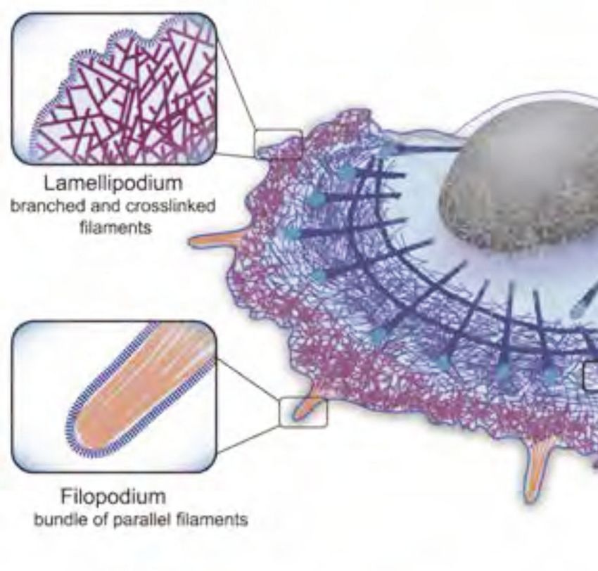
Cell size is set by co-regulation of cell
                                 division and cell growth

                                                                                                                        M

                                                                         Cell size                                                G2
                                    HeLa cell                                                                          Cell
                                                                                                                      cycle
                                                                                                               G1
               3,000             Mitotic volume                                                                               S
Volume (μm3)

                                     overshoot
               2,500 Short-timescale
                     fluctuations
               2,000                                                                                           EXIT

               1,500
                        0         5       10                15
                                      Time (h)                                            G1                                           S

                            C.Cadart L. Venkova, P. Recho, M. C.               Miriam B. Ginzberg et al. and M Kirschner. Science 348, (2015);
                            Lagomarsino and M. Piel Nature Physics             DOI: 10.1126/science.1245075
                            15: 993–1004 (2019)

                       Thomas LECUIT 2020-2021
                                                                     2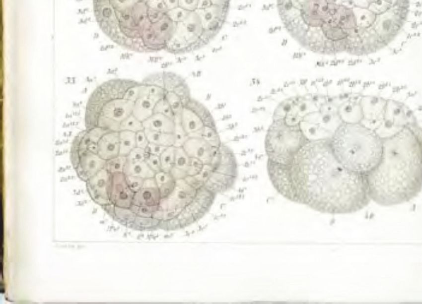
What do cells measure to control division?

• Sizer: cells divide at target size
        Deterministic vs Stochastic sizer

• Timer: cells divide after fixed duration       Trends in Genetics

• Adder: cells divide after fixed size increase
        Cell level vs population scale control

         Thomas LECUIT 2020-2021
                                   3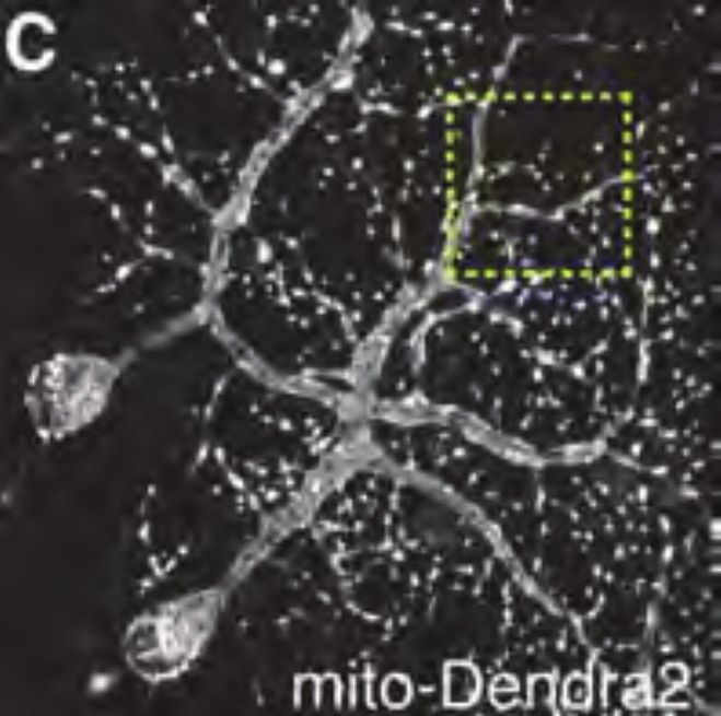
Scaling: Do cells measure their size?
                       Mechanisms that procure information about size

•   Ratio of two length scales: cell geometrical length and biochemical length scales
    — An inhibitor of cell division is inhibited from the poles. As cell size increases assembly of
    cytokinetic ring is possible beyond critical cell size.

                                                                           ex: MinCD in Bacteria
                                                                               Pom1 in S. pombe
                                                cell division

•   Indirect measure of cell volume:
    Production of a size-invariant negative regulator of cell cycle progression provides an indirect
    feedback information about size.

           Thomas LECUIT 2020-2021
                                                     4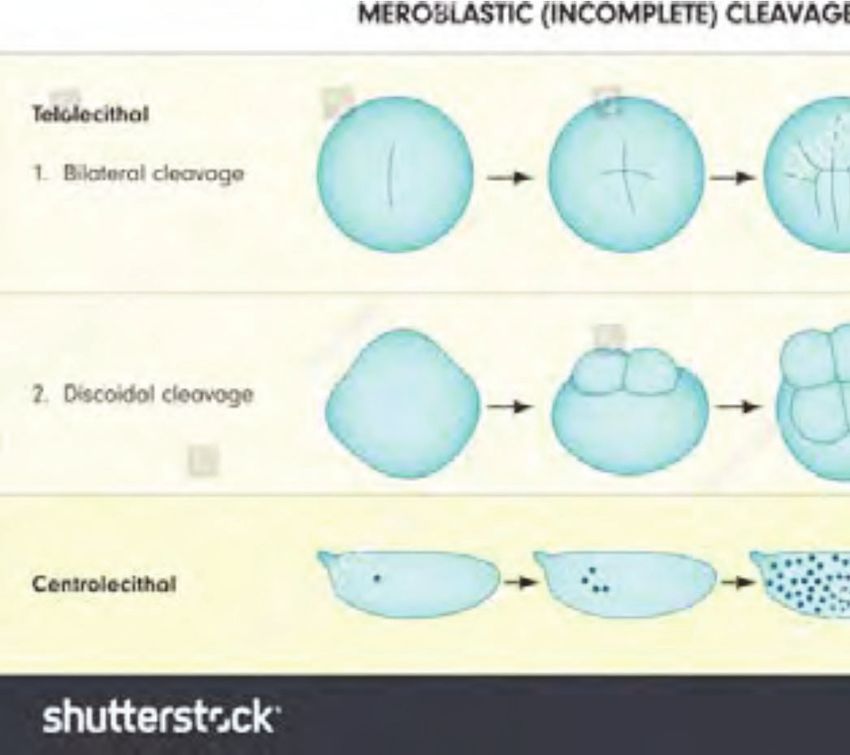
Scaling: How do cells adjust their proportions?

 Thomas LECUIT 2020-2021
                           5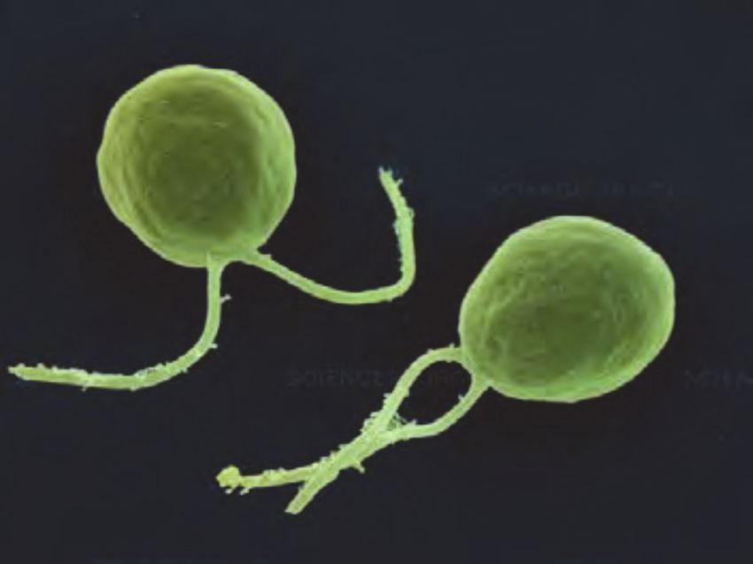
Scaling in living organisms
Scaling of body parts, internal (organs, skeleton) and external (limbs) in animals
• Size range spans 6 orders of magnitude in land vertebrates
50
30 Blue whale
20
Sperm
body length (L)
whale
length, l mass, m 10 1/3
Gray whale
5 White whale Bottle-nosed whale
Killer whale
Bottle-nosed dolphin
La Plata river Pygmy sperm whale
2 dolphin
Dall porpoise
1
10 20 50 100 200 500 1,000 2,000 5,000 10,000 50,000 100,000
body mass (M)
isometry
10
White rhinoceros African
elephant
Black rhinoceros
5
Bear
Julian S. Huxley Georges Teissier Camel
Hippopotamus
Zebra
Spotted hyena Moose Indian
2 Bison rhinoceros
Orangutan
Ass
Lynx leopard
body length (L)
Pygmy hippopotamus
1 Howler monkey
scaling law: f(x) = b xĮ
Man
•
Hare
Macaque
scaling invariance: f(cx) Ӗf(x) 0.5 Baboon
Giant rat
Gibbon
Wild rabbit
Giant squirrel
Lemur Beaver
0.2
allometry: Į > l or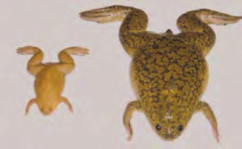
Cellular scaling between species
                               Cell internal organisation: number of organelles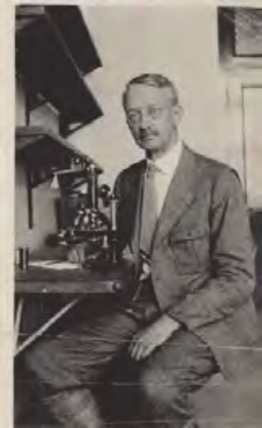
Cellular scaling between species…
                                      Cell internal organisation: size of organelles
1875: G. Gulliver’s observation of red blood cells in different vertebrate species
revealed approximate scaling of nuclei to cell size

…and within species
This was later further documented within a
given species:
1900’:
• Richard Hertwig (1850-1937): sea urchins
  and protists.
  Kern-plasma relation: ratio between nuclear
  and plasma (cytoplasm) volumes
       (1903) Uber Korrelation von Zell- und Kerngrosse und     R. Hertwig
       ihre Bedeutung für die geschlechtliche Differenzierung
       und die Teilung der Zelle. Biol. Centralb., Bd. 22.

•   Theodor Boveri (1862-1915): sea urchins
       (1905) Zellenstudien V. Uber die Abhangigkeit der
       Kerngrosse u. Zellenzahl der Ausgangszellen. Jena.

•   Edwin Conklin (1863-1952): Crepidula plana                               Gulliver, G. (1875). On the size and
                                                                             shape of red corpuscles of the blood of
        (1912) Conklin, E. J. Exp. Embryol. 12, 1–98.
                                                                             vertebrates. Proc. Zool. Soc. Lond.
                                                                             474–495.
                                                                T. Boveri

                        Thomas LECUIT 2020-2021
                                                                      8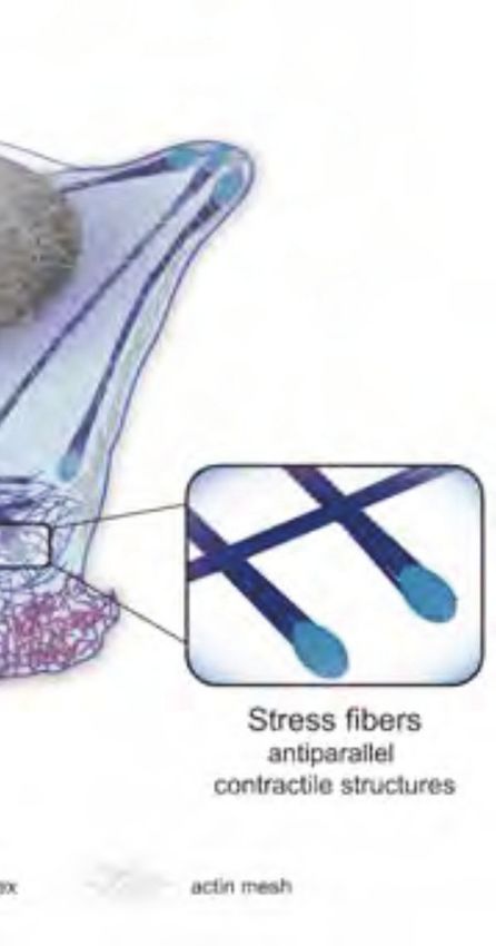
Cellular scaling within species
Cell internal organisation — Cell shape and external organisation
                                       Primary                     D                                                L3
                                       Secondary                                                                                                    (A)                                (B)                                         (C)

                                                              A          P
                                       Tertiary
                                       Quaternary                  V

                                                                                                                                                                       2 μm                                         10 μm                        20 μm

                                                                       L2                                                                           (D)                                      (E)                             (F)

           E17                                L1
                                                                                                                                                                                    20 μm                   40 μm                              50 μm
                                                                                                                                                    (G)                       (H)                             (I)                        (J)

     19hrsAEL±2                         24hrsAEL±3                48hrsAEL±3                                        72hrsAEL±3
                                                                                                                                        10μm
          19h                                 24h                                48h                                 72h                                       80 μm                               100 μm                   200 μm

                                                                                                                                                                                    Wallace Marshall
         late                               larva L1                   larva L2                                    larva L3
        embryo                                                                                                                                      Ciliates and other Protists: large cells
         E17
                                                                                                                                                    with complex morphologies (see
                                                                                                                                                    Lecture 1 — 2020)                                                                             5 mm

                    Number of branches at each order of complexity                     C             Relative distance between secondary branches
                      35
                                                                                                                              ns
   Primary                             30                                      E17                           0.4
                  Number of branches

                                                                               L1
   Secondary                           25
                                              ns         **                    L2
                                                                                           Relative length

                                                                               L3
   Tertiary                                             ns
                                                                                                             0.3
                                       20
   Quaternary
                                       15               ***                                                  0.2
                                       10
                                                                          *                                  0.1
                                                                          ns
                                       5
                                                                        ns
                                       0                                                                     0
                                            Secondary   Tertiary       Quaternary                                  E17   L1        L2   L3

A. Palavalli, N. Tizon-Escamilla, J-F. Rupprecht, T. Lecuit, Current Biology (2020),
https://doi.org/10.1016/j.cub.2020.10.054
                                                                                                                                                            Green alga — Micrasterias rotata

      Thomas LECUIT 2020-2021
                                                                                                                         9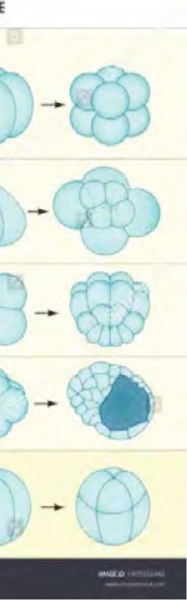
Cell size change during embryonic cleavage

Cells decrease exponentially their size (volume) during early steps of animal development
           First 10 hours of amphibian embryogenesis, cell diameter decreases around 100-fold, from a
           1.2 mm egg to 12 μm diameter blastomeres

         The cell in development and inheritance                         The Embryology of Crepidula (Conklin 1897)
         EB Wilson (1897)

          Thomas LECUIT 2020-2021
                                                    10
Cell size change during embryonic cleavage
Cells decrease exponentially their size (volume) during early steps of animal development
tissuee
tissue
Marine Mollusk Gasteropod
Thomas LECUIT 2020-2021
11Cell size change during embryonic cleavage
Cells decrease exponentially their size (volume) during early steps of animal development
Strongylocentrotus droebachiensis —Green urchin Sarsia sp. — Jellyfish
Georges von Dassow
http://www.gvondassow.com/
Thomas LECUIT 2020-2021
12Cell size change during embryonic cleavage
Cells decrease exponentially their size (volume) during early steps of animal development
Echinoderm
Amphioxus
Cephalopods
Annelids
Molluscs
Fish, Reptiles, Birds
Tunicates
Insects
Mammals
Arthropods www.shutterstock.com ID1459533926
Nematodes
Amphibians
www.shutterstock.com ID1459533440
Thomas LECUIT 2020-2021
13Cell size — scaling
Statement of the problem:
• Cell size is tightly regulated but varies strongly during embryonic
cleavage (by a factor of 2N for N divisions)
• Cells grow 2-fold before they divide or may grow while they increase
their ploidy
• As cells change their volume do internal organelles scale? What are the
mechanisms of scaling?
• When cells display symmetric structures (eg. flagella or cilia) how is the
size of these structures controlled
Q: Do internal cell structures and organelles scale with cell size?
Thomas LECUIT 2020-2021
14Cell size change during embryonic cleavage
Q: Do internal cell structures and organelles scale with cell size?
Cerebratulus marginatus
Georges von Dassow
http://www.gvondassow.com/
Thomas LECUIT 2020-2021
15Internal cell organisation
Organelles
Cytoskeletal structures
Cell Biology by the numbers. Ron Milo, Rob Phillips, illustrated by Nigel Orme.
Garland Science 2012
Thomas LECUIT 2020-2021
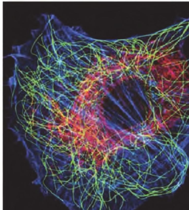
16Internal cell organisation
Cytoskeletal structures
Microtubules (green) Letort G, Ennomani H, Gressin L et al.
Actin filaments (blue) https://doi.org/10.12688/f1000research.6374.1
Intermediate filaments (red)
Thomas LECUIT 2020-2021
17External cell organisation
Appendages — eg. Flagella
• Protists: unicellular organisms, autotrophic or heterotrophic
(neither animals nor plants)
• Flagellates
A , Pharyngomonas kirbyi ; B , Percolomonas cosmopolitus ; C , Percolomonas
descissus ; D , Psalteriomonas lanterna ; E , Heteramoeba clara ; F , Pleurostomum
flabellatum ; G , Trimastigamoeba philippinensis ; H , Naegleria gruberi
I ,. Stephanopogon minuta .
Cf – cytopharynx; Cl – collar; CV – contractile vacuole; Gl – globule of
hydrogenosomes; Ro – rostrum.
Scale bars = 10 μm.
After Broers et al., 1990; Bovee, 1959; Brugerolle & Simpson, 2004; Droop, 1962;
Fenchel & Patterson, 1986; Page, 1967, 1988; Park et al., 2007; Park & Simpson,
2011; Yubuki & Leander, 2008.
Thomas LECUIT 2020-2021
18Within- and between-species scaling
Example: mitotic spindle
• Spindle size (and shape) vary among different species and cell types to ensure chromosome
segregation fidelity, and proper spindle positioning
• In nematodes, embryo size, and thus cell size, is subject to stabilising selection
• Stabilising selection on embryo size quantitatively predicts within-species and between-species variation of spindle
traits (eg. length)
Pole-to-pole distance (μm) Pole-to-pole distance (μm) Pole-to-pole distance (μm) Pole-to-pole distance (μm)
C. castelli (n=10) C. angaria
C C. angaria (n=22)
35
C. sp. 8 (n=10) 30
C. virilis (n=24)
25
Final spindle length (μm)
C. drosophila (n=10)
30 individual embryo C. sp. 2 (n=11) 20
C. portoensis (n=21)
natural isolate C. guadeloupensis (n=10) 15
C. afra (n=33) 10
C. imperialis (n=21)
0 100 200 300 400 500
C. yunquensis (n=10) Time (s)
26 C. nouraguensis (n=20)
C. macrosperma (n=30)
35
C. japonica
C. japonica (n=10) 30
50 C. kamaaina (n=10) 25
C. doughertyi (n=25)
C. brenneri (n=157) 20 species average
37
22
Spindle final length (μm)
C. tropicalis (n=39)
C. wallacei (n=31)
15 predicted
45 C. nigoni (n=31) 10 measured
C. briggsae (n=233) 0 100 200 300 400 500 31
C. sp. 5 (n=51) Time (s)
C. sp. 5
45 50 55 60
Centrosome size (μm2)
C. remanei (n=150) 35
C. elegans (n=2597)
25
40 Embryo size (μm) C. plicata (n=10)
C. sp. 1 (n=21)
30
25
Diploscapter sp. (n=10)
20
Rhabditis sp. (n=19) 19
R. axei (n=11) 15
35 P. typica (n=12)
O. tipulae (n=11) 10
O. dolichura (n=10) 0 100 200 300 400 500
13
C. cristata (n=12) P. maupasi Time (s) 35 45 55 65 75
35
P. pacificus (n=10) Embryo size (μm)
30 P. entomophagus (n=10) 30
P. maupasi (n=10)
P. strongyloides (n=33) 25
P. teres (n=20) 20
R. regina (n=12)
25 M. longespiculosa (n=10) 15
MY23
ED3005
JU258
PS2025
CB4857
JU751
MY18
CX11315
ED3040
MY16
JU847
JU830
JU1896
CB4856
ED3077
ED3052
JU323
JU360
JU1580
JU406
MY1
JU1568
JU1350
EG4349
DL238
LKC34
JU1246
CX11264
JU1561
JU1212
JU1581
JU363
QX1233
DL226
CX11314
CB4858
JU1652
CX11292
CX11307
JU1213
CB4932
JU394
CX11285
JU1172
CX11262
JU775
EG4724
ED3048
JU561
JU346
CB4854
N2
CB4853
JU440
JU1491
KR314
EG4725
JU1440
JU397
AB4
PB306
PX179
JU792
JU1409
JU311
JU1586
EG4946
CB4852
JU462
LSJ1
JU642
JU367
ED3011
WN2002
JT11398
JU1400
ED3017
CX11276
JU310
RC301
JU1200
CX11271
ED3073
JU1242
JU1088
MY10
JU778
DL200
AB1
ED3046
EG4347
JU774
ED3049
PB303
ED3012
JU393
JU398
T. palmarum (n=10) 10
0.1 substitutions/site 0 100 200 300 400 500
C. elegans natural isolates (97 strains)
Time (s)
D
Within-species analysis Between-species analysis
Farhadifar et al., and Marie Delattre and Dan. J Needleman. Current Biology 25, 732–740 (2015)
Thomas LECUIT 2020-2021
19Cellular scaling
Between species Between cell types
within species
100 µm
50 µm
Pancreatic Hepatocytes Keratinocytes Fibroblasts Adipocytes
beta cells
Within cell type
within species
• Organelles must scale with cell volume: exponential?
• Yet the genome is not scaling with cell size
Thomas LECUIT 2020-2021
20Cellular scaling
• Biochemical composition of cell:
Potential role of genes and gene regulation between species and cell types
X. laevis X. tropicalis
Katanin: Microtubule severing protein
reduces spindle size
Science 328, 633-636
Xenopus tropicalis Xenopus laevis In X. laevis, Katanin p60 is phosphorylated
by Aurora kinase B and inactivated
A single amino acid change renders X.
tropicalis refractory to AuroraB inactivation
R. Loughlin et al. F. Nédélec and R. Heald. Cell 147, 1397–1407 (2011)
Thomas LECUIT 2020-2021
21Cellular scaling
• Non genetic/biochemical adaptation to size:
— geometric cues, size of pool of constituents?
— how does biochemistry respond to geometry?
First 12 cleavage cell divisions. The cell radius decreases 16-fold with little change in biochemistry
T. Mitchison et al. Cold Spring Harb Perspect Biol 2015;7:a019182 (2015)
Thomas LECUIT 2020-2021
22Cellular scaling
• Number of organelles/subcellular structures
— mitochondria, Golgi, endosomes etc.
— fixed size
• Size of organelles/subcellular structures
— nucleus, endoplasmic reticulum, centrosomes, spindles, cilia etc.
— adaptation of size
Thomas LECUIT 2020-2021
23Scaling to cytoplasmic volume not cell dimension
• Centrifugation experiments:
centrosome
• Observation of Crepidula cleavage divisions
cytoplasm
nucleus
yolk
nucleus: 24μm 14μm spindle
• Due to centrifugation along the indicated axis
(arrows) during cleavage, cell size and the amount
of cytoplasm (dotted) and yolk (brown) are not
proportional in the two blastomeres.
• The size of organelles (nucleus, spindle and
centrosome) scales with cytoplasm volume but
NOT cell size stricto sensu.
Conklin, E. (1912). J. Exp. Embryol. 12, 1–98.
N. Goehring and A. Hyman Current Biology 22, R330–R339 (2012)
Thomas LECUIT 2020-2021
24Scaling to cytoplasmic volume not cell dimension
C Size Scaling In Vitro D Size Scaling In Vivo (Embryo)
45 Compostional
Spindle Size
•
Reg. ONLY
55
Encapsulation of Xenopus laevis extracts into 55
Spindle Length (μm)
Spindle Length (μm)
40
Spindle Length (μm)
droplets reveals 2 phases: linear size scaling of Reg. By
Cell Size 45 Max 45
spindles and «saturation» where the maximum 35 Droplet Size
35 35
size is set by extract composition (eg. 30 Linear
developmental stage, or species) 25
Min
Scaling 25
Stages 1-8
25
15 15
B 0 100 200 300 0 100 200 300
Cell-like Compartment 20 Droplet Diameter (μm) Cell Diameter (μm)
20 40 60 80
Boundary Cytoplasmic
Extract Droplet Diameter (μm)
Uncompressed PREDICTIONS - Compressed Drops
Droplets
Spindle Spindle
Same Size Elongates
Oil PEG30 -PHS
Tracks Tracks
Droplet Volume Droplet Diameter
• Disentangling the role of physical constraints
Uncompressed Uncompressed
(spatial extent of droplet) from the role of Compressed Compressed
cytoplasmic volume. 50 50
Spindle Length (μm)
Spindle Length (μm)
• Compression alters the droplet geometry but not 40 40
its volume.
•
30 30
Spindle length scales with droplet volume, not -14
p = 0.44 p < 10
geometry 20 20
10 -5 10 -4 10 -3 20 60 100 140 180
Droplet Volume (μl) Imaged Droplet Diameter (μm)
Matthew C. Good, et al and Rebecca Heald. Science 342, 856-860. (2013)
DOI: 10.1126/science.1243147
Thomas LECUIT 2020-2021 see also: J. Hazel et al. Science 342, 853-856. (2013)
25 DOI: 10.1126/science.1243110Size control mechanism of organelles
• The rate of biochemical reaction is typically a function of substrate concentration
• The assembly kinetics of organelles is proportional to the cytoplasmic concentration of constituents.
Timer Constraint Balance
A B C
assembly rate
Max
disassembly rate
Rate
Size
Size
t0 t1
Time Time Organelle size
target size
High concentration Low concentration
• Size could be set by the • Size could be limited by a • Size could be limited by the
duration of assembly. physical constraint such that balance between assembly
• Different concentrations despite different assembly and disassembly.
would yield different size of kinetics the maximum size is • Negative feedback between size
internal structures set and assembly rate:
N. Goehring and A. Hyman Current Biology 22, R330–R339 (2012)
Thomas LECUIT 2020-2021
26Limiting pool mechanism
Scaling without need to measure the size of the cell
D E F
assembly
f([monomer])
Rate
Rate
Rate
disassembly
Organelle size Organelle size Organelle size
• If the monomers form a finite,
• If the monomer concentration is • Competition for monomer in a
size-independent (ie. synthesis of common pool, and rapid diffusion
limiting pool, then assembly of the
monomer is proportional to cell has two consequences:
organelle consumes monomers
and monomer concentration
size), then a smaller cell decreases • increasing the number N of
the concentration of monomer organelles decreases their size as
decreases
faster than a larger cell because 1/N
• A s a re s u l t , a s s e m b ly r a t e the amount of monomer is lower. • The N different organelles have
decreases as the organelle size
the same size unless additional
increases up to a steady state
• A s a re s u l t , a s s e m b ly r a t e mechanisms are considered (in fact
decreases faster as organelle size not really, see later)
Ntot = Nmonomer + Norganelle increases and the target size is
smaller in smaller cell, hence
scaling
N. Goehring and A. Hyman Current Biology 22, R330–R339 (2012)
Thomas LECUIT 2020-2021
27Limiting pool mechanism
Centrosome scaling - Spindle scaling
• The centrosome nucleate microtubules
• The size of centrosome determines the size of the mitotic spindle
A C D
• 9 9 9
The size of centrosomes and P0 Wild-Type sas-4(RNAi) large
Half Spindle Length (μm)
sas-4(RNAi) small
Half Spindle Length (μm)
Half Spindle Length (μm)
8 P1 AB spd-2(RNAi) 8
P2 ABa 8
mitotic spindles are correlated 7
6
7 7
between cells and within cells 5 6 6
(asymmetric spindles) 4
3
5 5
4 4
2
1 3 slope = 4.48 ± 0.4 3 slope = 4.51 ± 0.5
0
0.5 1.0 1.5 2.0 0.5 1.0 1.5 2.0
Centrosome Diameter (μm) Centrosome Diameter (μm)
• TPXL-1 targets Aurora kinase to
MTs and controls spindle length. E
• TPXL1 concentration at A
2000
C
4.0
TPXL-1 TPXL-1
Fluorescence Intensity (A.U.)
centrosomes correlates with
TPXL-1::GFP Tubulin::mCh Merge
Wild-Type
3.5 tpxl-1(RNAi)
1600
spindle size 3.0
•
1200
λ (μm)
2.5
Centrosomes control spindle size 800 2.0
by regulating the length scale of a Tubulin::mCh
1.5 slope = 0.366 ± 0.04
400
gradient of TPXL-1 on TPXL-1::GFP
1.0
0 0.5
microtubules -15 -10 -5 0 5
Distance relative to Chromatin (μm)
10 15 2 3 4 5
Half Spindle Length (μm)
6 7 8
B D
Greenan, G., Brangwynne, C.P., Jaensch, S., Gharakhani, J., Jülicher, F., and Hyman, A.A. (2010). Curr. Biol. 20, 353–358.
TPX2, Aurora Kinase and spindle length: Bird, A.W., and Hyman, A.A. (2008). J. Cell Biol. 182, 289–300.
Thomas LECUIT 2020-2021
28Limiting pool mechanism
C
Fixed subunit pool
Centrosome scaling
loaded into embryos
by the mother
Subunit pool supports
two large centrosomes
and a long spindle
• The centrosome nucleate microtubules Disassembled
•
subunits partitioned at
Centrosome size is governed by a limiting pool mechanism cytokinesis
of centrosomal components that scale with cell size Centrosomes and
spindle scale with
inherited fraction of
subunit pool
A
10 P0 1-cell
• Centrosome volume scales with 8
cell size, and is independent of
Centrosome Volume (μm³)
AB P1 2-cell
6
cell fate. 4 4-cell
ABp
ABa P2
EMS
8-cell
2 16-cell
0
-450 -300 -150 0 150
Time (sec)
• The total volume of centrosome remains
constant over time as cells divide
• Increasing the number of centrosomes reduces
the size of individual centrosomes but the total
volume of centrosomes remains invariant
• The size of centrosomes is sensitive to total
SPD2 in a cell.
• This is consistent with a limiting pool of maternally
inherited SPD-2 governing centrosome size and
scaling
Decker, M., et al., and Hyman, A.A. (2011). Curr. Biol. 21, 1259–1267.
Thomas LECUIT 2020-2021
29Limiting pool mechanism
• Scaling of centrosome size by limiting pool of centrosomal components
• Scaling of mitotic spindles with centrosomes
A
D E F
assembly
disassembly
Rate
Rate
Rate
Organelle size Organelle size Organelle size
N. Goehring and A. Hyman Current Biology 22, R330–R339 (2012)
Thomas LECUIT 2020-2021
30Limiting pool mechanism
Microtubule Dynamics Scale with Cell Size to Set Spindle Length
Scaling of MT dynamics with
cell volume
can be explained by existence
of a limiting pool of a
regulator of MT dynamics
Lacroix et al., F. Nedelec and J. Dumont. 2018, Developmental Cell 45, 496–511
Thomas LECUIT 2020-2021
31Limiting pool mechanism
Microtubule Dynamics Scale with Cell Size to Set Spindle Length
1-cell stage 2-cell stage 4-cell stage 8-cell stage 16-cell stage
Growth rate
GFP-tubulin
0.8
Growth rate (μm/s)
Caenorhabditis elegans 0.6
Nematode 1-cell 2-cell 4-cell 8-cell 16-cell
0.4
ABp
0.2
P2 ABxx P1xx
P0 AB P1 ABa
EMS 0.0
1-cell 2-cell 4-cell 8-cell 16-cell
GFP-PH
Spindle MT growth rate
**
•
n.s.
Microtubule average length depends on 2 kinetic parameters, growth and shrinkage rates, and 0.8
**
**
Growth rate (μm/s)
2 switch probabilities (catastrophe and rescue) 0.6
• Growth rate is reduced over time while other parameters are unchanged 0.4
• The reduction in growth rate is due to a reduction in cell volume but not to change in cell 0.2
0.31 0.28 0.26 0.32 0.38
composition (fate) 0.0 ±0.07 ±0.06 ±0.05 ±0.06 ±0.08
)
)
AB
P0
P1
Ai
(ts
N
27 k-1
R
•
2-cell
1(
Spindle length scales with and is tuned by spindle microtubule growth rate.
cy
9.
D
Control
C
Spindle length vs Spindle length vs A
growth rate shrinkage rate GFP-tubulin
25 r =0.81
2 25 no corr.
Spindle length (μm)
1-cell control
2-cell control 20 20
4-cell control 15 15
8-cell control
10 10 Control C27D9.1(RNAi) cls-2(RNAi)
16-cell control
1-cell C27D9.1(RNAi) 5 5
1-cell cls-2(RNAi) 0 0
0.1 0.2 0.3 0.4 0.5 0.0 0.5 1.0 1.5
Growth rate (μm/s) Shrinkage rate (μm/s)
cyk-1(ts)
Thomas LECUIT 2020-2021
32 Lacroix et al., F. Nedelec and J. Dumont. 2018, Developmental Cell 45, 496–511Limiting pool mechanism
Microtubule Dynamics Scale with Cell Size to Set Spindle Length
A Early divisions in the sea urchin Paracentrotus lividus
1-cell 2-cell 4-cell 8-cell 16-cell 16-cell 32-cell >44-64-cell
Paracentrotus lividus
(micromeres,μ) Echinoderm
ATTO 565-tubulin
Spindle MT growth rate vs cell volume 1-cell Spindle length vs spindle MT growth rate
0.5 Growth rate 0.3 2-cell r2 = 0.93
Spindle length (μm)
24
Growth rate (μm/s)
Growth rate (μm/s)
0.4 4-cell
8-cell 20
0.3 0.2
16-cell
0.2 16
16-cell (μ)
0.1 0.1 32-cell 12
0.0 > 44-64-cell
8
ll
500 400 300 200 100 0 0.28 0.24 0.20 0.16 0.12 0.08
-c ell
-6 l
ce
32 )
2- l
4- l
8- l
16 ll
44 el
l
l
l
μ
ce
ce
ce
ce
l(
16 -c
-c
4-
1-
el
Cell volume (pL) Growth rate (μm/s)
>
Lacroix et al., F. Nedelec and J. Dumont. 2018, Developmental Cell 45, 496–511
Thomas LECUIT 2020-2021
33Limiting pool mechanism
Microtubule Dynamics Scale with Cell Size to Set Spindle Length
A
Spindle MT growth rate during
P. lividus and C. elegans
0.8 P. lividus C. elegans
embryo cleavage
1-cell 1-cell
Growth rate (μm/s)
• C.elegans embryos are about 20 times smaller than
0.6 2-cell
4-cell
2-cell
4-cell
P. lividus embryos. 0.4 8-cell 8-cell
•
16-cell 16-cell
Though spindle microtubule growth rate 0.2
16-cell (μ)
determines spindle size and scales with cell volume 0.0
32-cell
> 44-64-cell
in each species
c l
1- cel
el ll
4- l
l
2- ell
4- ell
8- ell
16 16 ell
2- ell
4- ell
8- ell
-c l
> 32 (μ)
-6 el
el
16 cel
-c -ce
44 -c
c
c
c
c
c
c
1-
l
• MT growth rate is not an absolute predictor of B C
spindle size: growth rate is different in cells of 0.4
Growth rate vs spindle length in
P. lividus and C. elegans 0.4
Growth rate vs cell volume in
P. lividus and C. elegans
different volume across species.
Growth rate (μm/s)
Growth rate (μm/s)
0.3 0.3
0.2 0.2
0.1 0.1
• However, cells of similar size across species have similar 0.0
20 15 10 5
0.0
500 400 300 200 100 0
average microtubule length and spindle length Spindle length (μm) Cell volume (pL)
• So the dynamic parameters of MT polymerisation scale of spindle MTs vs cell volume in
P. lividus and C. elegans
with cell volume Spindle length vs cell volume in 6
of spindle MTs (μm)
32 P. lividus and C. elegans
Spindle length (μm)
4
16
8
2
log2-log2
Small cells (Vol.Limiting pool mechanism
Microtubule Dynamics Scale with Cell Size to Set Spindle Length
• The dynamic parameters of MT polymerisation scale with cell volume
• This can be explained by Limiting pool model
• Tubulin is unlikely to be limiting and evidence indicate that it is not in many instances
• However, MT associated proteins, MAPs, could be limiting
• MAPs affect MT dynamics, in particular growth rate: CLS-2 (CLASP)
• CLS-2 titration reduces spindle length
E
Normalized CLS-2 level vs Spindle length vs duration of RNAi treatment Spindle length vs normalized CLS-2 level
12 duration of RNAi treatment 16
Normalized CLS-2 level (A.U.)
16
6hr control
Spindle length (μm)
Spindle length (μm)
14 14
8 9hr
17hr
12 12 12hr 20hr
4 15hr
10 10 24hr
0 8 8
0 4 8 12 16 20 24 control 6hr 9hr 12hr 15hr 17hr 20hr 24hr 10 8 6 4 2 0
Duration of RNAi treatment (hr) Duration of RNAi treatment (hr) Normalized CLS-2 level (A.U.)
G
Cell volume
Regulator of MT growth
Number
Microtubules
Microtubule (MT)
Regulator of MT growth
Chromosome Cell volume decrease
Lacroix et al., F. Nedelec and J. Dumont. 2018, Developmental Cell 45, 496–511
Thomas LECUIT 2020-2021
35Limiting pool mechanism
—Nuclear scaling
Nuclei scale to cell size: Kern-plasma relation from Hertwig
• Yeast • Shoot apical meristem
50 μm
1 Arabidopsis thaliana, 2 Lobularia maritime (Sweet Alison), 3 Hypericum
P. Jorgensen et al., Molecular Biology of the Cell, 18:3523, 2007
virginicum (Marsh St. John’s wort), 4 Cicer arietinum (chickpea), 5 Nelumbo
lutea, 6 Spinacia oleracea (spinach), 7 Cyanotis pilosa, 8 Anemone pulsatilla
Cell Biology by the numbers. Ron Milo, Rob Phillips, illustrated by Nigel Orme. Garland Science 2012 (Meadow Anemone), 9 Tradescania navicularis (day flower), 10 Convallaria
majalis (Lily of the valley), 11 Fritillaria laneeolata (chocolate lily), 12
Fritillaria camtschatcensis, 13 Lilium longiflorum (Easter lily)(4 x ), 14
Sprekelia formosissima (Aztec lily). (Adapted from H. J. Price et al.,
Thomas LECUIT 2020-2021 Experientia, 29:1028, 1973.)
36Limiting pool mechanism
—Nuclear scaling
Nuclei scale to cell size: nuclei grow to scale with cell size
• Injection of small nuclei into
larger cells (Hela cells) cause
nuclear growth
nucleus of hen
nucleus
erythrocyte Harris, H. (1967). The reactivation of the red cell nucleus. J. Cell Sci. 2, 23–32.
HeLa cell
B
• Injection of Hela cells nuclei into large Xenopus
oocytes causes nucleus enlargement.
Enlargement is reduced as the number of injected
nuclei increases.
• This is consistent with nuclei competing for a
limited component released by the germinal
vesicle. Competition between nuclei reduces
nuclear growth
Gurdon, J.B. (1976). Injected nuclei in frog oocytes: fate, enlargement, and chromatin dispersal. J. Embryol. Exp. Morphol. 36, 523–540.
Thomas LECUIT 2020-2021
37Limiting pool mechanism
—Nuclear scaling
Nuclei scale to cell size: nuclei grow to scale with cell size
A model of limiting pool of component of nuclear envelope growth is consistent with experimental observations
D E
Time
N. Goehring and A. Hyman Current Biology 22, R330–R339 (2012)
Thomas LECUIT 2020-2021
38A variant: Phase transition based scaling
• Proteins and RNAs in cells form liquid mixtures and so-called membrane-less organelles:
centrosomes, centrioles, P granules etc
• Proteins and RNA mixtures phase separate above a critical concentration.
phase
dilute
condensed phase
Nucleoli (and other RNP droplets) in — A solution of proteins/RNAs forms a homogeneous
nucleus of X. laevis oocyte
mixture when solutes don’t interact. Solutes distribute evenly
on average to maximise the entropy of the system.
— When proteins/RNAs interact, the free energy is
multimodal and the system can exist in 2 configurations
where the concentration is different but the chemical
potential is the same. At equilibrium there is no net flux
between the two phases despite rapid exchanges.
Phase separation occurs at the concentration of proteins/
RNAs where the solute-solute interactions overcome the
entropic tendency of the system to remain homogenous.
Brangwynne. J. Cell Biol. 203:875–881 (2013) Banani, Lee, Hyman and Rosen. Nat Rev Mol Cell Biol 18(5):285-298. (2017)
Hyman, Weber, Jülicher. Annu. Rev. Cell Dev. Biol. 30:39–58 (2014)
Thomas LECUIT 2020-2021
39A variant: Phase transition based scaling
• Scaling of liquid-liquid de-mixing phases
Scaling of nucleoli, nuclei
and cells in dorsal root
• Proteins and RNA mixtures phase separate ganglia neurons
above a critical concentration. Berciano et al.
J. Struct. Biol. 158:410–420. (2007)
20μm
• As a cell grows, if the protein/RNA synthesis does not • If concentration is kept constant (scaling of
synthesis to volume as commonly observed, see
scale with cell size, the concentration of solutes
Lecture 2, «What sets cell volume?»), then
decreases, and liquid condensates dissolve when the
scaling occurs
concentration goes below the critical concentration
• This results in the absence of scaling with cell size • the size of the condensate is set by the size of
the pool of solutes if it is limiting. When
concentration will drop below the critical
concentration, growth of the condensates ceases
Brangwynne. J. Cell Biol. 203:875–881 (2013)
Thomas LECUIT 2020-2021
40A variant: Phase transition based scaling
—Nucleolus scaling is highly relevant to cell growth
• Nucleoli are the site of ribosome biogenesis
• Ribosome synthesis and assembly is limiting for cell growth due to limits on the synthesis of rRNAs
(See Lecture 2: «What sets cell volume?»)
• Nucleolar size scales directly with cell size during development (in C. elegans) and in dorsal root ganglia neurons
A C
• However, comparison of 8-cell stage cells of different size (using RNAi conditions), shows that smaller cells
have brighter nucleoli, indicating inverse scaling.
• In each condition however, nucleoli scale directly with cell size during development…!
A
A B
Thomas LECUIT 2020-2021 Weber & Brangwynne, 2015, Current Biology 25, 641–646
41A variant: Phase transition based scaling
—Nucleolus scaling is highly relevant to cell growth
• Nucleoli assembly depends on the concentration of components
• This concentration is higher in smaller embryos (and smaller cells)
• Components are loaded maternally in oocytes independent of the size of oocytes
• As a result, smaller embryos inherit a higher concentration of nucleolar
components
• A phase separation based model explains quantitatively both:
—Direct size scaling during development
—Inverse scaling across embryo size
Weber & Brangwynne, 2015, Current Biology 25, 641–646
Thomas LECUIT 2020-2021
42Scaling by surface to volume ratio sensor
Nuclear size Is regulated by Importin α
and Ntf2 in Xenopus
• Xenopus laevis and tropicalis have different size.
• X. laevis is tetraploid while X. tropicalis is diploid
Science 328, 633-636
A B
• Difference in nuclear size in 2 species
Different kinetics of growth of the nuclear
envelope (NE) are observed in extracts.
• Mixed extracts indicate that titratable
cytoplasmic factors are responsible for C D
determining nuclear size C
• Compositional differences in extracts:
Importinα2 and Ntf2 levels are different in 2 species
(Importin α2 promotes nuclear import of NLS-
containing proteins such as Lamins which are required
for nuclear growth)
fold change in NE area
• Functional tests:
• Adding Importin α2 to X. tropicalis extracts that
formed nuclei increases nuclear size
Thomas LECUIT 2020-2021
43 D. Levy and R. Heald. Cell 143, 288–298 (2010)Scaling by surface to volume ratio sensor
Nuclear scaling is regulated by Importin α and Ntf2 in Xenopus
Xenopus embryonic cleavage
A
• Nuclear scaling:
• The nuclei reduce their size during progressive
embryonic cleavage, together with cell size
• This correlates with a reduction in the
concentration of Importin α2 and Nft2 in nuclei
• Injection of Importing α2 increases nuclear size
during developmental cleavage and scaling
Thomas LECUIT 2020-2021
44 D. Levy and R. Heald. Cell 143, 288–298 (2010)Scaling by surface to volume ratio sensor
—Nuclear scaling
Importin α partitioning to the plasma membrane regulates intracellular scaling
Question: how to couple change in biochemical composition and change in size?
• Change in surface to volume ratio couples cell size to cytoplasmic Importin α
• Smaller cells titrate cytoplasmic Importin α to the plasma membrane, thereby reducing nuclear size
• Importin α is a surface area-to-volume sensor that scales intracellular structures to cell size.
(NB: this model cannot apply to cells with very different geometry than sphere)
Į
cytoplasmic
Surface/Volume ∝1/R Importin-a
cell size
nucleus
C. Brownlee and R. Heald. Cell 176, 805–815 (2019)
Thomas LECUIT 2020-2021
45Scaling by surface to volume ratio sensor
—Nuclear scaling
Importinα partitioning to the plasma membrane regulates intracellular scaling
Imp α WT Imp α WT Imp α NP
palmostatin
palmostatin
• Importin a is palmitoylated and partitions to the plasma membrane
• Palmitoylation of Importin a reduces nuclear size
ȝ
• And reduces nuclear recruitment of LaminB3 (an NLS containing protein)
Lamin B3 intensity
Imp αWT Imp α NP Imp αWT Imp α NP
+ palmostatin
$ $)
$( )+*
Imp α WT Imp α NP Imp αWT Imp α NP
+ palmostatin
C. Brownlee and R. Heald. Cell 176, 805–815 (2019)
Thomas LECUIT 2020-2021
46Scaling by surface to volume ratio sensor
Importin a partitioning to the plasma membrane regulates intracellular scaling
A *-,.0(+Į
*-,.0(+ Į#30,-)!/*(##,+%+0.!0(,+
**
** #,+#%+0.!0(,+ *-,.0(+Į#30,-)!/*(##,+#%+0.!0(,+
• Model of membrane versus cytoplasmic
.%!,##1-(%$"3
(*-,.0(+Į +*
partitioning of Importin a as a function of cell size. *
• Non linear decrease of Importin a.
*
'' 0!'% 0!'%
%&0,2%.
#30,-)!/*(#
(*-,.0(+Į
%$1#0(,+(+
#30,-)!/*(#
(*-,.0(+Į
%))$(!*%0%.
palmitoylation
B inhibitor C
• Scaling of nuclear size to cell size is lost when
!)*,/0!0(+ +0
1#)%!.$(!*%0%. *
palmitoylation is inhibited in embryos.
%))%$'%#30,-)!/*
+0%+/(03
!0(,
*-,.0(+Į
!)* +0
stage 7 embryo
!)*,/0!0(+
+0
%))$(!*%0%.*
C. Brownlee and R. Heald. Cell 176, 805–815 (2019)
Thomas LECUIT 2020-2021
47Scaling by surface to volume ratio sensor
Importin a partitioning to the plasma membrane regulates spindle size
Cell-like Compartment
Boundary Cytoplasmic
• Importin-a binds the kinesin Kif2 which reduces spindle size.
Extract
A Į Į
• Membrane recruitment of Importin-a increases Kif2 binding to spindles.
• Hence in smaller cells, where cytoplasmic Importin a is lowest, Kif2
reduces spindle size Oil PEG30 -PHS
Į Į
B
spindle
!#
!
%"
!
#
!%
Į
cytoplasmic
Surface/Volume ∝1/R Importin-a
!
# !
#!
cell size
A
nucleus
Į
Į
Į
Thomas LECUIT 2020-2021 C. Brownlee and R. Heald. Cell 176, 805–815 (2019)
48Scaling by surface to volume ratio sensor
Importin a partitioning to the plasma membrane regulates spindle size
• The kinesin Kif2 is an NLS-containing protein which binds mitotic spindles in stage 8 but not in stage 3 embryos
• Importin-a binds and inhibits Kif2 microtubule depolymerising activity
• Importin a antagonises Kif2 dependent reduction of spindle size
• Hence in smaller cells, where cytoplasmic Importin a is lowest, Kif2 reduces spindle size
B
A B
Thomas LECUIT 2020-2021 Wilbur and Heald. eLife;2:e00290. DOI: 10.7554/eLife.00290 (2013)
49Scaling mechanisms — Summary
• Limiting pool mechanism
• Sensor of surface to volume ratio:
tunes the effective cytoplasmic concentration of
regulator of organelle size
These mechanisms rely on non local sensing:
geometry of environment
• Let’s consider local size sensing mechanisms
Thomas LECUIT 2020-2021
50Limits of « Limiting pool mechanism »
Controlling the size of multiple organelles - Cilia αβ
tubulin
dimer
C G H%TQUUUGEVKQPQHCOQVKNGEKNKWO ȯCIGNNWO
%GPVTCNRCKT 4CFKCNURQMG
+PPGT
UJGCVJ
D
+PPGTF[PGKPCTO
0GZKPNKPM 1WVGTF[PGKPCTO
I%TQUUUGEVKQPQHCRTKOCT[EKNKWO
#ZQPGOG 1WVGTOKETQVWDWNG
FQWDNGV
#VWDWNG
E $VWDWNG
%KNKWO
%KNKCT[OGODTCPG
F
10μm
https://www.sciencesource.com/archive/Chlamydomonas--SEM--SS2526322.html 2NCUOCOGODTCPG
Chlamydomonas reinhardtii %KNKCT[RQEMGV
Green algae 6TCPUKVKQP\QPG
6TCPUKVKQPȮDTG
$CUCNDQF[
cilium microtubule
H Ishikawa and WF Marshall Nature Reviews Mol Cell Biol 12: 222-234 (2011)
Thomas LECUIT 2020-2021
51Limits of « Limiting pool mechanism »
Controlling the size of multiple organelles - Cilia
Pharyngomonas kirbyi
Protist, 162, 691–709 (2011)
A , Pharyngomonas kirbyi ; B , Percolomonas cosmopolitus ; C , Percolomonas
descissus ; D , Psalteriomonas lanterna ; E , Heteramoeba clara ; F , Pleurostomum
flabellatum ; G , Trimastigamoeba philippinensis ; H , Naegleria gruberi
I ,. Stephanopogon minuta .
Cf – cytopharynx; Cl – collar; CV – contractile vacuole; Gl – globule of
hydrogenosomes; Ro – rostrum.
Scale bars = 10 μm.
After Broers et al., 1990; Bovee, 1959; Brugerolle & Simpson, 2004; Droop, 1962;
Fenchel & Patterson, 1986; Page, 1967, 1988; Park et al., 2007; Park & Simpson,
2011; Yubuki & Leander, 2008.
Thomas LECUIT 2020-2021
52Limits of « Limiting pool mechanism »
Controlling the size of multiple organelles - Cilia
C
retinal pigment
epithelial (RPE1) cells
D
inner medullary
collecting duct
(IMCD 3) cells.
E
%
mouse nodal cilia
F hair cells inner ear David Furness
mouse tracheal
motile cilia
H Ishikawa and WF Marshall Nature Reviews Mol Cell Biol 12: 222-234 (2011)
Thomas LECUIT 2020-2021
53Limits of « Limiting pool mechanism »
Controlling the growth of multiple organelles
• The limiting-pool mechanism of size
control successfully assembles one
structure of a well defined size
• Multiple structures assembled from
a common limiting pool exhibit
large size fluctuations
• Assembly of multiple structures
exhibits three characteristic
timescales
Mohapatra et al. and Jane Kondev, Cell Systems 4, 559–567 (2017)
Thomas LECUIT 2020-2021
54Limits of « Limiting pool mechanism »
Controlling the growth of multiple organelles
A B
• Single nucleating center
d pðl; tÞ 0
= k + ðN l + 1Þpðl 1; tÞ + k pðl + 1; tÞ
dt (Equation 1)
0
k + ðN lÞpðl; tÞ k pðl; tÞ:
—determine p(l,t) at steady state
given detailed balance: pðlÞk + ðN lÞ = pðl + 1Þk
0
C D
• Two nucleating center
d pðl1 ; l2 ; tÞ 0
= k + ðN l1 l2 + 1Þðp ðl1 1; l2 ; tÞ
dt
+ p ðl1 ; l2 1; tÞÞ + k pðl1 + 1; l2 ; tÞ
0
+ k pðl1 ; l2 + 1; tÞ 2 k + ðN l1 l2 Þ + k pðl1 ; l2 ; tÞ:
(Equation 2)
• So the growth of multiple cilia of same length is evidence that other
mechanisms than limiting pool mechanism are involved
Mohapatra et al. and Jane Kondev, Cell Systems 4, 559–567 (2017)
Thomas LECUIT 2020-2021
55« Antenna » model of length control
1D length control: how filaments control their length
Feedback of length on local dynamics
• Short microtubules grow fast Actin filament Microtubule
• Long microtubules grow slowly
• Above critical length growth is arrested
r _ +r
+ – γ
Idealized filament
γ
depolymerisation rate r
+
γ
+1
Length-change kinetics
polymerisation rate
S. cerevisiae S. pombe
Interphase
filament length
steady state
length
polymerisation rate
Mitosis
depolymerisation rate
filament length
F-actin cable or filament Actin patch Actomyosin ring
Spindle pole body Spindle
Thomas LECUIT 2020-2021 Mishra et al FEMS Microbiol Rev 38 (2014) 213–227
56« Antenna » model of length control
Feedback of length on dynamics
[Kip3p]
(1) the total rate at which Kip3p molecules land on a microtubule is proportional to the microtubule’s
length
(2) Kip3p is a highly processive motor over a wide range of Kip3p densities on the microtubule lattice
(3) new Kip3p arriving at the + end of MT induces dissociation of stalled Kip3p
(4) each dissociation of Kip3 removes 1-2 tubulin dimers
(5) the rate of depolymerization is proportional to the flux of Kip3p molecules to the microtubule end
• The antenna model predicts well the length dependent depolymerisation rate
• Negative feedback of length on MT depolymerisation.
• Energetic cost: few 1000 ATPs for dissociation of 1-2 dimers!!
energy used to collectively «compute» MT length and
control depolymerisation rate
Varga, V., Leduc, C., Bormuth, V., Diez, S., and Howard, J. (2009). Cell 138, 1174–1183.
Thomas LECUIT 2020-2021
57« Antenna » model of length control
Antenna Mechanism of Length Control of Actin Cables.
• «Antenna mechanism” involves three key proteins:
—Formins, polymerize actin, Formin
—Smy1 proteins, bind formins and inhibit actin polymerization,
—Myosin motors, deliver Smy1 to formins, Actin patch
• This leads to a length-dependent actin polymerization rate.
Actin cable
Lee. Nature Cell Biology 4: E29–E30(2002)
Smy1 binding affinity to formins Smy1 concentration
• Steady state cable length distributions depend on
Smy1 concentration and binding affinity of Smy1 to
formins
kon(l) = wl
w :: Smy1 concentration
Thomas LECUIT 2020-2021 Mohapatra L, Goode BL, Kondev J (2015)
58 PLoS Comput Biol 11(6): e1004160. doi:10.1371/journal.pcbi.1004160Design principles of length control
of cytoskeletal structures
• Size invariant assembly and disassembly: no characteristic length scale
a Length-independent rates
Disassembly
r
+
γ
Rate
+1
Assembly
• stochastic dynamics of filaments
• What are the principles of length control? Length
Actin filament Microtubule
• Size dependent assembly and disassembly: characteristic length at steady state
r
+ – γ _ +r • Negative feedback: increased polymerisation in shorter filaments and/or
Idealized filament
γ
increased disassembly in longer filaments
r
+
γ
+1 b Length-dependent rates
Length-change kinetics
Assembly
r( )
+
γ( )
Rate
+1
Disassembly
* Length
L. Mohapatra, B. Goode, P. Jelenkovic, R. Phillips and Jane Kondev. Annu. Rev. Biophys. 45:85–116 (2016)
doi: 10.1146/annurev-biophys-070915-094206
Thomas LECUIT 2020-2021
59Design principles of length control
of cytoskeletal structures
a b Transitions Probability
1 r
+ rΔt
Filament 1 –1
Probability
γ
+ γΔt
2 +1
Filament 2 γ
+ γΔt
–1
r
+ rΔt
N
Length of a filament +1
(monomers)
Filament N
P (l, t) : probability that filament c r γ
r γ
is of length l at time t
Probability
ℓ–1 ℓ ℓ+1 ℓ–1 ℓ ℓ+1 ℓ–1 ℓ ℓ+1 ℓ–1 ℓ ℓ+1
Length of a filament Length of a filament Length of a filament Length of a filament
(monomers) (monomers) (monomers) (monomers)
dP(ℓ,t) dP(ℓ,t) dP(ℓ,t) dP(ℓ,t)
0 0
dt dt dt dt
dP(l, t)
= r P(l − 1, t) − r P(l, t) + γ P(l + 1, t) − γ P(l, t).
dt
Calculate steady state distribution with detailed balance: r(l)P(l) = γ (l + 1)P(l + 1)
• The rates of growth r and shrinkage γ need not be constant
• In fact, their dependency on length l will give rise to interesting properties
P (l) = f (r(l), r(l − 1), . . . r(0), γ (l), γ (l − 1), . . . γ (0))P (0).
L. Mohapatra, B. Goode, P. Jelenkovic, R. Phillips and Jane Kondev. Annu. Rev. Biophys. 45:85–116 (2016)
Thomas LECUIT 2020-2021 doi: 10.1146/annurev-biophys-070915-094206
60Design principles of length control
of cytoskeletal structures
Length control by assembly
• Finite/limiting pool model
a Finite subunit pool b
Assembly
Rate (monomer/s)
80
Probability distribution
Disassembly l
γ
40
r N t!
r P(l) = P(0)
γ (N t − l)!
0
0 50 ℓ* 150
Length (monomers)
r = r′Nt
c
0.04
Mean length: l = N t − rγ
Probability
0.02
h
Limited by Nt : length l with size of pool (Nt)
increases
Higher dissociation reduces size
γ
0.00
0 50 100 150
Variance
r
Length (monomers)
L. Mohapatra, B. Goode, P. Jelenkovic, R. Phillips and Jane Kondev. Annu. Rev. Biophys. 45:85–116 (2016)
doi: 10.1146/annurev-biophys-070915-094206
Thomas LECUIT 2020-2021
61Design principles of length control
of cytoskeletal structures
Length control by assembly
• Antenna model: negative feedback by length dependent recruitment of assembly inhibitor
a Transported dampers b
0.8
a Possible transitions b Diffusing damper Assembly
Rate (μm/s)
0.6
0.4
r
ON
γ 0.2 Disassembly
ℓ ℓ+1
ℓ
0
0 2 4 ℓ* 6 8
koff kon koff kon Length (μm)
ℓ
c Transported damper c
OFF 0.0012
γ
Probability
ℓ ℓ+1
0.0008
ℓ
0.0004
0.0
0 2 4 6 8
Length (μm)
koff r
Mean length: l = −1
w γ
Increases as affinity of damper decreases (increased dissociation koff)
Increases as concentration of damper decreases
L. Mohapatra, B. Goode, P. Jelenkovic, R. Phillips and Jane Kondev. Annu. Rev. Biophys. 45:85–116 (2016)
doi: 10.1146/annurev-biophys-070915-094206
Thomas LECUIT 2020-2021
62Design principles of length control
of cytoskeletal structures
Length control by assembly
• Antenna model: negative feedback by active transport of monomer
• microtubules grow at the tip of cilia a Active transport of monomers b
• diffusion of monomers is slow (10s to reach tip of
10μm long cilila) Assembly
Rate (monomer/s)
4
• active transport of monomers to the tip controls the Cilia
vr r
flow of monomers: 10s of monomers every second – +
2
• The number of IFT molecules that link monomer to va
ℓ γ
Disassembly
motor is fixed and independent of cilia length.
•
IFT 0
time of transport of monomers increases with length kinesin
1 200 400 ℓ * 600
Length (monomers)
of filament Cell body dynein
• the assembly rate decreases with filament length c
• longer filaments grow slowly and shorter grow faster
Probability
0.01
0.00
• mean length l = r
γ
+1 1 200 400
Length (monomers)
600
r = αN v
Increases with the number of transporters (IFT)
htt // i /
10
hi /
Chlamydomonas reinhardtii L. Mohapatra, B. Goode, P. Jelenkovic, R. Phillips and Jane Kondev. Annu. Rev. Biophys. 45:85–116 (2016)
doi: 10.1146/annurev-biophys-070915-094206
Thomas LECUIT 2020-2021
63Design principles of length control
of cytoskeletal structures
• Statistical features of different models • Condition for scaling
a
Mechanism
Finite subunit pool
Probability
distribution
Mean Variance
with cell size
Probability
r γ
monomer pool scales with cell size
Length [Free monomers] [Free monomers]
b Diffusing dampers
kon
Probability
• Length control by assembly r γ
Length [Damper] [Damper]
c Transported dampers
Probability
v kon
0
r γ
Length Elongator-damper Elongator-damper
binding strength binding strength
d Active transport of monomers
transporter number scales with
Probability
v r
– +
γ
cell size
Length [Transporters] [Transporters]
e Depolymerizers
Probability
kon v r
•
γ
Length control by disassembly
Length [Depolymerizer] [Depolymerizer]
f Severing
s
Probability
r
Length [Severing protein] [Severing protein]
L. Mohapatra, B. Goode, P. Jelenkovic, R. Phillips and Jane Kondev. Annu. Rev. Biophys. 45:85–116 (2016)
Thomas LECUIT 2020-2021 doi: 10.1146/annurev-biophys-070915-094206
64Cellular scaling and cell size control
Article
Cellular Allometry of Mitochondrial Functionality
Establishes the Optimal Cell Size
Graphical Abstract Authors
Teemu P. Miettinen, Mikael Björklund
Correspondence
mikael.bjorklund.lab@gmail.com
In Brief Highlights
Organelle content scales linearly with cell d Mitochondrial functionality is highest in intermediate-sized
size. Miettinen and Björklund investigate cells in a population
how this relates to organelle function and d Mitochondrial membrane potential changes with cell size, not
show that mitochondrial functionality and cell cycle
cellular fitness are highest at intermediate
d Evidence for an optimal cell size, whereby functionality and
cell sizes, suggesting the existence of an fitness are maximized
mitochondrial membrane potential optimal cell size. The mevalonate
d Mitochondrial dynamics and mevalonate pathway required
pathway contributes to cell size scaling of
for the optimal cell size
mitochondrial function.
Miettinen & Björklund, 2016, Developmental Cell 39, 370–382
Thomas LECUIT 2020-2021
65Conclusion
How is biological size encoded?
Coupling scales: development arrest of growth
tissue homeostasis
Tissue/organism scale Cellular scale
2019 2020
Thomas LECUIT 2020-2021
66• Motor, Constraints and Regulation of Growth
Cell growth
Sizer
Feedback
Adder
Energy Metabolism Cell division
supply
carbohydrates
amino-acids Synthesis: ATP, mRNAs, proteins, ribosomes
ions etc lipids, carbohydrates
y
Feedback
Osmotic flow: ions, H2O kidney cell
Energy Kidney,
y liver
Feedback #3
uptakee
coordination
Cell growth
Energy Metabolism Organ growth
Energgy
Energy Celldivision Sizer
delivery
deliver
ry Gut, muscle, bones,
bones …
Feedbacks: #1 Patterning, #2 Mechanics
Thomas LECUIT 2020-2021
67 44 !%+
&%+)"%+
*
+'#*+"
"+4,
&,)*
,
4-
#&''
$
%+
+
#84-&#,+"&%
Constraints and plasticity in Development and Evolution
-
%"*,&,#
!")
2-&#,+"&%
* 4%&$
*
+
4-
#&''
$
%+
3-4 Juin 2021 — 9h-18h
"1.
%
$4
""/"
%44$*37/6
$98$" %"4'#
Detlev Arendt (EMBL Heidelberg)
Virginie Courtier-Orgogozo (Paris)
Stanislas Dehaene (Collège de France)
Claude Desplan (NYU)
Organisateurs: Caroline Dean (John Innes Center)
Denis Duboule (chaire: Evolution des Liam Dolan (Oxford)
Hopi Hoekstra (Harvard)
génomes et développement)
Laurent Keller (Univ. Lausanne)
Thomas Lecuit (chaire: Dynamiques du Natacha Kurpios (Cornell Ithaca)
vivant) Shigeru Kuratani (Kobe)
L. Mahadevan (Harvard)
Marie Manceau (Collège de France)
Nipam Patel (Woods Hole)
Olivier Pourquié (Harvard)
Luis Quitana-Murci (Pasteur & Collège de France)
Eric Siggia (Rockefeller University)
Vikas Tervidi (EMBL Barcelona)
Elly Tanaka (IMP Vienna)
Günter Wagner (Yale Univ.)
68You can also read