Autophagy suppresses resveratrol induced apoptosis in renal cell carcinoma 786 O cells
←
→
Page content transcription
If your browser does not render page correctly, please read the page content below
ONCOLOGY LETTERS 19: 3269-3277, 2020

                       Autophagy suppresses resveratrol‑induced
                      apoptosis in renal cell carcinoma 786‑O cells
                                        HONGWEI YAO, MIN FAN and XIAOZHOU HE

    Department of Urology, The Third Affiliated Hospital of Soochow University, Changzhou, Jiangsu 213003, P.R. China

                                      Received April 10, 2019; Accepted November 12, 2019

                                                     DOI: 10.3892/ol.2020.11442

Abstract. As a polyphenolic compound, resveratrol (Res)               has been made in targeted molecular therapy for the treatment
is widely distributed in a variety of plants. Previous studies        of metastatic RCC, their medicinal performance remains less
have demonstrated that Res can inhibit various different types        than satisfactory.
of tumor growth. However, its role in renal cell carcinoma                Resveratrol (Res), a polyphenolic compound, is widely
(RCC) remains largely unknown. The present study first                distributed in a variety of plants (4). Since the first study
demonstrated that Res inhibited cell viability and induced            reported its inhibitory effect on carcinogenesis in a mouse
apoptosis in RCC 786‑O cells. Further experiments revealed            skin cancer model in 1997 (5), a large number of studies
that Res damaged the mitochondria and activated caspase 3.            have demonstrated that Res can inhibit multiple types of
In contrast, Z‑VAD‑FMK, a pan‑caspase inhibitor, suppressed           cancer in vitro. Furthermore, Res also possesses antitumor
Res‑induced apoptosis. Reactive oxygen species (ROS)                  effects in vivo (6). However, the antitumor effect of Res on
were involved in the process of Res‑induced apoptosis, and            RCC remains largely unknown due to its complex pharmaco-
antioxidant N‑acetyl cysteine could significantly attenuate           logical activities. The present study aimed to investigate the
this. Furthermore, Res activated c‑Jun N‑terminal kinase via          underlying molecular mechanism of Res in RCC.
ROS to induce autophagy, whereas inhibition of autophagy                  The 786‑O cell line possesses numerous characteristics
with chloroquine or Beclin 1 small interfering RNA aggra-             of RCC, including mutations in the VHL gene (7) and high
vated Res‑induced apoptosis, indicating that autophagy served         activation of vascular endothelial growth factor (VEGF) (8),
as a pro‑survival mechanism to protect 786‑O cells from               and is widely used in RCC research. The present study
Res‑induced apoptosis. Therefore, a combination of Res and            revealed that in 786‑O cells, Res damaged mitochondria,
autophagy inhibitors could enhance the inhibitory effect of           activated caspase 3 and induced apoptosis through reactive
Res on RCC.                                                           oxygen species (ROS). Furthermore, Res activated c‑Jun
                                                                      N‑terminal kinase (JNK) via ROS to induce autophagy, while
Introduction                                                          inhibition of autophagy further exacerbated Res‑induced
                                                                      apoptosis.
Renal cell carcinoma (RCC) is the most common subtype
of kidney cancer that accounts for 3% of all malignancies in          Materials and methods
adults in the United States (1). RCC incidence rates among
men in 2012 varied from ~1 in Africa to >15 in Europe (cases          Reagents and antibodies. Res was purchased from Selleck
per 100,000 standard population) (2). RCC is resistant to radio-      Chemicals. A Cell Counting Kit‑8 (CCK‑8) was obtained
therapy and chemotherapy, and surgical resection remains              from Dojindo Molecular Technologies, Inc. Z‑VAD‑FMK
the primary therapeutic technique for early‑localized RCC.            was purchased from Santa Cruz Biotechnology, Inc.
Between 25‑30% of patients have metastatic disease at the             Chloroquine (CQ) was supplied by Enzo Life Sciences, Inc.
time of RCC diagnosis, and patients with metastatic RCC have          N‑acetyl cysteine (NAC) and 2',7'‑dichlorofluorescin‑diacetate
a very poor prognosis (3). Although a great deal of progress          (DCFH‑DA) were purchased from Beyotime Institute of
                                                                      Biotechnology. SB203580 and SP600125 were obtained from
                                                                      MedChemExpress. Antibodies against PARP (1:1,000; catalog
                                                                      no. 9532), GAPDH (1:2,000; catalog no. 5714), AMPKα
                                                                      (1:1,000; catalog no. 5831), p‑AMPKα (1:1,000; catalog
Correspondence to: Dr Xiaozhou He, Department of Urology, The
Third Affiliated Hospital of Soochow University, 185 Juqian Street,   no. 2535), S6 (1:1,000; catalog no. 2317), p‑S6 (1:1,000; catalog
Changzhou, Jiangsu 213003, P.R. China                                 no. 4858), p38 (1:1,000; catalog no. 8690), p‑p38 (1:1,000;
E‑mail: fnmong@hotmail.com                                            catalog no. 4511), JNK (1:1,000; catalog no. 9252), p‑JNK
                                                                      (1:1,000; catalog no. 4668), ERK (1:1,000; catalog no. 4695),
Key words: resveratrol, apoptosis, reactive oxygen species,           p‑ERK (1:1,000; catalog no. 4370), BCL2 (1:1,000; catalog
autophagy                                                             no. 4223) and p‑BCL2 (1:1,000; catalog no. 2827) were all
                                                                      purchased from Cell Signaling Technology, Inc. LC3B anti-
                                                                      body (1:1,000; catalog no. ab192890) was purchased from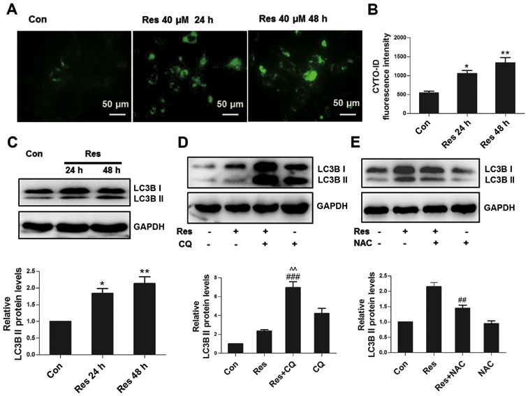
3270 YAO et al: AUTOPHAGY SUPPRESSES RESVERATROL-INDUCED APOPTOSIS IN 786-O CELLS
Abcam, and Beclin 1 antibody (1:500; catalog no. sc‑48341) Cells were re‑washed twice with PBS and harvested, and
was purchased from Santa Cruz Biotechnology, Inc. ΔΨm was determined via the aforementioned flow cytometry
method. ΔΨm was calculated as the ratio of red to green
Cell culture. The 786‑O cell line was purchased from the fluorescence.
Shanghai Institute of Cell Biology, Chinese Academy of
Sciences. Cells were maintained in RPMI‑1640 medium Cyto‑ID autophagy detection assay. A CYTO‑ID autophagy
(HyClone; GE Healthcare Life Sciences) supplemented detection kit (Enzo Life Sciences, Inc.) was utilized in the
with 10% FBS (Gibco; Thermo Fisher Scientific, Inc.) and present study. Briefly, cells were washed twice with PBS and
1% penicillin and streptomycin, at 37˚C in a humidified then incubated in PBS containing CYTO‑ID probe and 5%
atmosphere containing 5% CO2 until they reached 80‑90% FBS at 37˚C for 20 min in the dark. Following the incuba-
confluence. tion, cells were re‑washed twice with PBS and observed
under a fluorescence microscope (Olympus Corporation;
Cell viability assay. Cell viability assay was performed using magnification, x200). In order to evaluate autophagy with flow
CCK‑8 reagent (Dojindo Molecular Technologies), according cytometry, cells were harvested following incubation with
to the manufacturer's protocol. The 786‑O cells were seeded CYTO‑ID probe at 37˚C for 20 min in the dark. Subsequently,
at a density of 4x10³ cells/well into 96‑well plates. Following autophagy was determined via the aforementioned flow
overnight incubation at 37˚C, the cells were treated with the cytometry method.
indicated concentrations of Res (10, 20, 40 and 80 µM) for
24 or 48 h. Following Res treatment, CCK‑8 reagent was Western blotting. Cells were treated with 40 µM Res for 24 or
added into every well, followed by incubation at 37˚C for 1 h 48 h. For some experiments, 10 mM NAC, 20 µM SP600125
in the dark. Subsequently, the optical density was determined or 20 µM SB203580 were used. In order to evaluate autophagic
using a microplate reader (Bio‑Rad Laboratories, Inc.), at a flux, cells were treated with 40 µM Res for 48 h in the presence
wavelength of 450 nm. or absence of 50 µM CQ.
Cells were lysed using a total protein extraction kit (Nanjing
Cell apoptosis assay. Cell apoptosis was assessed using an KeyGen Biotech Co., Ltd.), according to the manufacturer's
AnnexinV‑FITC‑propidium iodide (PI) double staining kit protocol. Protein concentrations were determined using a
(MultiSciences Biotech, Co., Ltd.), according to the manufac- bicinchoninic acid assay kit (Nanjing KeyGen Biotech Co.,
turer's protocol. Briefly, cells were treated with 10, 20 µM Res Ltd.) and 30 µg protein/lane was separated via SDS‑PAGE on
for 48, and 40 µM of Res for 24 or 48 h. For some experiments, a 10‑12% gel. The separated proteins were subsequently trans-
cells were treated with 40 µM Res for 48 h in the presence ferred onto a polyvinylidene difluoride membrane and blocked
or absence of 50 µM Z‑VAD‑FMK, 10 mM NAC or 50 µM with 5% BSA 1 h at room temperature. The membranes were
CQ. Following treatment, cells were harvested and washed incubated with the aforementioned primary antibodies, over-
twice with PBS. Subsequently, cells were incubated in buffer night at 4˚C. The membranes were then washed three times
containing Annexin V‑FITC and PI at room temperature for with TBST and incubated with corresponding anti‑rabbit or
5 min in the dark. Apoptotic cells were identified using a BD anti‑mouse HRP‑conjugated secondary antibodies (1:5,000;
FACSCanto II flow cytometer (BD Biosciences) and data catalog no. GAR007 and GAM007; MultiSciences Biotech,
were analyzed using FACSDiVa software (version 7.0; BD Co., Ltd.) at room temperature for 1 h. Protein bands were
Biosciences). visualized using an ECL system (EMD Millipore) and densito-
metric analysis was performed using ImageJ software (version
ROS assay. Cells were harvested, washed twice with PBS, and 1.48v; National Institutes of Health).
then incubated in serum‑free RPMI‑1640 medium containing
DCFH‑DA at 37˚C for 20 min. Cells were re‑washed twice Small interfering (si)RNA and transfection. Beclin 1
with PBS and intracellular ROS was detected via the (catalog no. sc‑29797) and scrambled control siRNA (catalog
aforementioned flow cytometry method. no. sc‑37007) were purchased from Santa Cruz Biotechnology,
Inc. The Beclin 1 siRNA sequences were as follows: Forward,
Caspase 3 activity assay. Caspase 3 activity was determined 5'‑CAGCUCAACGUCACUGAAATT‑3' and reverse, 5'‑UUU
using a caspase 3 activity assay kit (ApexBio Technology), CAG U GA C GU U GA G CU G TT‑3'. The control siRNA
according to the manufacturer's protocol. Briefly, cells were sequences were not available. Briefly, 100 nM of Beclin 1 or
washed twice with PBS and incubated in staining buffer control siRNA was transfected into cells using Lipofectamine®
containing FITC‑DEVD‑FMK probe at 37˚C for 30 min. Cells 2000 reagent (Invitrogen; Thermo Fisher Scientific, Inc.),
were re‑washed twice with PBS and harvested, and caspase 3 according to the manufacturer's protocol, and non‑transfected
activity was detected via the aforementioned flow cytometry cells were set as a blank control. After 24 h, cells were treated
method. with 40 µM Res for an additional 48 h and subsequently
analyzed via western blotting as previously described.
Mitochondrial membrane potential (ΔΨm) assay. The ΔΨm
assay was performed using a JC1 mitochondrial membrane Statistical analysis. All experiments were performed in
potential assay kit (Beijing Solarbio Science & Technology triplicate, and the data are presented as the mean ± standard
Co., Ltd.), according to the manufacturer's protocol. In brief, deviation. Unpaired Student's t‑test was used for comparisons
cells were washed twice with PBS and incubated in fresh between two groups. One‑way analysis of variance followed
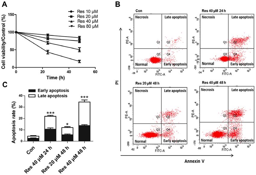
RPMI‑1640 medium containing JC1 regent at 37˚C for 30 min. by post hoc comparisons using Tukey's test was used forONCOLOGY LETTERS 19: 3269-3277, 2020 3271 Figure 1. Res decreases cell viability and induces apoptosis in 786‑O cells. (A) Cell viability was analyzed using a Cell Counting Kit‑8 assay. (B) Representative images of the flow cytometric apoptosis assay. (C) Quantitative analysis of apoptosis detected by flow cytometry. *P
3272                     YAO et al: AUTOPHAGY SUPPRESSES RESVERATROL-INDUCED APOPTOSIS IN 786-O CELLS

Figure 2. Res damages mitochondria and activates caspase to execute apoptosis. (A) Representative images of flow cytometric assay for ΔΨm. (B) Quantitative
analysis of JC1 red/green fluorescence value as detected by flow cytometry. (C) Quantitative analysis of caspase 3 activity detected by flow cytometry.
(D) Following treatment with 40 µM Res for 24 or 48 h, a western blot analysis to detect changes in PARP was performed. (E) Following treatment with
40 µM Res in the presence or absence of 50 µM Z‑VAD‑FMK for 48 h, quantitative analysis of apoptosis detected by flow cytometry was performed. *P
ONCOLOGY LETTERS 19: 3269-3277, 2020                                                             3273

Figure 3. ROS are responsible for apoptosis induced by Res. (A) Following treatment with 40 µM Res for 24 or 48 h, representative images of flow cytometric
assay for ROS. (B) Following treatment with 40 µM Res for 24 or 48 h, quantitative analysis of ROS detected by flow cytometry was performed. Following
treatment with 40 µM Res in the presence or absence of 10 mM NAC for 48 h, quantitative analysis of (C) ROS, (D) JC1 red/green fluorescence value,
(E) caspase 3 activity, (F) apoptosis and (G) western blot analysis of PARP expression were detected by flow cytometry. *P
3274                    YAO et al: AUTOPHAGY SUPPRESSES RESVERATROL-INDUCED APOPTOSIS IN 786-O CELLS

Figure 5. JNK activated by ROS is required for Res‑induced autophagy. Following treatment with 40 µM Res for 24 h or 48 h, (A) western blot analysis of
p‑AMPK and p‑S6, and (B) p‑ERK, p‑p38, p‑JNK, ERK, p38 and JNK was performed. (C) Following treatment with 40 µM Res in the presence or absence of
10 mM NAC for 48 h, western blot analysis of p‑ERK, p‑p38 and p‑JNK was performed. (D) Following treatment with 40 µM Res in the presence of 20 µM
SB203580 or 20 µM SP600125 for 48 h, western blot analysis of LC3B was performed. (E) Following treatment with 40 µM Res in the presence or absence of
20 µM SP600125 for 48 h, western blot analysis of p‑JNK and p‑BCL2. NS, *P
ONCOLOGY LETTERS 19: 3269-3277, 2020                                                               3275

Figure 6. Inhibition of autophagy enhances Res‑induced apoptosis. Following treatment with 40 µM Res in the presence or absence of 50 µM CQ for 48 h, quantita-
tive analysis of (A) apoptosis and (B) caspase 3 activity were detected by flow cytometry, and (C) via western blot analysis of PARP and LC3B. (D) Cells were
transfected with 100 nM Beclin 1 siRNA or control siRNA, and non‑transfected cells were set as a blank control. After 24 h, cells were treated with 40 µM Res for
an additional 48 h, and western blotting was used for the analysis of Beclin 1, LC3B and PARP expressions. NS vs. control group; #P
3276 YAO et al: AUTOPHAGY SUPPRESSES RESVERATROL-INDUCED APOPTOSIS IN 786-O CELLS
increased amount of BCL2 phosphorylation, suggesting that Patient consent for publication
JNK was involved in the autophagic process. It was also noted
that Res inhibited ERK activity. ERK can activate HIF‑1α to Not applicable.
promote VEGF transcription (36). Notably, high expression
of VEGF is a common feature of RCC, and the association Competing interests
between Res and ERK deserves further investigation.
Under oxidative stress, the association between autophagy The authors declare that they have no competing interests.
and apoptosis is complicated. On the one hand, autophagy
can capture damaged proteins and organs (such as damaged References
mitochondria) for degradation, maintaining cell survival. On
the other hand, extreme autophagy promotes cell death (37). 1. Simard EP, Ward EM, Siegel R, Jemal A: Cancers with increasing
In the experiments performed in the present study, autophagy incidence trends in the United States: 1999 through 2008. CA
Cancer J Clin 62: 118‑128, 2012.
inhibitor CQ and Beclin 1 siRNA further promoted apoptosis, 2. Znaor A, Lortet‑Tieulent J, Laversanne M, Jemal A and Bray F:
demonstrating that autophagy exerted a protective effect on International variations and trends in renal cell carcinoma inci-
Res‑induced apoptosis in 786‑O cells. To the best of our knowl- dence and mortality. Eur Urol 67: 519‑530, 2015.
3. Motzer RJ, Agarwal N, Beard C, Bhayani S, Bolger GB,
edge, only one study (38) has reported the role of Res‑induced Carducci MA, Chang SS, Choueiri TK, Hancock SL,
autophagy in RCC, demonstrating that Res induces autophagy Hudes GR, et al: Kidney cancer. J Natl Compr Canc Netw 9:
via the AMPK‑mTOR signaling pathway and that autophagy, 960‑977, 2011.
4. Novelle MG, Wahl D, Diéguez C, Bernier M and de Cabo R:
in turn, promotes apoptosis. This difference may be attributed Resveratrol supplementation: Where are we now and where
to the different cell lines used. Furthermore, the previous study should we go? Ageing Res Rev 21: 1‑15, 2015.
only detected the expression levels of autophagy‑associated 5. Jang M, Cai L, Udeani GO, Slowing KV, Thomas CF, Beecher CW,
Fong HH, Farnsworth NR, Kinghorn AD, Mehta RG, et al:
proteins and genes to ascertain their effect on Res‑mediated Cancer chemopreventive activity of resveratrol, a natural product
apoptosis. In the absence of autophagic flux studies, their derived from grapes. Science 275: 218‑220, 1997.
evidence remains unconvincing. 6. Carter LG, D'Orazio JA and Pearson KJ: Resveratrol and cancer:
Focus on in vivo evidence. Endocr Relat Cancer 21: R209‑R225,
Overall, ROS was involved in the process of Res‑induced 2014.
apoptosis in 786‑O cells. On the one hand, ROS damaged 7. Iliopoulos O, Kibel A, Gray S and Kaelin WG Jr: Tumour
mitochondria and activated caspase to execute apoptosis. suppression by the human von Hippel‑Lindau gene product. Nat
Med 1: 822‑826, 1995.
On the other hand, it induced autophagy through JNK, and 8. Kucejova B, Peña‑Llopis S, Yamasaki T, Sivanand S, Tran TA,
autophagy suppressed apoptosis. Therefore, a combination Alexander S, Wolff NC, Lotan Y, Xie XJ, Kabbani W, et al:
of Res and autophagy inhibitor could enhance the inhibitory Interplay between pVHL and mTORC1 pathways in clear‑cell
renal cell carcinoma. Mol Cancer Res 9: 1255‑1265, 2011.
effect of Res on RCC. 9. Khan MA, Chen HC, Wan XX, Tania M, Xu AH, Chen FZ and
Zhang DZ: Regulatory effects of resveratrol on antioxidant
Acknowledgements enzymes: A mechanism of growth inhibition and apoptosis
induction in cancer cells. Mol Cells 35: 219‑225, 2013.
10. Chen LB: Mitochondrial membrane potential in living cells.
Not applicable. Annu Rev Cell Biol 4: 155‑181, 1988.
11. Dröge W: Free radicals in the physiological control of cell func-
tion. Physiol Rev 82: 47‑95, 2002.
Funding 12. Dewaele M, Maes H and Agostinis P: ROS‑mediated mecha-
nisms of autophagy stimulation and their relevance in cancer
The present study was funded by the Natural Science Foundation therapy. Autophagy 6: 838‑854, 2010.
13. Chan LL, Shen D, Wilkinson AR, Patton W, Lai N, Chan E,
of Jiangsu Province (grant no. BK20151180), the Applied Basic Kuksin D, Lin B and Qiu J: A novel image‑based cytometry
Research of Changzhou City (grant no. CJ20159014) and the method for autophagy detection in living cells. Autophagy 8:
Major Science and Technology Project of Changzhou Health 1371‑1382, 2012.
14. Pajares M, Jiménez‑Moreno N, Dias IHK, Debelec B, Vucetic M,
Bureau (grant no. ZD201405). Fladmark KE, Basaga H, Ribaric S, Milisav I and Cuadrado A:
Redox control of protein degradation. Redox Biol 6: 409‑420,
Availability of data and materials 2015.
15. Xu J, Wu Y, Lu G, Xie S, Ma Z, Chen Z, Shen HM and Xia D:
Importance of ROS‑mediated autophagy in determining apop-
The datasets used and/or analysed in the present study are totic cell death induced by physapubescin B. Redox Biol 12:
198‑207, 2017.
available from the corresponding author upon reasonable 16. Xie X, Le L, Fan Y, Lv L and Zhang J: Autophagy is induced through
request. the ROS‑TP53‑DRAM1 pathway in Response to mitochondrial
protein synthesis inhibition. Autophagy 8: 1071‑1084, 2012.
17. Hung AC, Tsai CH, Hou MF, Chang WL, Wang CH, Lee YC,
Authors' contributions Ko A, Hu SC, Chang FR, Hsieh PW and Yuan SS: The synthetic
β‑nitrostyrene derivative CYT‑Rx20 induces breast cancer cell
HY and MF interpreted the data and drafted the initial manu- death and autophagy via ROS‑mediated MEK/ERK pathway.
Cancer Lett 371: 251‑261, 2016.
script. XH designed the experiments and revised the initial 18. Tsao CC and Corn PG: MDM‑2 antagonists induce p53‑
manuscript and HY performed the experiments.All authors dependent cell cycle arrest but not cell death in renal cancer cell
read and approved the final manuscript lines. Cancer Biol Ther 10: 1315‑1325, 2010.
19. Wei Y, Pattingre S, Sinha S, Bassik M and Levine B:
JNK1‑mediated phosphorylation of Bcl‑2 regulates starva-
Ethics approval and consent to participate tion‑induced autophagy. Mol Cell 30: 678‑688, 2008.
20. Mariño G, Niso‑Santano M, Baehrecke EH and Kroemer G:
Self‑consumption: The interplay of autophagy and apoptosis. Nat
Not applicable. Rev Mol Cell Biol 15: 81‑94, 2014.ONCOLOGY LETTERS 19: 3269-3277, 2020 3277
21. Gurkar AU, Chu K, Raj L, Bouley R, Lee SH, Kim YB, Dunn SE, 31. Yan HW, Hu WX, Zhang JY, Wang Y, Xia K, Peng MY and
Mandinova A and Lee SW: Identification of ROCK1 kinase as a Liu J: Resveratrol induces human K562 cell apoptosis, erythroid
critical regulator of Beclin1‑mediated autophagy during meta- differentiation, and autophagy. Tumour Biol 35: 5381‑5388, 2014.
bolic stress. Nat Commun 4: 2189, 2013. 32. Alayev A, Sun Y, Snyder RB, Berger SM, Yu JJ and Holz MK:
22. Otera H, Ohsakaya S, Nagaura Z, Ishihara N and Mihara K: Resveratrol prevents rapamycin‑induced upregulation of
Export of mitochondrial AIF in response to proapoptotic stimuli autophagy and selectively induces apoptosis in TSC2‑deficient
depends on processing at the intermembrane space. EMBO J 24: cells. Cell Cycle 13: 371‑382, 2014.
1375‑1386, 2005. 33. Alayev A, Berger SM, Kramer MY, Schwartz NS and Holz MK:
23. Brenner C and Kroemer G: Apoptosis. Mitochondria‑the death The combination of rapamycin and resveratrol blocks autophagy
signal integrators. Science 289: 1150‑1151, 2000. and induces apoptosis in breast cancer cells. J Cell Biochem 116:
24. Muqbil I, Beck FW, Bao B, Sarkar FH, Mohammad RM, 450‑457, 2015.
Hadi SM and Azmi AS: Old wine in a new bottle: The warburg 34. Simon HU, Friis R, Tait SW and Ryan KM: Retrograde signaling
effect and anticancer mechanisms of resveratrol. Curr Pharm from autophagy modulates stress responses. Sci Signal 10:
Des 18: 1645‑1654, 2012. pii: eaag2791, 2017.
25. Miki H, Uehara N, Kimura A, Sasaki T, Yuri T, Yoshizawa K 35. de la Cruz‑Morcillo MA, Valero ML, Callejas‑Valera JL,
and Tsubura A: Resveratrol induces apoptosis via ROS‑triggered Arias‑González L, Melgar‑Rojas P, Galán‑Moya EM,
autophagy in human colon cancer cells. Int J Oncol 40: García‑Gil E, García‑Cano J and Sánchez‑Prieto R: P38MAPK
1020‑1028, 2012. is a major determinant of the balance between apoptosis and
26. Gu S, Chen C, Jiang X and Zhang Z: ROS‑mediated endoplasmic autophagy triggered by 5‑fluorouracil: Implication in resistance.
reticulum stress and mitochondrial dysfunction underlie apop- Oncogene 31: 1073‑1085, 2012.
tosis induced by resveratrol and arsenic trioxide in A549 cells. 36. Masoud GN and Li W: HIF‑1α pathway: Role, regulation and
Chem Biol Interact 245: 100‑109, 2016. intervention for cancer therapy. Acta Pharm Sin B 5: 378‑389,
27. Kim C, Baek SH, Um JY, Shim BS and Ahn KS: Resveratrol 2015.
attenuates constitutive STAT3 and STAT5 activation through 37. Kaminskyy VO and Zhivotovsky B: Free radicals in cross talk
induction of PTPε and SHP‑2 tyrosine phosphatases and potenti- between autophagy and apoptosis. Antioxid Redox Signal 21:
ates sorafenib‑induced apoptosis in renal cell carcinoma. BMC 86‑102, 2014.
Nephrol 17: 19, 2016. 38. Liu Q, Fang Q, Ji S, Han Z, Cheng W and Zhang H:
28. Moriyama M, Moriyama H, Uda J, Kubo H, Nakajima Y, Goto A, Resveratrol‑mediated apoptosis in renal cell carcinoma via
Morita T and Hayakawa T: BNIP3 upregulation via stimula- the p53/AMP‑activated protein kinase/mammalian target of
tion of ERK and JNK activity is required for the protection of rapamycin autophagy signaling pathway. Mol Med Rep 17:
keratinocytes from UVB‑induced apoptosis. Cell Death Dis 8: 502‑508, 2018.
e2576, 2017.
29. Wang X, Luo F and Zhao H: Paraquat‑induced reactive
oxygen species inhibit neutrophil apoptosis via a p38 This work is licensed under a Creative Commons
MAPK/NF‑κ B‑IL‑6/TNF‑ α positive‑feedback circuit. PLoS Attribution-NonCommercial-NoDerivatives 4.0
One 9: e93837, 2014. International (CC BY-NC-ND 4.0) License.
30. Kumar B, Iqbal MA, Singh RK and Bamezai RN: Resveratrol
inhibits TIGAR to promote ROS induced apoptosis and
autophagy. Biochimie 118: 26‑35, 2015.You can also read