Track distance runners exhibit bilateral differences in the plantar fascia stiffness - Nature
←
→
Page content transcription
If your browser does not render page correctly, please read the page content below
www.nature.com/scientificreports
OPEN Track distance runners exhibit
bilateral differences in the plantar
fascia stiffness
Hiroto Shiotani1,2, Ryo Yamashita3, Tomohiro Mizokuchi3, Natsuki Sado4,
Munekazu Naito2,5 & Yasuo Kawakami1,2*
Human steady-state locomotion modes are symmetrical, leading to symmetric mechanical function
of human feet in general; however, track distance running in a counterclockwise direction exposes
the runner’s feet to asymmetrical stress. This may induce asymmetrical adaptation in the runners’
foot arch functions, but this has not been experimentally tested. Here, we show that the plantar
fascia (PF), a primary structure of the foot arch elasticity, is stiffer for the left than the right foot
as a characteristic of runners, via a cross-sectional study on 10 track distance runners and 10
untrained individuals. Shear wave velocity (index of tissue stiffness: SWV) and thickness of PF and
foot dimensions were compared between sides and groups. Runners showed higher PF SWV in their
left (9.4 ± 1.0 m/s) than right (8.9 ± 0.9 m/s) feet, whereas untrained individuals showed no bilateral
differences (8.5 ± 1.5 m/s and 8.6 ± 1.7 m/s, respectively). Additionally, runners showed higher left to
right (L/R) ratio of PF SWV than untrained men (105.1% and 97.7%, respectively). PF thickness and
foot dimensions were not significantly different between sides or groups. These results demonstrate
stiffer PF in the left feet of runners, which may reflect adaptation to their running-specific training that
involves asymmetrical mechanical loading.
During human locomotion, the medial longitudinal arch of the foot is lowered while being stretched out in
response to weight-bearing, and then recoils as the load is removed. Such a spring-like property of the foot arch
helps to attenuate impact forces and store/release elastic strain energy leading to energy saving in running1,2.
Previous studies indicate that the foot arch elasticity is attributed to the plantar fascia (PF)1,3,4. PF behaves vis-
coelastically under load5,6, and its resistive tension helps to prevent the lengthening and lowering of the foot
arch. During each foot contact of running, PF is repetitively loaded with the tension reaching as high as 0.6–3.7
times bodyweight with its longitudinal strain up to 6%7–10. Such sizable stress concentrates around the proximal
site of PF11–13, which may be associated with the heterogeneity of mechanical and morphological properties (e.g.,
stiffness and thickness) of PF14–16 as well as the occurrence of plantar fasciitis17,18.
The localized stiffness of PF can be quantitatively assessed as the shear wave velocity (SWV) in vivo14–16. PF
has higher SWV (i.e., stiffer) at the proximal site than middle and distal sites14,16. Additionally, long-distance
running induced a transient decrease of SWV at the proximal site of PF while long-distance runners showing
smaller changes in SWV than untrained individuals15, suggesting that runners had built up a more resilient
PF. These findings are evidence of PF adaptability to site-specific and chronic mechanical stress, which can be
reflected in its stiffness and morphology.
Human steady-state locomotion modes are symmetrical, leading to symmetric mechanical function of human
feet in general; however, track distance running is performed always in a counterclockwise direction, i.e., the left
leg being inside during curve running. In this phase, runners are required to generate greater forces with their
left legs19,20 to exert centripetal force21. This is associated with the greater load on the left foot, resulting in the
lowering of the left foot arch, and thus leading to an increase of mechanical stress to PF. Therefore, runners’ feet
can be exposed to asymmetrical stress during running. This may induce asymmetrical adaptation in runners’ PF
stiffness and morphology. Although PF SWV and thickness, and the foot dimensions were comparable between
left and right sides in a healthy and untrained population14, this may not be true for track distance runners. If
1
Faculty of Sport Sciences, Waseda University, Saitama, Japan. 2Human Performance Laboratory, Comprehensive
Research Organization, Waseda University, Tokyo, Japan. 3School of Sport Sciences, Waseda University, Saitama,
Japan. 4Faculty of Health and Sport Sciences, University of Tsukuba, Ibaraki, Japan. 5Department of Anatomy, Aichi
Medical University, Aichi, Japan. *email: ykawa@waseda.jp
Scientific Reports | (2021) 11:9260 | https://doi.org/10.1038/s41598-021-88883-4 1
Vol.:(0123456789)www.nature.com/scientificreports/
Figure 1. Bilateral differences in SWV and thickness of runners and untrained men. Data are shown as
means ± SD.
the bilateral differences in runner’s feet can be confirmed, this provides an indication of a threshold of mechani-
cal stress that causes adaptation of PF and foot arch functions. A profound understanding of PF adaptability is
essential for improvements in their performance as well as prevention of plantar fasciitis.
Therefore, the purpose of this study was to investigate the bilateral differences in mechanical and morpho-
logical properties of PF and foot dimensions in track distance runners, as contrasted to untrained individuals.
We hypothesized that track distance runners have bilateral differences in PF SWV, thickness, and the foot arch
height, and that runners show sizable differences in the left to right (L/R) ratios of measured variables as com-
pared to untrained individuals.
Results
In runners, SWV at the proximal site was significantly higher in left (9.4 ± 1.0 m/s) than right foot (8.9 ± 0.9 m/s)
(p = 0.021, d = 0.813), but not at the middle (p = 0.782, d = 0.073) or distal sites (p = 0.554, d = 0.138) (Fig. 1). Even
in a lefty runner (n = 1), PF SWV at the proximal site was higher for his left (10.0 m/s) than right foot (9.1 m/s).
PF SWV at the proximal site was also higher for the left than the right feet both in rearfoot strike (n = 7, left:
9.0 ± 1.0 m/s and right: 8.6 ± 0.8 m/s, respectively) and forefoot strike runners (n = 3, left: 10.2 ± 0.3 m/s and right:
9.6 ± 0.5 m/s, respectively). In untrained men, SWV at each measurement site was not significantly different
between left and right feet (p ≥ 0.222, d ≤ 0.264). PF thickness at each measurement site was not significantly dif-
ferent between left and right feet in runners (p ≥ 0.327, d ≤ 0.141) or untrained men (p ≥ 0.411, d ≤ 0.305) (Fig. 1).
Foot dimensions were not significantly different between left and right feet in either of runners or untrained
men (Table 1).
The L/R ratio of SWV at the proximal site was significantly higher in runners than untrained men (p = 0.027,
d = 1.076), but not at the middle (p = 0.815, d = 0.107) or distal sites (p = 0.421, d = 0.369). The L/R ratios of thick-
ness at any of the measurement sites or foot dimensions were not significantly different between groups (Table 2).
Age, body height, body mass, BMI, and fractions of leg dominance and foot strike pattern were not signifi-
cantly different between runners and untrained men (Table 3). All participants were healthy and free from injury
of the lower extremity in the past 12 months and had no present or past history of plantar fasciitis. The runners
had kept habitual running of at least 10 km/week for the past year, mainly on a running track, and their running
experiences ranged between 9 and 16 years. Their personal best time of 5000 m ranged from 14′ 15 to 15′ 30.
The untrained participants were either sedentary or lightly active, and none of them had been involved in any
structured training program or continuous sports participation at least 12 months before the measurements. All
participants used conventional running shoes rather than minimalist, high cushion, or high motion control shoes.
Scientific Reports | (2021) 11:9260 | https://doi.org/10.1038/s41598-021-88883-4 2
Vol:.(1234567890)www.nature.com/scientificreports/
Runners (n = 10) Untrained men (n = 10)
p value p value
Left Right Cohen’s d Left Right Cohen’s d
p = 0.520 p = 0.445
Foot length (mm) 245.3 ± 7.3 245.9 ± 8.6 247.4 ± 8.7 248.2 ± 8.1
d = 0.075 d = 0.107
p = 0.760 p = 0.671
Dorsal height (mm) 61.1 ± 4.4 60.8 ± 4.2 60.5 ± 5.5 61.1 ± 4.0
d = 0.070 d = 0.125
p = 0.080 p = 0.507
Navicular height (mm) 43.3 ± 6.2 41.9 ± 6.8 41.3 ± 4.8 40.9 ± 5.4
d = 0.215 d = 0.078
p = 0.064 p = 0.400
Arch height ratio (%) 17.7 ± 2.7 17.1 ± 3.0 16.7 ± 1.8 16.4 ± 1.9
d = 0.210 d = 0.162
Table 1. Bilateral differences in foot dimensions of runners and untrained men. Data are shown as
means ± SD.
p value
Variable Runners Untrained men Cohen’s d
Shear wave velocity
p = 0.027
Proximal 105.1 ± 6.2 97.7 ± 7.6
d = 1.076
p = 0.815
Middle 101.8 ± 12.9 100.2 ± 17.2
d = 0.107
p = 0.421
Distal 104.6 ± 13.7 99.7 ± 12.8
d = 0.360
Thickness
p = 0.610
Proximal 101.3 ± 6.4 99.6 ± 8.0
d = 0.232
p = 0.756
Middle 97.8 ± 5.9 98.8 ± 8.7
d = 0.141
p = 0.429
Distal 102.3 ± 14.1 97.6 ± 11.7
d = 0.362
p = 0.843
Foot length 99.8 ± 1.1 99.7 ± 1.3
d = 0.083
p = 0.583
Dorsal height 100.5 ± 3.9 99.1 ± 6.3
d = 0.267
p = 0.270
Navicular height 103.9 ± 5.8 101.2 ± 4.4
d = 0.525
p = 0.329
Arch height ratio 104.1 ± 6.0 101.6 ± 5.1
d = 0.449
Table 2. Left/right ratios (%) of individual parameters in runners and untrained men. Data are shown as
means ± SD. Bold fonts indicate significant difference between runners and untrained men (p < 0.05) with a
“large” effect size (d ≥ 0.8).
Variable Runners Untrained men p value
n 10 10 –
Age (years) 22.0 ± 0.7 22.5 ± 1.4 0.309
Body height (m) 1.68 ± 0.04 1.70 ± 0.05 0.392
Body mass (kg) 55.5 ± 4.2 58.4 ± 5.6 0.062
BMI (kg/m2) 19.6 ± 1.2 20.3 ± 1.7 0.113
Dominant leg (Lefty:Righty) 1:9 1:9 1.000
Foot strike pattern (RFS:FFS) 7:3 10:0 0.060
Running experience (years) 11.0 ± 2.2 – –
Running distance (km/week) 43.7 ± 35.4 – –
Table 3. Physical characteristics of participants. Data are shown as means ± SD. BMI body mass index, RFS
rear foot strikers, FFS forefoot strikers. Age, body height, body mass, BMI, and fractions of leg dominance and
foot strike pattern were not significantly different between runners and untrained men.
Scientific Reports | (2021) 11:9260 | https://doi.org/10.1038/s41598-021-88883-4 3
Vol.:(0123456789)www.nature.com/scientificreports/
Discussion
To the best of our knowledge, this is the first study to investigate bilateral differences in the mechanical and
morphological properties of PF in track distance runners. The most striking finding of the present study was
that track distance runners showed stiffer PF at the proximal site in their left than the right feet, unlike the
untrained participants. A number of previous studies addressing running m echanics22,23 and mechanical and
15,24
morphological properties of the musculotendinous and fascial t issues in runners have focused on unilateral
leg by assuming bilateral symmetry. Our findings however suggest that asymmetry in runners is the major issue
that needs to be carefully considered.
The greater load on the left foot during curve running19,20 can induce an increase of mechanical stress on
PF. We previously revealed that running causes a decrease in PF SWV at its proximal site15, and this coincides
with the simulation of stress distribution along P F11–13. The bilateral differences in PF stiffness at the proximal
site in runners may reflect the adaptation to such stress accumulation in this region of the left foot during track
running, regardless of the lateral dominance or foot strike patterns. The fact that PF stiffness in the right feet of
runners was comparable to that of both feet in untrained individuals suggests a threshold of mechanical stress
that causes adaptation of PF stiffness, which is side-specific for track runners. The finding that PF can be stiffened
in response to a sufficient load is valuable for the general population toward injury prevention and rehabilitation
as well as improvements in human locomotor performance.
Since the runners in this study had no history of plantar fasciitis, they might have been successful examples
who had been optimally adapted to their running-specific training. However, plantar fasciitis is one of the most
common injuries in long-distance runners, regardless of their performance levels25,26. This injury frequently
occurs around the proximal site of P F17,18 where the mechanical stress is c oncentrated11–13. Thus, there is a clinical
implication that the left foot of track runners can suffer from a higher incidence of this injury if their training
adaptation does not work well. The bilateral imbalances in strength, morphology, and running mechanics are
considered to be risk factors for injury of r unners27,28. Interactions between these factors for the occurrence of
plantar fasciitis are worth examining in future studies.
No bilateral differences in PF thickness of runners and untrained individuals are consistent with previous
findings that PF thickness was not different between recreational runners and untrained individuals15, and that
PF thickness was not influenced by physical a ctivity29. These results, together with our findings, discard the pos-
sibility of PF adaptability in terms of its thickness for reducing mechanical stress induced by distance running.
No bilateral difference in the foot arch dimensions suggests that the arches of both feet of runners can fulfill their
imposed roles through different mechanical properties with comparable morphology.
We could not obtain the running mechanics and the foot arch deformation during running. This is one of the
limitations of the present study. As our findings suggest a threshold of mechanical stress that causes adaptation of
PF stiffness, quantifying the mechanical stress applied to bilateral feet during track running will lead to a better
understanding of the nature of PF adaptability. Additionally, the runners who participated in the present study
can be categorized as recreational level30. Runners of different performance levels (e.g., competitive and elite
runners) show different running m echanics31,32 and fatigue r esponses33,34. Thus, competitive and elite runners
have the possibility to exhibit different signs of adaptation in PF and foot morphology. Comparisons between
runners in different performance levels should be incorporated in these future studies. Moreover, there is a vari-
ation in the training volume of runners (Table 3). As we previously reported that long-distance running induced
transient decreases of PF S WV15, the training volume can be a potential factor that affects PF properties and their
adaptation. In addition, the material and mechanical properties of the running surface (e.g., rubber, asphalt,
or grass) as well as shoe sole have the possibility to affect the magnitude of stress to the f oot35–38. Future studies
addressing the chronic effects of training volume and environment on PF adaptation as well as foot arch functions
are needed. Lastly, it can be assumed that sprinters, participating in the event of 200 and 400 m in particular, and
long/high jumpers may also apply asymmetrical stress to their feet with a greater magnitude of stress compared
to distance runners. Further investigation of bilateral differences in PF characteristics and foot dimensions in
other events and sports athletes can be an option of the future theme in understanding PF adaptability.
In conclusion, this study showed bilateral differences in the mechanical but not in the morphological proper-
ties of PF and foot arch dimensions in track distance runners as compared to untrained individuals. PF SWV at
the proximal site was higher in the left feet of track distance runners while their right feet showing comparable
values to that of untrained individuals. These results demonstrate stiffer proximal PF in the left feet of runners,
which may reflect adaptation to their running-specific training that involves asymmetrical mechanical loading.
Methods
Study design and participants. A cross-sectional study was conducted at Waseda University (Tokoro-
zawa campus) in Japan from August to November 2017. This study was approved by the Human Research Ethics
Committee of Waseda University (reference number: 2016-310) and was carried out in accordance with the
Declaration of Helsinki. Written informed consent was obtained from all participants before data collection.
The necessary sample size was calculated from our preliminary results (n = 6 in each group; total = 12). A
priori power analysis (G*Power v3.1, Heinrich Heine-Universität Dusseldorf, Germany) with an assumed type
1 error of 0.05 and a statistical power of 0.80 was conducted to find significant differences in PF SWV between
left and right feet of runners and between groups, respectively. The critical sample sizes were estimated to be
at least 7 runners and 9 in each group (total = 18), respectively. Thus, 10 track distance male runners and 10
untrained men were recruited in this study (Table 1). Twelve runners were eligible for participation in this study.
Of these, 2 runners met the exclusion criteria of history of plantar fasciitis and operative treatment of the lower
limb (Fig. 2). Finally, 10 runners and 10 untrained men who matched the baseline physical characteristics with
those of runners were successfully recruited in this study.
Scientific Reports | (2021) 11:9260 | https://doi.org/10.1038/s41598-021-88883-4 4
Vol:.(1234567890)www.nature.com/scientificreports/
Figure 2. Flow diagram depicting participant selection. 12 runners were eligible for participation in this study.
Two runners met the exclusion criteria of history of plantar fasciitis and operative treatment of the lower limb.
Thus, 10 runners and 10 untrained men who matched the baseline physical characteristics with those of runners
were included in this study.
Before the main measurements, the profiles including age, body height, body mass, dominant leg, athletic
experiences, exercise habits for the past year, foot strike pattern, history of injuries and operative treatment, and
model of their running shoes were collected from all participants. The dominant leg was determined according
to the participant’s favorite leg for kicking a ball. The foot strike pattern (rearfoot or forefoot strikers) of partici-
pants was visually confirmed on another o ccasion15. Additionally, the training environment (e.g., affiliation and
running surfaces), personal best time of 5000 m, and running volume/week for the year were asked for runners.
To avoid any confounding factors, we recruited participants attending the same university, and attempted to
match the baseline physical characteristics of untrained participants with those of runners. Participants were
not allowed to perform any strenuous exercises for at least 24 h before the measurement.
Ultrasound measurements. The supersonic shear imaging (SSI) and B-mode ultrasonography techniques
with an Aixplorer ultrasound scanner (version 6.4, Supersonic Imagine, Aix-en-Provence, France) and a linear
array probe (SL 15-4, Supersonic Imagine, Aix-en-Provence, France) were used to measure the mechanical and
morphological properties of PF. SSI is a valid and reliable technique to evaluate the stiffness of skeletal muscles,
tendons, and fasciae in vivo14,39–41. In principle, SSI uses multiple push pulses to generate the shear waves propa-
gating within the soft tissues and measures their velocity (i.e., SWV). Since SWV is related to Young’s modulus
and shear modulus of the soft tissues, it can be used as an index of stiffness42,43.
Details of SSI measurement and data processing were based on our previous published w ork14,15. During
ultrasound measurements, participants were requested to rest in a supine position on the examination bed with
their knee fully extended. Additionally, their ankle and toe digits were secured to a custom-made fixture at the
neutral position. PF was scanned at three different sites along the longitudinal line between the medial calcaneal
tubercle and the second toe. The locations of measurement sites were that at the proximal (in the proximity to
the calcaneus), middle (the level of navicular tuberosity), and distal (proximity to the second metatarsal head)
(Fig. 3). The longitudinal line of the foot and the locations of the transducer were marked on the skin surface
using a waterproof marker. The scanning head of the probe was coated with transmission gel. An acoustic standoff
pad (Gelpad for StatUS, Enraf–Nonius, Rotterdam, Netherland) was used to avoid applying excessive compres-
sion on the skin surface. Three images were obtained at each measurement site, and used for further analysis.
After data collection, SWV at each measurement site was measured as the mean value within the region of
interest (ROI) which was manually traced over the fascial boundaries of PF using a measurement tool included
in the Aixplorer software (i.e., Q-box Trace). PF thickness at each measurement site was measured the distance
between the superficial and deep fascial boundaries was measured to determine thickness using a measurement
Scientific Reports | (2021) 11:9260 | https://doi.org/10.1038/s41598-021-88883-4 5
Vol.:(0123456789)www.nature.com/scientificreports/
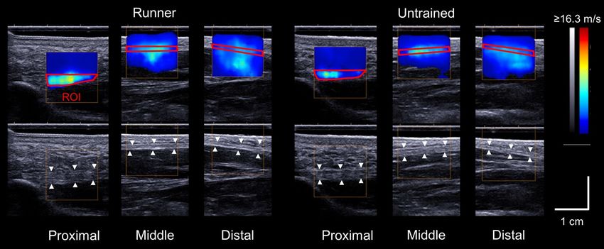
Figure 3. Representative shear wave and ultrasound B-mode images of the plantar fascia at the proximal,
middle, and distal sites in a runner and an untrained participant. The region of interest (ROI) is bounded in red.
tool (i.e., Distance). For SWV and thickness at each measurement site, three images were analyzed at each meas-
urement site, then the three values were averaged to obtain the representative value.
Measurements of the foot dimensions. A foot scanner (JMS-2100CU, Dream GP, Osaka, Japan) was
used to obtain three-dimensional foot shape data. Details of measurement and data processing were based on
our previous study using the same system15. Participants were requested to stand in a relaxed position with their
feet approximately shoulder-width apart. The longitudinal axis of their feet, which is the line connecting between
the most posterior point of the heel and the head of the second toe, aligned parallel with the guidelines drawn
on the footplate in the foot scanner. A laser scanner moved around the foot in an oval trajectory, measuring the
foot dimensions and the anatomical marker positions based on laser line triangulation. After the scanning, foot
length, dorsal height, and navicular height were measured. The foot length was defined as the length projected
on the longitudinal axis between the most posterior point of the heel and the head of the first or second toe,
whichever was longer. The dorsal height was defined as the height of the highest point from the floor at 55%
of the length of the foot from the heel. The navicular height was defined as the height of the most medial point
of the navicular bone from the floor. Additionally, the arch height ratio was calculated as the navicular height
normalized to the foot length.
Statistical analysis. The normality of the data was assessed using a Shapiro–Wilk test. After the normal-
ity was confirmed, the difference in physical characteristics between groups were compared using an unpaired
t-test. The fraction of dominant legs within each group was compared with a Pearson chi-squared test. Com-
parisons of measured variables between left and right feet in each group were performed using a paired t-test.
The L/R ratios were calculated for the measured variables, and were compared using an unpaired t-test between
groups. Cohen’s d was calculated as a measure of effect size. For the within-subject factor,√ it was corrected for
dependence between mean values using the following equation: d = Mdiff /SDpooled 2(1 − r) , where Mdiff is
mean difference between conditions, SDpooled is pooled SD, and r is correlation between mean values44. Effect
size is interpreted as trivial (d < 0.2), small (0.2 ≤ d < 0.5), medium (0.5 ≤ d < 0.8) and large effect (d ≥ 0.8)45. Sta-
tistical significance was set at α = 0.05. Statistical analysis was performed using SPSS software (SPSS Statistics 25,
IBM, Armonk, USA).
Received: 17 November 2020; Accepted: 19 April 2021
References
1. Ker, R. F., Bennett, M. B., Bibby, S. R., Kester, R. C. & Alexander, R. M. The spring in the arch of the human foot. Nature 325,
147–149 (1987).
2. Stearne, S. M. et al. The foot’s arch and the energetics of human locomotion. Sci. Rep. 6, 19403 (2016).
3. Cifuentes-De la Portilla, C., Larrainzar-Garijo, R. & Bayod, J. Analysis of the main passive soft tissues associated with adult acquired
flatfoot deformity development: A computational modeling approach. J. Biomech. 84, 183–190 (2019).
4. Huang, C. K., Kitaoka, H. B., An, K. N. & Chao, E. Y. Biomechanical evaluation of longitudinal arch stability. Foot Ankle 14, 353–357
(1993).
5. Pavan, P. G., Stecco, C., Darwish, S., Natali, A. N. & De Caro, R. Investigation of the mechanical properties of the plantar aponeu-
rosis. Surg. Radiol. Anat. 33, 905–911 (2011).
6. Guo, J., Liu, X., Ding, X., Wang, L. & Fan, Y. Biomechanical and mechanical behavior of the plantar fascia in macro and micro
structures. J. Biomech. 76, 160–166 (2018).
7. Chen, T. L., Wong, D. W., Wang, Y., Lin, J. & Zhang, M. Foot arch deformation and plantar fascia loading during running with
rearfoot strike and forefoot strike: A dynamic finite element analysis. J. Biomech. 83, 260–272 (2019).
8. Giddings, V. L., Beaupr, G. S., Whalen, R. T. & Carter, D. R. Calcaneal loading during walking and running. Med. Sci. Sports Exerc.
32, 627–634 (2000).
Scientific Reports | (2021) 11:9260 | https://doi.org/10.1038/s41598-021-88883-4 6
Vol:.(1234567890)www.nature.com/scientificreports/
9. McDonald, K. A. et al. The role of arch compression and metatarsophalangeal joint dynamics in modulating plantar fascia strain
in running. PLoS ONE 11, e0152602 (2016).
10. Wager, J. C. & Challis, J. H. Elastic energy within the human plantar aponeurosis contributes to arch shortening during the push-
off phase of running. J. Biomech. 49, 704–709 (2016).
11. Chen, Y. N., Chang, C. W., Li, C. T., Chang, C. H. & Lin, C. F. Finite element analysis of plantar fascia during walking: A quasi-static
simulation. Foot Ankle Int. 36, 90–97 (2015).
12. Cheng, H. Y., Lin, C. L., Wang, H. W. & Chou, S. W. Finite element analysis of plantar fascia under stretch-the relative contribution
of windlass mechanism and achilles tendon force. J. Biomech. 41, 1937–1944 (2008).
13. Lin, S. C. et al. Stress distribution within the plantar aponeurosis during walking—A dynamic finite element analysis. J. Mech.
Med. Biol. 14, 145005 (2014).
14. Shiotani, H., Yamashita, R., Mizokuchi, T., Naito, M. & Kawakami, Y. Site- and sex-differences in morphological and mechanical
properties of the plantar fascia: A supersonic shear imaging study. J. Biomech. 85, 198–203 (2019).
15. Shiotani, H., Mizokuchi, T., Yamashita, R., Naito, M. & Kawakami, Y. Acute effects of long-distance running on mechanical and
morphological properties of the human plantar fascia. Scand. J. Med. Sci. Sports 30, 1360–1368 (2020).
16. Shiotani, H., Maruyama, N., Kurumisawa, K., Yamagishi, T. & Kawakami, Y. Human plantar fascial dimensions and shear wave
velocity change in vivo as a function of ankle and metatarsophalangeal joint positions. J. Appl. Physiol. 130, 390–399 (2021).
17. Wearing, S. C., Smeathers, J. E., Urry, S. R., Hennig, E. M. & Hills, A. P. The pathomechanics of plantar fasciitis. Sports Med. 36,
585–611 (2006).
18. League, A. C. Current concepts review: Plantar fasciitis. Foot Ankle Int. 29, 358–366 (2008).
19. Chang, Y. H. & Kram, R. Limitations to maximum running speed on flat curves. J. Exp. Biol. 210, 971–982 (2007).
20. Churchill, S. M., Trewartha, G., Bezodis, I. N. & Salo, A. I. Force production during maximal effort bend sprinting: Theory vs
reality. Scand. J. Med. Sci. Sports 26, 1171–1179 (2016).
21. Greene, P. R. Running on flat turns: Experiments, theory, and applications. J. Biomech. Eng. 107, 96–103 (1985).
22. Bazuelo-Ruiz, B., Dura-Gil, J. V., Palomares, N., Medina, E. & Llana-Belloch, S. Effect of fatigue and gender on kinematics and
ground reaction forces variables in recreational runners. PeerJ 6, e4489 (2018).
23. Sado, N., Yoshioka, S. & Fukashiro, S. A biomechanical study of the relationship between running velocity and three-dimensional
lumbosacral kinetics. J. Biomech. 94, 158–164 (2019).
24. Fletcher, J. R. & MacIntosh, B. R. Changes in achilles tendon stiffness and energy cost following a prolonged run in trained distance
runners. PLoS ONE 13, e0202026 (2018).
25. Taunton, J. E. et al. A retrospective case-control analysis of 2002 running injuries. Br. J. Sports Med. 36, 95–101 (2002).
26. van Gent, R. N. et al. Incidence and determinants of lower extremity running injuries in long distance runners: A systematic review.
Br. J. Sports Med. 41, 469–480 (2007).
27. Zifchock, R. A., Davis, I. & Hamill, J. Kinetic asymmetry in female runners with and without retrospective tibial stress fractures.
J. Biomech. 39, 2792–2797 (2006).
28. Rauh, M. J., Koepsell, T. D., Rivara, F. P., Rice, S. G. & Margherita, A. J. Quadriceps angle and risk of injury among high school
cross-country runners. J. Orthop. Sports Phys. Ther. 37, 725–733 (2007).
29. Uzel, M., Cetinus, E., Ekerbicer, H. C. & Karaoguz, A. The influence of athletic activity on the plantar fascia in healthy young adults.
J. Clin. Ultrasound 34, 17–21 (2006).
30. Clermont, C. A., Benson, L. C., Osis, S. T., Kobsar, D. & Ferber, R. Running patterns for male and female competitive and recrea-
tional runners based on accelerometer data. J. Sports Sci. 37, 204–211 (2019).
31. Anderson, T. Biomechanics and running economy. Sports Med. 22, 76–89 (1996).
32. Williams, K. R. & Cavanagh, P. R. Relationship between distance running mechanics, running economy, and performance. J. Appl.
Physiol. 63, 1236–1245 (1987).
33. Willwacher, S., Sanno, M. & Bruggemann, G. P. Fatigue matters: An intense 10 km run alters frontal and transverse plane joint
kinematics in competitive and recreational adult runners. Gait Posture 76, 277–283 (2019).
34. Sanno, M., Willwacher, S., Epro, G. & Bruggemann, G. P. Positive work contribution shifts from distal to proximal joints during
a prolonged run. Med. Sci. Sports Exerc. 50, 2507–2517 (2018).
35. Birch, J. V., Kelly, L. A., Cresswell, A. G., Dixon, S. J. & Farris, D. J. Neuromechanical adaptations of foot function to changes in
surface stiffness during hopping. J. Appl. Physiol. 130, 1196 (2021).
36. Tessutti, V., Ribeiro, A. P., Trombini-Souza, F. & Sacco, I. C. Attenuation of foot pressure during running on four different surfaces:
Asphalt, concrete, rubber, and natural grass. J. Sports Sci. 30, 1545–1550 (2012).
37. Kelly, L. A., Lichtwark, G. A., Farris, D. J. & Cresswell, A. G. Shoes alter the spring-like function of the human foot during running.
J. R. Soc. Interface 13, 20160174 (2016).
38. Roy, J. P. & Stefanyshyn, D. J. Shoe midsole longitudinal bending stiffness and running economy, joint energy, and emg. Med. Sci.
Sports Exerc. 38, 562–569 (2006).
39. Aubry, S. et al. Viscoelasticity in achilles tendonopathy: Quantitative assessment by using real-time shear-wave elastography.
Radiology 274, 821–829 (2015).
40. Lacourpaille, L., Hug, F., Bouillard, K., Hogrel, J. Y. & Nordez, A. Supersonic shear imaging provides a reliable measurement of
resting muscle shear elastic modulus. Physiol. Meas. 33, N19-28 (2012).
41. Otsuka, S., Shan, X. & Kawakami, Y. Dependence of muscle and deep fascia stiffness on the contraction levels of the quadriceps:
An in vivo supersonic shear-imaging study. J. Electromyogr. Kinesiol. 45, 33–40 (2019).
42. Eby, S. F. et al. Validation of shear wave elastography in skeletal muscle. J. Biomech. 46, 2381–2387 (2013).
43. Helfenstein-Didier, C. et al. In vivo quantification of the shear modulus of the human achilles tendon during passive loading using
shear wave dispersion analysis. Phys. Med. Biol. 61, 2485–2496 (2016).
44. Morris, S. B. & DeShon, R. P. Combining effect size estimates in meta-analysis with repeated measures and independent-groups
designs. Psychol. Methods 7, 105–125 (2002).
45. Cohen, J. A power primer. Psychol. Bull. 112, 155–159 (1992).
Acknowledgements
This study was part of research activities of the Human Performance Laboratory, Comprehensive Research
Organization, Waseda University. This study was supported by JSPS KAKENHI Grant Numbers 19J14912 and
16H01870. The authors express their gratitude to Dr. Pavlos Evangelidis, Dr. Takaki Yamagishi and Mr. Hidetaka
Hayashi for grammatical corrections of the manuscript.
Author contributions
H.S., R.Y., T.M. and Y.K. designed the research. H.S., R.Y. and T.M. corrected the data. H.S. analyzed the data.
H.S., R.Y., T.M., N.S., M.N. and Y.K. interpreted the results of experiments. H.S. wrote the main manuscript
text and prepared Figures and Tables. H.S., N.S., M.N. and Y.K. revised the manuscript. All authors read and
approved the final version of the manuscript.
Scientific Reports | (2021) 11:9260 | https://doi.org/10.1038/s41598-021-88883-4 7
Vol.:(0123456789)www.nature.com/scientificreports/
Competing interests
The authors declare no competing interests.
Additional information
Correspondence and requests for materials should be addressed to Y.K.
Reprints and permissions information is available at www.nature.com/reprints.
Publisher’s note Springer Nature remains neutral with regard to jurisdictional claims in published maps and
institutional affiliations.
Open Access This article is licensed under a Creative Commons Attribution 4.0 International
License, which permits use, sharing, adaptation, distribution and reproduction in any medium or
format, as long as you give appropriate credit to the original author(s) and the source, provide a link to the
Creative Commons licence, and indicate if changes were made. The images or other third party material in this
article are included in the article’s Creative Commons licence, unless indicated otherwise in a credit line to the
material. If material is not included in the article’s Creative Commons licence and your intended use is not
permitted by statutory regulation or exceeds the permitted use, you will need to obtain permission directly from
the copyright holder. To view a copy of this licence, visit http://creativecommons.org/licenses/by/4.0/.
© The Author(s) 2021
Scientific Reports | (2021) 11:9260 | https://doi.org/10.1038/s41598-021-88883-4 8
Vol:.(1234567890)You can also read