Insights into extrinsic foot muscle activation during a 75 min run using T2 mapping - Nature
←
→
Page content transcription
If your browser does not render page correctly, please read the page content below
www.nature.com/scientificreports
OPEN Insights into extrinsic foot muscle
activation during a 75 min run
using T2 mapping
Grischa Bratke1*, Steffen Willwacher2, Florian Siedek1, David Maintz1, Daniela Mählich2,
Kilian Weiss3, Tilman Hickethier4 & Gert‑Peter Brüggemann2,5
The extrinsic foot muscles are essentially for controlling the movement path but our knowledge of
their behavior during prolonged running is still very limited. Therefore, this study analyzed the time-
course of muscle activation using T2 mapping during 75 min of running. In this prospective study, 19
recreational active runners completed 75 min of treadmill running at a constant speed. Interleaved
T2 mapping sequences were acquired and segmented at timepoints 0, 2.5, 5, 10, 15, 45, and 75 min.
ANOVA for repeated measurements followed by a Tukey post hoc test and Pearson correlation
between running speed and initial signal increase at 2.5 min were calculated. All muscles showed
a significant signal increase between baseline and 2.5 min (e.g. medial gastrocnemius: + 15.48%;
p < 0.01). This was followed by a plateau phase till 15 min for all but the extensor digitorum longus
muscle and a significant decrease at 45 or 75 min for all muscles (all p < 0.05). Correlation between
running speed and signal increase was negative for all muscles and significant for both gastrocnemii
(e.g. medial: r = − 0.57, p = 0.0104) and soleus (r = − 0.47, p = 0.0412). The decrease of relaxation times
times in the later running phases was less pronounced for faster runners (≥ 10 km/h). T2 relaxation
times do not only decrease after cessation of exercise but already during prolonged running. The lesser
initial increase and later decrease in faster runners may indicate training induced changes.
Millions of recreational runners aim for longer distances such as marathons by pushing their limits with a rela-
tively high risk for injuries. Van Middelkoop et al. reported that 28% of runners suffer an injury in the month
before or during a m arathon1 with most running associated injuries located in the knee or lower leg with 41.7%
and 27.9%, r espectively2. The main reasons for injuries are training errors and overuse (about 72%)3 followed
by extrinsic and intrinsic risk factors such as previous injuries, body size, anatomical alignment, and level of
competition4.
Running is a complex interaction of activation and relaxation of the involved muscles, starting with muscle-
tuning even before heel strike5. The central nervous system gains input over the ground reaction forces and tunes
the muscles accordingly to a predefined and cost-effective personal preferred motion p ath6. The extrinsic foot
muscles (gastrocnemius medialis, gastrocnemius lateralis, soleus, tibialis anterior, tibialis posterior) play an essen-
tial role in controlling the movement path as they act as foot invertors and prevent pronation of the foot during
running. Electromyography is a widely used technique for the detection of muscle activation. However, surface
EMG is only suitable for assessing large muscle groups without good spatial resolution, and even the invasive
method of intramuscular EMG is still affected by "cross-talk" between closely adjacent m uscles7. Furthermore,
both methods are highly dependent on the placement of the electrodes8. Another non-invasive way of muscle
analysis is by means of ultrasound. The focus here is on morphological changes, which, however, can only be
determined focally in the context of a defined movement for individual muscles or as long-term changes9–11.
In 1988, Fleckenstein et al.12 were the first to show a correlation between activation patterns of skeletal muscles
before and after exercise with an increase of the T2 signal using magnetic resonance imaging (MRI). Using MRI
allows near real-time, non-invasive observation of muscle activation with excellent spatial resolution. Although
the exact physiological mechanism remains unclear it is believed that most of the effect is due to intracellular
events with an increased microcirculation in the active muscle, accumulation of osmolytes (phosphate, lactate,
and sodium) and intracellular acidification resulting in increased intramuscular water content and volume of
1
Department of Diagnostic and Interventional Radiology, University of Cologne, Kerpenerstraße 62,
50937 Cologne, Germany. 2Institute of Biomechanics and Orthopaedics, German Sport University Cologne,
Am Sportpark Müngersdorf 6, 50933 Cologne, Germany. 3Philips Healthcare Germany, Röntgenstraße 24,
22335 Hamburg, Germany. 4Radiologie Heinrichsallee, Heinrichsallee 50‑54, 52062 Aachen, Germany. 5True Motion
Running GmbH, Hermann‑Sudermann‑Str. 3, 48155 Münster, Germany. *email: grischa.bratke@uk-koeln.de
Scientific Reports | (2021) 11:7331 | https://doi.org/10.1038/s41598-021-86810-1 1
Vol.:(0123456789)www.nature.com/scientificreports/
the muscle13–15. Multiple studies were able to detect muscle activation in lower extremities using T2 mapping
scans after exercise, resulting in increased relaxation times13,16–18 and showing good correlation between muscle
activation and exercise i ntensity19–21 as well as glucose u
ptake22. Jenner et al. reported relaxation times increas-
ing to a maximum after only a few repetitions of dynamic ankle dorsiflexion exercise with a following plateau
phase for the next 15 min18 and Fisher et al. described a return to the initial level about 20 min after cessation
of the e xercise23. While the existing literature covers direct pre- and post-exercise comparisons and the starting
phase (first 15 min), we still lack information about the reaction and adoption of muscle activation to prolonged
running.
Therefore, this study is the first to describe the time-course of muscle activation based on T2 mapping during
a prolonged run of 75 min to test for the hypothesis of a consistent plateau phase after initial activation.
Materials and methods
Participants. This prospective study was approved by the local institutional review board of the University
of Cologne and registered in the German Clinical Trials Registry (DRKS00011152) on the 02/07/2018. Informed
consent was obtained from all study participants and all procedures were carried out in accordance with relevant
guidelines. Participants and corresponding imaging data were acquired from December 2016 until August 2018.
Participants needed to be recreationally active, between 18 and 40 years old and without any known neurological
disorder, cardiovascular disease, or sport-related injuries at the time of the study (Fig. 1). Dividing the subjects
in terms of running speed was based on the previously described correlation between load intensity and muscle
activation19–21. The 10 km/h were chosen in order to obtain two groups as equal as possible.
Exercise protocol. Each volunteer tested and subsequently chose the highest possible running speed that
he or she was able to maintain for 75 min on the provided treadmill (F85, Sole Fitness, Neu-Ulm, Germany).
The treadmill was positioned directly in front of the scanner room to minimize time and potential influences
between running and scanning. Based on the results from Fisher et al.23, all volunteers completed an initial rest-
ing phase of 30 min on the moveable MR table outside the scanner room to neutralize any exercise or muscle
activation before the experiment. The volunteers laid supine on the table in a relaxed position with slightly
bent knees (about 20°) and were brought to the MR room to avoid any movement of the leg before starting the
baseline scan. The running phases were divided into different sections with interposed MR scans to allow a high
temporal resolution of muscle activity changes. The measuring timepoints were 0, 2.5, 5, 10, 15, 45 and 75 min
to account for the previously described rapid increase of T2 times and the following plateau. The effective times
for MRI scanning, running on the treadmill, and switching between the scanner and the treadmill were recorded
using a stopwatch. All runners were asked whether they felt exhausted or could continue running with the same
running speed.
MRI examination. All scans were performed on a whole-body 3.0 T MRI system (Philips Ingenia 3.0 T,
Philips Healthcare, Best, the Netherlands) equipped with a dedicated 16-channel transmit and receive knee
coil. To ensure full consistency between all scans for the individual participant, a custom-made MR-compatible
board fixed to the MR table was used to position the feet at the same spot after each bout. For each participant,
the coil base was kept fixed to the MRI table while the right calf was positioned in the center of the coil base.
The initial scan included T1-weighted sequences in coronal and axial direction for optimal anatomical plan-
ning. A Carr-Meibom-Purcell-sequence identical to the one described by Willwacher et al.24 and comparable to
Schuermans et al.25 was used to obtain T2 maps after acquisition of 16 echoes ranging from 10 to 160 ms with
a repetition time of TR = 2000 ms (flip angle: 90 degrees, SENSE factor: 1.7, voxel size: 1.5 × 1.5 × 5 mm). The
relaxation time was calculated by the scanner based on the exponential decay of the signal intensity. Ten slices
with a 15 mm gap were acquired, resulting in a FOV with a craniocaudal length of 185 mm starting from the
fibula head and with a transversal dimension of 150 × 197 mm.
Data analysis. Due to field inhomogeneities or a poor signal-to-noise ratio in some scans, the first and
last two slices were excluded with six slices remaining for analysis. On each slice and for all time points the
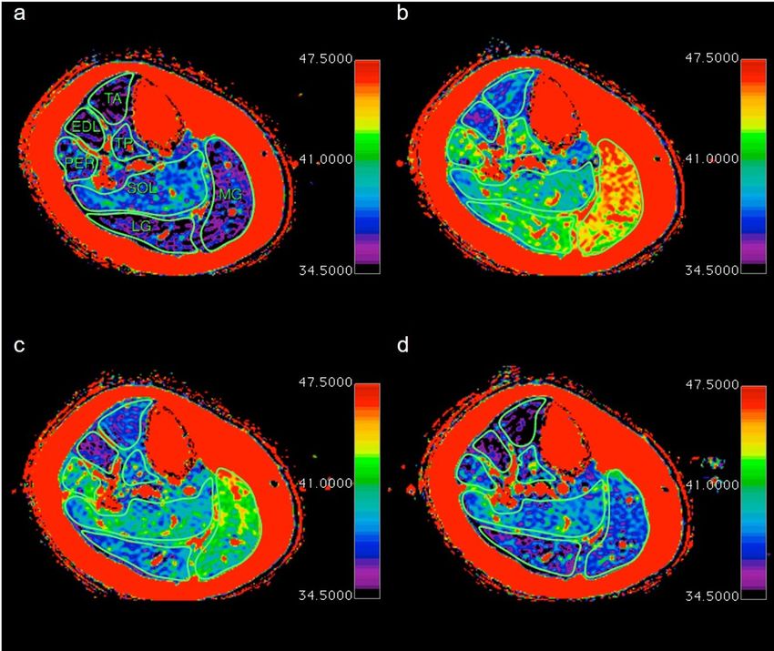
medial (MG) and lateral gastrocnemius (LG), soleus (SO), peroneus longus (PER), tibialis posterior (TP), tibi-
alis anterior (TA) and extensor digitorum longus (EDL) were manually segmented by a radiologist specialized
in musculoskeletal imaging (7 years of experience) using HOROS viewer 3.3.2 (The Horos Project, Annapolis,
MD, USA). Segmentation was performed on the first echo (TE = 10 ms) due to the best delineation of anatomi-
cal structures and copied to the calculated T2 map. Comparable to previous studies, vessels and intramuscular
fat were excluded25. The average T2 relaxation time for each muscle for a given time point was calculated as the
sum of multiplying the relaxation time for each slice with the cross-sectional area (CSA) averaged to the whole
measured cross-sectional area of the muscle (ACSA), comparable to previous work by Ploutz-Snyder et al.13:
6
T2 relaxation timemuscle = slice CSAslice x
slice 1 relaxation timeslice x × CSAall six slices
This procedure ensures that the relaxation time for each slice is included according to the total percent-
age of muscle volume. Muscle volumes for 18 of the volunteers in this study had been reported previously by
Wilwacher et al.24.
Statistical analysis. Statistical analyses were performed using GraphPad Prism Version 8.4.1 (GraphPad
Software Inc., San Diego, California, USA). All data are reported as the mean ± standard deviation (SD). The data
was checked for normal distributaion using the Kolmogorov–Smirnov test. The continuous data of the relaxa-
Scientific Reports | (2021) 11:7331 | https://doi.org/10.1038/s41598-021-86810-1 2
Vol:.(1234567890)www.nature.com/scientificreports/
Figure 1. Flow diagram for study participants with exercise protocol and subgroup analysis.
tion times were tested for significant differences with a one-way analysis of variance (ANOVA) for repeated
measurements followed by a post hoc analysis using the Tukey test to correct for multiple comparisons. The
times for the individual running, changing, and measuring segments were tested using ANOVA. Correlations
of the running speed with the increase in relaxation times between the baseline and after the initial increase at
2.5 min were assessed by calculating Pearson’s correlation coefficient. Differences between fast and slow runners
were assessed with the unpaired t test. A p value of < 0.05 was considered statistically significant.
Scientific Reports | (2021) 11:7331 | https://doi.org/10.1038/s41598-021-86810-1 3
Vol.:(0123456789)www.nature.com/scientificreports/
n = 20 N = 19 Fast Slow
28.75 ± 3.18 28.68 ± 3.25
Age [years] 29.33 ± 2.49 28.10 ± 3.70
(23–33) (23–33)
176.10 ± 7.94 176.42 ± 8.02
Height [cm] 180.56 ± 9.74 172.70 ± 2.72*
(162–198) (162–198)
70.20 ± 9.74 70.74 ± 9.70
Body mass [kg] 73.33 ± 11.16 68.40 ± 7.43
(51–95) (51–95)
Sex 11 male/9 female 11 male/8 female 8 male/1 female 3 male/ 7 female*
2.68 ± 0.39 2.68 ± 0.40
speed [km/h] 3.04 ± 0.25 9.66 ± 1.40*
(2.08–3.61) (2.08–3.61)
Table 1. Demographics of study participants before and after dropout of one volunteer due to an incomplete
run as well as subgroups for fast (≥ 10 km/h or 2.78 m/s) and slow (< 10 km/h) runners. The subgroups differed
significantly with regards to height, sex and speed (marked with *).
0 min 2.5 min 5 min 10 min 15 min 45 min 75 min
44.67 ± 2.89 44.36 ± 2.63 44.14 ± 2.63 43.88 ± 2.74 42.06 ± 1.68 40.79 ± 1.22
MG [ms] 38.68 ± 1.52 (100%)–/*
(115.48%)*/* (114.68%)*/* (114.11%)*/* (113.44%)*/* (108.73%)*/* (105.44%)*/–
42.40 ± 2.69 42.25 ± 2.84 42.25 ± 3.40 42.27 ± 3.78 41.06 ± 3.65 39.50 ± 1.92
LG [ms] 37.10 ± 1.01 (100%)–/*
(114.28%)*/* (113.70%)*/* (113.88%)*/* (113.92%)*/* (110.67%)*/– (106.46%)*/–
39.70 ± 1.82 39.25 ± 1.69 39.26 ± 1.89 39.22 ± 1.79 39.51 ± 1.76 38.74 ± 1.24
TP [ms] 37.36 ± 1.13 (100%)–/*
(106.27%)*/– (105.06%)*/– (105.09%)*/– (104.97%)*/– (105.76%)*/* (103.70%)*/–
39.16 ± 1.18 40.92 ± 1.46 40.68 ± 1.34 40.64 ± 1.37 40.45 ± 1.17 40.23 ± 0.85 39.70 ± 0.89
SOL [ms]
(100%)–/– (104.49%)*/* (103.87%)*/* (103.76%)*/* (103.29%)*/* (102,72%)*/* (101.36%)–/–
40.63 ± 3.10 40.61 ± 2.96 40.99 ± 3.21 40.89 ± 3.08 40.38 ± 2.22 39.41 ± 1.69
PER [ms] 37.49 ± 2.67 (100%)–/*
(108.36%)*/– (108.32%)*/– (109.32%)*/– (109.07%)*/– (107.70%)*/* (105.11%)*/–
38.66 ± 2.19 38.10 ± 2.29 38.27 ± 2.45 38.05 ± 2.42 37.35 ± 1.20 36.67 ± 1.04
EDl [ms] 35.79 ± 0.86 (100%)–/*
(108.02%)*/* (106.45%)*/– (106.93%)*/* (106.33%)*/– (104.37%)*/* (102.46%)*/–
39.04 ± 2.08 38.66 ± 2.38 39.07 ± 2.55 38.69 ± 2.29 37.61 ± 1.38 36.93 ± 0.96
TA [ms] 35.50 ± 0.77 (100%)–/*
(109.95%)*/* (108.90%)*/* (110.05%)*/* (108.97%)*/* (105.94%)*/– (104.02%)*/–
Table 2. Absolute and relative changes of the T2 relaxation times during prolonged running. */* marking
significant difference (p < 0.05) compared to baseline (0 min) (* before diagonal slash) or 75 min timepoint (*
after diagonal slash).
Results
Eleven male and nine female healthy young volunteers participated in this study (Table 1). One female volunteer
was unable to complete the 75 min run at the chosen speed and was excluded from the following data analysis
(n = 19). The average running speed was 9.66 ± 1.40 km/h or 2.68 ± 0.40 m/s, and all runners felt exhausted,
fulfilling the criteria for maximal possible running speed over the 75 min period. Normality test was passed for
all timepoints with all p > 0.1.
The effective time for the individual running sessions were almost identical to the targeted times with
2:31,18 ± 0:01,58, 5:00,99 ± 0:03,02 and 30:01,97 ± 0:03,27 min for the 2.5, 5, and 30 min running segments,
respectively. The running sessions were interrupted in total by 3:45,70 ± 0:46,40 min consisting of the average
time within the scanner with 2:17,23 ± 0:27,54 min, 0:48,40 ± 0:15,87 min when switching from the scanner to the
treadmill and 0:41,23 ± 0:07,95 min for switching from the treadmill back to the scanner. The time from leaving
the treadmill until the end of the scan as a potential confounding factor for the relaxation times ranged from
2:55,55 to 3:04,23 min without significant differences between the individual running segments (all p > 0.05).
The initial ANOVA showed statistical significance for all muscles with R squared ranging from 0.80 to 0.45
(MG: 0.80; LG: 0.78; TP: 0.49; SOL: 0.45; PER: 0.63; EDL: 0.55; TA: 0.58). All segmented muscles showed a
statistical significant increase of relaxation times between baseline and 2.5 min (MG: + 15.48% ; LG: + 14.28%;
TP: + 6.27%; SOL: + 4.49%; PER: + 8.36%; EDL: + 8.02%; TA: + 9.95% (all p < 0.01)) (Table 2) (Fig. 2). The initial
steep increase was followed by a plateau phase, including time points 2.5, 5, 10, and 15 min. In this phase, there
were no significant differences between any of these time points (all p > 0.05) except for the EDL with a decline
of T2 relaxation times from 38.66 to 38.10 ms (p = 0.03) between 2.5 and 5 min. The initial increase and plateau
were followed by reduced relaxation times in the later running phases (Fig. 3). As a result, the relaxation times
of all muscles were significantly lower at 75 min than for at least one previous timepoint (all p < 0.05) (Fig. 4).
MG, LG, SOL, and TA showed significantly higher relaxation times for all time points during the plateau phase
(2.5–15 min) compared to the 75 min time point (all p < 0.05). The plateau phase was extended for TP and PER
without any significant difference between 2.5 and 45 min (all p > 0.05), while both muscles showed a significant
decrease from 45 to 75 min (TP: p = 0.01; PER: p = 0.02).
In the study population, there was a significant negative correlation between running speed as a measure
of exercise intensity and the increase of relaxation times for MG (r = − 0.57, p = 0.01), LG (r = − 0.46, p = 0.04)
Scientific Reports | (2021) 11:7331 | https://doi.org/10.1038/s41598-021-86810-1 4
Vol:.(1234567890)www.nature.com/scientificreports/
Figure 2. Mean relaxation times with standard deviation for the extrinsic foot muscle.
and SOL (r = − 0.47, p = 0.04). For example, the initial increase in MG for the fastest male (13 km/h) and female
(11 km/h) were 2.80 ms and 3.33 ms while they were 7.96 ms (8 km/h) and 8.51 ms (7.5 km/h) for the slowest
male and female, respectively. The correlation coefficients for all other muscles were negative as well but missed
statistical significance (TP: r = − 0.26, p = 0.27; PER: r = − 0.17, p = 0.48; EDL: r = − 0.28, p = 0.24; TA: r = − 0.38,
p = 0.11). The whole participant collective could be divided into nine faster and ten slower runners using a run-
ning speed threshold of 10 km/h (Table 3). The groups differed significantly regarding speed (p = 0.01), height
(p = 0.03) and sex (p = 0.01). Based on this subdivision, there was a smaller increase of relaxation times and a
smaller decrease in the later phases for the faster compared to the slower runners. Compared to faster runners,
in slower runners a significantly higher relaxation time was measured for all muscles (p < 0.05), except the PER,
at least once before the end of the run (75 min). In contrast, this was only true for EDL (5 min; p = 0.04) and TA
(2.5 min, p = 0.01) for the faster runners at one time point each. The difference was particularly pronounced for
MG, LG and SOL (Fig. 5), where at least three of the first four time points had significantly higher relaxation
Scientific Reports | (2021) 11:7331 | https://doi.org/10.1038/s41598-021-86810-1 5
Vol.:(0123456789)www.nature.com/scientificreports/
Figure 3. Relative changes of all muscles over time.
Figure 4. Time course for the relaxation times with the initial increase between baseline (a) and 2.5 min (b)
with the following plateau tille 15 min (c) and the later decrease at 75 min (d).
times compared to 75 min in the slower running group (all p < 0.05). This was not the case at any timepoint for
the faster runners.
Discussion
Overuse-related running injuries represent a severe problem for the individual runner, recreational or elite
level, and the community with high insurance costs and lost workdays. Despite a clear trend with more people
running longer distances (e.g., + 31.61% for marathon finishers in the United States between 2004 and 2 01626),
there is a gap of knowledge regarding physiological changes during prolonged running. This study extends the
current knowledge about short-term effects of muscle activation of the extrinsic foot muscles (< 15 min) with
changes during a 75 min run using the established T2 mapping. This is the first study to prove that T2 relaxation
Scientific Reports | (2021) 11:7331 | https://doi.org/10.1038/s41598-021-86810-1 6
Vol:.(1234567890)www.nature.com/scientificreports/
2.5 min (%) 5 min (%) 10 min (%) 15 min (%) 45 min (%) 75 min (%)
Fast 113.71 112.87 112.34 112.08 108.84 105.79
MG
Slow 117.08* 116.32* 115.72* 114.67* 108.63 105.13
Fast 112.22 111.63 111.38 111.58 110.17 107.17
LG
Slow 116.12* 115.54* 116.09* 116.00* 112.11 105.83
Fast 105.17 103.98 104.55 104.74 105.97 103.87
TP
Slow 107.25 106.04 105.57 105.17 105.57* 103.55
Fast 103.35 102.30 102.10 102.23 102.31 100.77
SOL
Slow 105.51* 105.28* 105.24* 104.24 103.08 101.88
Fast 107.42 108.07 108.60 108.86 108.10 106.46
PER
Slow 109.17 108.53 109.95 109.25 107.35 103.92
Fast 105.98 104.68* 105.71 105.39 105.19 102.65
EDl
Slow 109.84* 108.04 108.02 107.17 103.64 102.28
Fast 107.71* 106 .87 108.19 108.27 107.18 103.91
TA
Slow 111.93* 110.69 111.71 109.59 104.83 104.12
Table 3. Relative changes for fast and slow runners. The initial increase is lower for the fast runners. At the
same time, however, there is also a smaller drop in the later running phases. * marking significant difference
(p < 0.05) compared to 75 min timepoint (* after diagonal slash).
times do not remain at a plateau but significantly decrease already during exercise for all muscles. Additionally,
the increase of relaxation times is negatively correlated with the running speed of the participants, and faster
runners experience a lower drop of relaxation times during later running phases.
In line with previous studies19,27,28, we recognized significantly increased relaxation times after 2.5 min for
all measured muscles ranging from 4.49% for the SOL and 15.48% for the MG or 14.28% for LG. While these
three muscles work synergistically, the SOL mainly consists of slow-twitching type I muscle fi bers29 with lower
relaxation times30 and is always activated to counteract g ravity31, which explains the longest relaxation time at
baseline and smallest increase during exercise in our study. Absolute T2 relaxation times for baseline as well as
for the plateau phase are in the range of pre- and post-treadmill exercise results from Varghese et al.28, especially
when considering magnet-field strength dependency with a 3 T magnet used in this study32. Based on these first
insights into muscular processes during prolonged running, this newly discovered signal decrease might be a
novel, objective marker for physiological adaptation due to osmolyte changes, especially when considering the
evidence for a fast increase of blood lactate followed by a plateau phase and decrease between 15 and 30 min33.
On the other hand, it might indicate muscular fatigue with reduced activation of the extrinsic foot muscles
and changed movement patterns, which are known to lead to increased and altered bone s train34 and therefore
increasing the risk for running-related injuries such as fatigue fractures or tibia anterior syndrome. In line with
this, Kellis et al. found increased quadriceps muscle activity after ankle fatigue35, and Sanno et al. described a
shift of the joint work from distal to proximal during prolonged running with a continuous decrease in joint work
at the ankle through the plantar flexors reaching significance at 5 km36, a distance completed by our volunteers
between 15 and 45 min. Moreover, assuming all other parameters remain constant, a faster running speed results
in higher mechanical work37. The negative correlation between running speed and signal increase as well as the
reduced signal drop for faster runners in the later phases for MG, LG and SOL, therefore, implies either an altered
muscle fiber composition and metabolism due to training effects or a more efficient motion pattern for better
trained runners resulting in a reduced muscular strain. This would be in line with Costill et al. who found sig-
nificantly higher rates of type I fibers in long-distance runners38 or with Sanno et al. who described smaller shifts
of joint work in trained athletes compared to recreational r unners36 and Chapman et al. with significant longer
activations in EMG for the extrinsic foot muscles for less trained compared to elite level runners39, respectively.
This study has several limitations especially lacking physiological markers such as intramuscular/venous lac-
tate concentrations, which might have provided evidence for the underlying physiological processes. Accordingly,
additional studies should include blood samples and T2 mapping of other muscle groups for further differen-
tiation of the effect. Furthermore, the number of participants was rather small. However, the initial increase of
relaxation times, plateau phase, and final decrease were present in all participants, male and female, and reached
statistical significance for all muscles. Faster imaging after every running segment might be realized with a MR-
compatible treadmill, as Varghese et al.28 used, faster positioning of the volunteers in a standardized position or
with better acceleration techniques including compressed sensing40. However, since the duration of the pause
for running, resulting from scanning and changing times, did not show any statistical differences between the
individual run segments, these changes should only alter the absolute values for the specific times and should
not have any systematic effect. Another potential confounding factor is the sex as it is well established that
muscle metabolism differs between male and female r unners41. We tried to represent both groups in this study
but distribution is significantly unequally for the running speed subgroups. Additionally, the movement pattern
might be differet between male and female runners which might influence activation of individual muscles.
All participants were recreational athletes in various disciplines (soccer, basketball, triathlon, track and field).
Accordingly, the results may differ for non-active participants or for specific subgroups with specific l oads42.
Scientific Reports | (2021) 11:7331 | https://doi.org/10.1038/s41598-021-86810-1 7
Vol.:(0123456789)www.nature.com/scientificreports/
Figure 5. In the fast group, there is initially a smaller increase of the T2 relaxation times and at the same time a
less pronounced drop, exemplarily with the plantar flexors MG, LG and SOL.
In conclusion, significantly reduced T2 relaxation times during later running phases (≥ 45 min) in all calf
muscles provide evidence for either metabolic adoption or muscular fatigue during prolonged exercise after an
initial increase and plateau phase. Additionally, faster runners show a lower initial increase and later decrease in
Scientific Reports | (2021) 11:7331 | https://doi.org/10.1038/s41598-021-86810-1 8
Vol:.(1234567890)www.nature.com/scientificreports/
relaxation times, which strengthens the hypothesis that relaxation times are not only based on exercise intensity
but highly depend on training level and muscle fiber composition as a possible candidate for performance testing.
Data availability
The datasets generated during and/or analysed during the current study are available from the corresponding
author on reasonable request.
Competing intersts.
The author Kilian Weiss currently works for Philips Healthcare and Gert-Peter Brüggemann for the running shoe
company True Motion Running GmbH. The remaining authors have no potential conflict of interest to declare.
Received: 27 September 2020; Accepted: 18 March 2021
References
1. Middelkoop, M. V. et al. Risk factors for lower extremity injuries among male marathon runners. Scand. J. Med. Sci. Sports 18,
691–697 (2008).
2. Clement, D. B., Taunton, J. E., Smart, G. W. & McNicol, K. L. A survey of overuse running injuries. Phys. Sportsmed. 9, 47–58
(1981).
3. Lysholm, J. & Wiklander, J. Injuries in runners. Am. J. Sports Med. 15, 168–171 (1987).
4. Murphy, D. F. Risk factors for lower extremity injury: a review of the literature. Br. J. Sports Med. 37, 13–29 (2003).
5. Nigg, B. M. & Wakeling, J. M. Impact forces and muscle tuning: a new paradigm. Exerc. Sport Sci. Rev. 29, 37–41 (2001).
6. Nigg, B. M. The role of impact forces and foot pronation: a new paradigm. Clin. J. Sport Med. 11, 2–9 (2001).
7. Ivanenko, Y. P., Poppele, R. E. & Lacquaniti, F. Five basic muscle activation patterns account for muscle activity during human
locomotion. J. Physiol. 556, 267–282 (2004).
8. Martín-Fuentes, I., Oliva-Lozano, J. M. & Muyor, J. M. Electromyographic activity in deadlift exercise and its variants. A systematic
review. PLoS ONE 15, e0229507 (2020).
9. De-la-Cruz-Torres, B., Navarro-Flores, E., López-López, D. & Romero-Morales, C. Ultrasound imaging evaluation of textural
features in athletes with soleus pathology—a novel case-control study. Int. J. Environ. Res. Public Health 18, 1983 (2021).
10. Romero-Morales, C. et al. M-mode ultrasound examination of soleus muscle in healthy subjects: intra- and inter-rater reliability
study. Healthcare (Basel) 8, 555 (2020).
11. De-la-Cruz-Torres, B. et al. Does function determine the structure? Changes in Flexor Hallucis longus muscle and the associated
performance related to dance modality: a cross-sectional study. Medicina (Kaunas) 56, 186 (2020).
12. Fleckenstein, J., Canby, R., Parkey, R. & Peshock, R. Acute effects of exercise on MR imaging of skeletal muscle in normal volunteers.
Am. J. Roentgenol. 151, 231–237 (1988).
13. Ploutz-Snyder, L. L., Convertino, V. A. & Dudley, G. A. Resistance exercise-induced fluid shifts: change in active muscle size and
plasma volume. Am. J. Physiol. 269, R536–R543 (1995).
14. Saab, G., Thompson, R. T. & Marsh, G. D. Effects of exercise on muscle transverse relaxation determined by MR imaging and
in vivo relaxometry. J. Appl. Physiol. 88, 226–233 (2000).
15. Damon, B. M. et al. Intracellular acidification and volume increases explainR2 decreases in exercising muscle. Magn. Reson. Med.
47, 14–23 (2002).
16. Ploutz-Snyder, L. L., Yackel-Giamis, E. L., Rosenbaum, A. E. & Formikell, M. Use of muscle functional magnetic resonance imaging
with older individuals. J. Gerontol. A Biol. Sci. Med. Sci. 55, B504–B511 (2000).
17. Kinugasa, R., Kawakami, Y. & Fukunaga, T. Quantitative assessment of skeletal muscle activation using muscle functional MRI.
Magn. Reson. Imaging 24, 639–644 (2006).
18. Jenner, G., Foley, J. M., Cooper, T. G., Potchen, E. J. & Meyer, R. A. Changes in magnetic resonance images of muscle depend on
exercise intensity and duration, not work. J. Appl. Physiol. (Bethesda, MD, 1985) 76, 2119–2124 (1994).
19. Kinugasa, R. & Akima, H. Neuromuscular activation of triceps surae using muscle functional MRI and EMG. Med. Sci. Sports
Exerc. 37, 593–598 (2005).
20. Adams, G. R., Duvoisin, M. R. & Dudley, G. A. Magnetic resonance imaging and electromyography as indexes of muscle function.
J. Appl. Physiol. (Bethesda, MD: 1985) 73, 1578–1583 (1992).
21. Yue, G. et al. Sensitivity of muscle proton spin-spin relaxation time as an index of muscle activation. J. Appl. Physiol. 77, 84–92
(1994).
22. Haddock, B. et al. Assessment of muscle function using hybrid PET/MRI: comparison of 18F-FDG PET and T2-weighted MRI
for quantifying muscle activation in human subjects. Eur. J. Nucl. Med. Mol. Imaging 44, 704–711 (2017).
23. Fisher, M. J., Meyer, R., Adams, G. R., Foley, J. M. & Potchen, E. J. Direct relationship between proton T2 and exercise intensity in
skeletal muscle MR images. Investig. Radiol. 25, 480–485 (1990).
24. Willwacher, S. et al. The time course of calf muscle fluid volume during prolonged running. Physiol. Rep. 8, e14414 (2020).
25. Schuermans, J., Tiggelen, D. V., Danneels, L. & Witvrouw, E. Biceps femoris and semitendinosus—teammates or competitors?
New insights into hamstring injury mechanisms in male football players: a muscle functional MRI study. Br. J. Sports Med. 48,
1599–1606 (2014).
26. U.S. marathon finishers 2004–2016. Statista https:// w ww. s tati s ta. c om/ s tati s tics/ 2 80458/ number- of- m arat h on- f inis
hers-united-states/.
27. Price, T. B. et al. Comparison of MRI with EMG to study muscle activity associated with dynamic plantar flexion. Magn. Reson.
Imaging 21, 853–861 (2003).
28. Varghese, J. et al. Rapid assessment of quantitative T1, T2, and T2* in lower extremity muscles in response to maximal treadmill
exercise. NMR Biomed 28, 998–1008 (2015).
29. Johnson, M. A., Polgar, J., Weightman, D. & Appleton, D. Data on the distribution of fibre types in thirty-six human muscles: an
autopsy study. J. Neurol. Sci. 18, 111–129 (1973).
30. Kuno, S. et al. Relationship between MR relaxation time and muscle fiber composition. Radiology 169, 567–568 (1988).
31. Gambara, G. et al. Gene expression profiling in slow-type calf soleus muscle of 30 days space-flown mice. PLoS ONE 12, e0169314
(2017).
32. Duewell, S. H. et al. Musculoskeletal MR imaging at 4 T and at 1.5 T: comparison of relaxation times and image contrast. Radiology
196, 551–555 (1995).
33. van Hall, G. Lactate kinetics in human tissues at rest and during exercise. Acta Physiol. 199, 499–508 (2010).
34. Yoshikawa, T. et al. The effects of muscle fatigue on bone strain. J. Exp. Biol. 188, 217–233 (1994).
35. Kellis, E. & Liassou, C. The effect of selective muscle fatigue on sagittal lower limb kinematics and muscle activity during level
running. J. Orthop. Sports Phys. Ther. 39, 210–220 (2009).
Scientific Reports | (2021) 11:7331 | https://doi.org/10.1038/s41598-021-86810-1 9
Vol.:(0123456789)www.nature.com/scientificreports/
36. Sanno, M., Willwacher, S., Epro, G. & Brüggemann, G.-P. Positive work contribution shifts from distal to proximal joints during
a prolonged run. Med. Sci. Sports Exerc. 50, 2507–2517 (2018).
37. Lacour, J.-R. & Bourdin, M. Factors affecting the energy cost of level running at submaximal speed. Eur. J. Appl. Physiol. 115,
651–673 (2015).
38. Costill, D., Fink, W., Flynn, M. & Kirwan, J. Muscle fiber composition and enzyme activities in elite female distance runners*. Int.
J. Sports Med. 08, S103–S106 (1987).
39. Chapman, A. R., Vicenzino, B., Blanch, P. & Hodges, P. W. Is running less skilled in triathletes than runners matched for running
training history?. Med. Sci. Sports Exerc. 40, 557–565 (2008).
40. Huang, C., Graff, C. G., Clarkson, E. W., Bilgin, A. & Altbach, M. I. T2 mapping from highly undersampled data by reconstruction
of principal component coefficient maps (REPCOM) using compressed sensing. Magn. Reson. Med. 67, 1355–1366 (2012).
41. Devries, M. C. Sex-based differences in endurance exercise muscle metabolism: impact on exercise and nutritional strategies to
optimize health and performance in women. Exp. Physiol. 101, 243–249 (2016).
42. López-López, D. et al. Women’s foot health-related quality of life in ballet dancers and nondancers. Sports Health 12, 347–351
(2020).
Author contributions
G.B. and G.P.B. wrote the main manuscript text. G.B. prepared all figures. D.M. and G.B. collected the experi-
mental data. All authors reviewed the manuscript.
Funding
Open Access funding enabled and organized by Projekt DEAL.
Competing interests
The author Kilian Weiss currently works for Philips Healthcare and Gert-Peter Brüggemann for the running shoe
company True Motion Running GmbH. The remaining authors have no potential conflict of interest to declare.
Additional information
Correspondence and requests for materials should be addressed to G.B.
Reprints and permissions information is available at www.nature.com/reprints.
Publisher’s note Springer Nature remains neutral with regard to jurisdictional claims in published maps and
institutional affiliations.
Open Access This article is licensed under a Creative Commons Attribution 4.0 International
License, which permits use, sharing, adaptation, distribution and reproduction in any medium or
format, as long as you give appropriate credit to the original author(s) and the source, provide a link to the
Creative Commons licence, and indicate if changes were made. The images or other third party material in this
article are included in the article’s Creative Commons licence, unless indicated otherwise in a credit line to the
material. If material is not included in the article’s Creative Commons licence and your intended use is not
permitted by statutory regulation or exceeds the permitted use, you will need to obtain permission directly from
the copyright holder. To view a copy of this licence, visit http://creativecommons.org/licenses/by/4.0/.
© The Author(s) 2021
Scientific Reports | (2021) 11:7331 | https://doi.org/10.1038/s41598-021-86810-1 10
Vol:.(1234567890)You can also read