Inconsistency in using early differentiation markers of human pluripotent stem cells
←
→
Page content transcription
If your browser does not render page correctly, please read the page content below
International Research Journal of Medicine and Biomedical Sciences Vol.6 (2),pp. 11-18, May 2021
Available online at http://www.journalissues.org/IRJMBS/
https://doi.org/10.15739/irjmbs.21.003
Copyright © 2021 Author(s) retain the copyright of this article ISSN 2488-9032
Review
Inconsistency in using early differentiation markers of
human pluripotent stem cells
Received 15 February, 2021 Revised 4 April, 2021 Accepted 15 April, 2021 Published 8 April, 2021
Hassan H Kaabi1* Advances in the field of human pluripotent stem cells (hPSC) have prompted
researchers to advocate for the increased development of dependable
1Department of Oral Medicine therapies to cure degenerative diseases and replace damaged tissues. hPSCs
and Diagnostic Sciences,College have a one-of-a-kind ability to differentiate into all cell types in the body. The
of Dentistry, King Saud ability to characterise homogeneous primary cell populations, such as
University, P.O. Box 60169, pluripotent stem cells and germ layer cells, is required for the efficient
Riyadh 11545, Saudi Arabia. generation of adult cells. Several in vitro differentiation protocols for germ
layer lineages have been extensively researched. There is, however, no
Author’s Email: standard set of markers that can be used to separate endoderm, ectoderm,
hhkaabi@ksu.edu.sa and mesoderm populations from hPSC differentiation cultures. This review
discusses the inconsistency among studies in identifying endodermal,
mesodermal, and ectodermal cells using markers. The search was restricted
to markers used in the last 5 years to identify differentiated cells of the three
germ layers from hPSCs. The focus of this review, however, is on the most
commonly used early differentiation markers.
Keywords: Differentiation markers, ectoderm markers, mesoderm markers,
endoderm markers, Early differentiation.
INTRODUCTION
Human pluripotent stem cells (hPSCs) are unspecialized hPSCs is preceded by the formation of ectoderm, mesoderm
cells that can undergo indefinite self-renewal and or endoderm cells. Detecting the expression of cell type-
differentiate into all cell types of a human body, excluding specific markers is a critical tool for confirming the
extra-embryonic tissues. The main types of hPSCs include differentiation status of hPSCs (Liu and Zheng, 2019).
embryonic stem cells (ESCs) and induced pluripotent stem Therefore, it is essential to validate the quality of germ
cells (iPSCs) (Romito and Cobellis, 2016). The remarkable layer differentiation to generate efficient adult cells, such as
capacity of hPSCs to differentiate into all the cell lineages functional hepatocytes (Zakrzewski et al., 2019). In the last
renders them an attractive method for replacing damaged 5 years, a wide range of markers have been used in several
tissues, testing the toxicity of drugs and studying the studies to identify induced early ectoderm, endoderm or
mechanisms of diseases (Dakhore et al., 2018). During the mesoderm cells from hPSCs (Tables 1, 2 and 3). However,
earliest developmental stages of the human embryo, an there are inconsistencies between these studies due to the
important process known as gastrulation takes place lack of cross-study validation protocols. This review
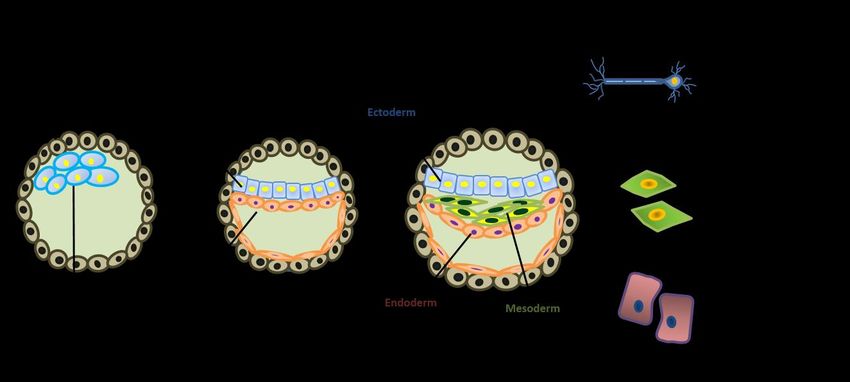
(Figure 1). Gastrulation involves the formation of three describes the inconsistency among studies in using the
germ layers (ectoderm, endoderm and mesoderm) from markers for identifying endodermal, mesodermal, and
pluripotent epiblast cells. Each layer, which is no longer ectodermal cells. Figure 2 represents the detailed
pluripotent, will later give rise to specific body organs and methodology used to select the early differentiation
tissues (Muhr and Ackerman, 2020). markers of hPSCs that have been used in the last 5 years in
In vitro generation of any differentiated cell type from the literature. However, the focus of this review is directedInt. Res. J. Med. Biomed. Sci. 12
Figure 1: Schematic diagram showing the process of gastrulation. After implantation, the inner cell mass differentiates to
form the bilaminar germ disc, which consists of the epiblast and the hypoblast. The bilaminar germ disk then
differentiates further into a trilaminar embryo consisting of the three distinct germ layers: ectoderm, mesoderm, and
endoderm. Cells of each germ layer eventually give rise to specific tissue types in the human body.
Figure 2. Methodology used to select the early differentiation markers of human pluripotent stem cells.Baba et al. 13
Table 1. The ectoderm markers cited in scientific literature in the
last 5 years
Marker Official gene name
CDH1 Cadherin 1 (E-cadherin)
DCX Doublecortin
FGF5 Fibroblast growth factor 5
IGF1 Insulin like growth factor 1
NCAM1 Neural cell adhesion molecule 1
NES Nestin
NEUROD1 Neuronal differentiation 1
OTX1 Orthodenticle homeobox 1
OTX2 Orthodenticle homeobox 2
PAX6 Paired box 6
SOX1 SRY-box transcription factor 1
TUBB3 Tubulin beta 3 class III
ZIP2 Zinc finger E-box binding homeobox 2
to the most commonly used markers. markers, only PAX6 was shown to be reliably used as an
ectoderm differentiation marker (Kuang et al., 2019).
Ectoderm Markers
Nestin (NES)
Ectoderm is the outermost layer of the three germ layers,
which goes on to form the central and peripheral nervous NES is an essential intermediate filament protein, which is
systems (CNS and PNS respectively), the epidermis of the mainly expressed by neural progenitor cells in the
skin and neural crest derivatives (Kiecker et al., 2016). mammalian CNS (Neradil and Veselska, 2015). NES is
Early in embryogenesis, the anterior part of the epiblast among the most commonly used ectoderm differentiation
differentiates to form the ectoderm germ layer. Of note is markers (Table 1). Before neurogenesis, NES is expressed
the fact that the primitive streak does not traverse this in almost all ectodermal neuroepithelial cells (Neradil and
region, unlike epiblast cells that form mesoderm and Veselska, 2015). NES, however, is not exclusively expressed
endoderm. Therefore, the culture medium used for the in neuroectodermal cells. A recent study found a similarity
induction of ectoderm is devoid of serum and any primitive in the level of NES expression between iPSCs and
streak inducers; hence, the process is known as the default differentiated ectoderm cells. Accordingly, NES was
pathway. Inhibitors of ectoderm formation in media include regarded as an unreliable early ectoderm differentiation
the Activin/Nodal, bone morphogenetic protein (BMP) and marker (Kuang et al., 2019).
WNT molecules (Leung et al., 2013; Liu et al., 2018;
Williams et al., 2012). These signaling molecules are SRY-box Transcription Factor 1 (SOX1)
switched off during ectoderm induction in the developing
embryo. The differentiating pluripotent stem cells produce SOX1 is a member of the sex-determining region Y-box B1
endogenous fibroblast growth factors (FGFs) on which the (SOXB1) subfamily (Feng et al., 2014). SOX1 has been used
cells depend to induce neuroectoderm (Zheng et al., 2010). as an early marker of ectoderm cells in several studies
However, when ectoderm is formed, the activation of the (Table 1). The in vivo expression of SOX1 is associated with
BMP-signaling pathway promotes epidermal derivatives, the proliferation of neuroepithelial cells, whereas the exit of
whereas it blocks neural subtype specifications (Bertero et neural cells from mitosis correlates with the subsequent
al., 2015, Feng et al., 2014). Several markers have been used downregulation of SOX1 (Imai et al., 2017). A strong
to identify induced ectoderm cells from hPSCs in the last 5 correlation between the level of SOX1 expression and
years. However, PAX6, NES, SOX1 and TUBB3, which are neural linage specification has also been demonstrated in
discussed below, have been the most commonly used human iPSCs. Researchers successfully isolated cells with a
markers for ectoderm differentiation (Table 1). neural phenotype after inhibiting BMP and observed a
significant increase in SOX1 levels (Zhang et al., 2018).
Paired Box Protein 6 (PAX6)
Cytoskeletal Tubulin Beta 3 Class III Protein (TUBB3)
PAX6, also called oculorhombin, is a highly conserved
transcription factor which in humans is encoded by the TUBB3 is widely used as a marker for early neuroectoderm
PAX6 gene (Thakurela et al., 2016). PAX6 is a pivotal differentiation (Table 1). TUBB3 expression, however, is
differentiation marker for the ectoderm germ layer lineage not limited to ectoderm and neuronal cells. It has been
(Cvekl and Callaerts, 2017), and has been used as an reported that TUBB3 is highly expressed in the developing
ectoderm marker in several studies (Table 1). A recent neural crest cells-derived melanocytes (Sebastian et al.,
study characterized different cell types with commonly 2017). Based on transcriptomic analyses, researchers found
used pluripotent and lineage specific markers. Among these no significant increase of TUBB3 transcripts in ectodermInt. Res. J. Med. Biomed. Sci. 14
Table 2. The endoderm markers cited in scientific literature in the last
5 years
Marker Official gene name
CXCR4 C-X-C motif chemokine receptor 4
CDX2 Caudal type homeobox 2
ECD Ecdysoneless cell cycle regulator
EOMES Eomesodermin
FOXA2 Forkhead box A2
GATA4 GATA binding protein 4
GATA6 GATA binding protein 6
GSC Goosecoid homeobox
HNF1B HNF1 homeobox B
KIT KIT proto-oncogene, receptor tyrosine kinase
MIXL1 Mix paired-like homeobox
SOX17 SRY-box transcription factor 17
SOX7 SRY-box transcription factor 7
AFP Alpha fetoprotein
differentiated cells compared to pluripotent stem cells associated with abnormal endoderm formation (Ogaki et
(Daily et al., 2017; Kuang et al., 2019). Accordingly, it was al., 2016).
recommended that TUBB3 might not be a reliable ectoderm
marker in stem cell trilineage validation studies (Kuang et Forkhead Box A2 (FOXA2)
al., 2019). Therefore, careful assessment of TUBB3
expression as an ectoderm marker or a neuronal marker The DNA-binding protein FOXA2 belongs to the forkhead
should be considered. box superfamily (Li et al., 2017). FOXA2 is highly expressed
in the endoderm cells of the developing embryo (Gosalia et
Endoderm Markers al., 2015). Several studies have used FOXA2 as an endoderm
marker during the in vitro differentiation of hESCs (Table
Endoderm is the mass of cells that is located internally 2). Besides, FOXA2 is necessary for the formation of
within ectoderm and mesoderm germ layers. Definitive numerous human tissues of endoderm origin. For instance,
endoderm differentiates into multiple organs during the role of FOXA2 during human pancreas development has
embryo development, including urinary, respiratory and been established, where FOXA2 knockout pluripotent stem
gastrointestinal systems, along with many glands in the cells failed to differentiate to pancreatic cells (Lee et al.,
endocrine system (Kiecker et al., 2016). 2019). Moreover, a recent study uncovered a novel role of
The efficiency of endoderm induction from hPSCs is FOXA2 at the early stages of embryogenesis. The
monitored through changes in gene expression patterns researchers found that FOXA2, hepatocyte nuclear factor 4
and/or expression of cell surface markers. Induction of alpha (HNF4A) and E1A binding protein p300 (EP300) are
efficient definitive endoderm lineage is the first step for the three most important genes for the first division of the
efficient differentiation into functional endoderm fertilized egg (Godini and Fallahi, 2019). In adults, FOXA2
derivatives (Holtzinger et al., 2015). In culture, endoderm has been detected in tissues derived from endoderm (liver)
cell derivation from human embryonic stem cells (hESCs) (Warren et al., 2020) and mesoderm (uterus) (Kelleher et
depends on Activin signaling (Wang et al., 2015). al., 2017). Since FOXA2 is expressed early in the dividing
Translating these findings from the developing embryo has zygote and also in non-endodermal derived tissues, the
allowed researchers to differentiate Activin-induced reliability of FOXA2 as an early endoderm marker needs
endoderm cells to respiratory- and digestive-related organs more attention.
such as the liver, lungs, stomach and pancreas (Luo et al.,
2017, Yiangou et al., 2018). SOX17, FOXA2, CXRC4 and C-X-C Motif Chemokine Receptor 4 (CXCR4)
GATA4 have been the most commonly used endoderm
markers (Table 2). CXCR4 is another marker commonly used for assessing the
efficiency of endoderm induction from hPSCs (Table 1).
SRY-box Transcription Factor 17 (SOX17) Multiple studies depend on CXCR4 expression to identify
proper endoderm populations, whereas additional studies
SOX17 is a pioneer marker of generated definitive evaluate endoderm induction by co-expression of CXCR4
endoderm cells from a variety of hPSCs (Table 2). The with other markers, mainly CD117 and epithelial cell
expression of SOX17 is critical for inducing stable definitive adhesion molecule (EPCAM) (Diekmann et al., 2019,
endoderm cells from hESCs (Irie et al., 2015). Expression of Holtzinger et al., 2015, Zhong et al., 2017). A study that
SOX17 has been detected in hESCs differentiating toward identified HDE1 as an endoderm-specific antibody
endodermal fates following the treatment of the cells with indicated that enrichment of definitive endoderm from
Activin A (Luo et al., 2017). Mutations in SOX17 are mixed-lineage populations could not be obtained by onlyBaba et al. 15
Table 3. The mesoderm markers cited in scientific literature in the last 5
years
Markers Official gene name
BMP4 Bone morphogenetic protein 4
BRA Brachyury
CDX2 Caudal type homeobox 2
DCN Decorin
DES Desmin
EOMES Eomesodermin
GATA2 GATA binding protein 2
GATA4 GATA binding protein 4
GSC Goosecoid homeobox
HAND1 Heart and neural crest derivatives expressed 1
IGF2 Insulin like growth factor 2
KDR Kinase insert domain receptor
MESP1 Mesoderm posterior bHLH transcription factor 1
MIXL1 Mix paired-like homeobox
MSX1 Msh homeobox 1
NCAM1 Neural cell adhesion molecule 1
NODAL Nodal growth differentiation factor
PDGFB Platelet derived growth factor subunit B
PDGFRA Platelet derived growth factor receptor alpha
SMN1 Survival of motor neuron 1
TBXT T-box transcription factor T
TNNT2 Troponin T2, cardiac type
TWIST1 Twist family bHLH transcription factor 1
WT1 WT1 transcription factor
using CXCR4. This indication was based on the analysis, (Kiecker et al., 2016). BMP4 activates FGF and
which revealed higher levels of non-endodermal genes, TGFB/Activin/Nodal pathways; the inhibition of these
such as POU class 5 homeobox 1 (OCT4) (pluripotency), Mix signaling cascades results in repression of the BMP4
paired-like homeobox (MIXL1) (primitive streak) function to induce mesoderm (Gordeeva, 2019).
and CD56 (mesoderm) in the HDE1-CXCR4+ cells than in the Manipulation of different pathways can induce
HDE1+CXCR4+cells (Holtzinger et al., 2015). Taken subpopulations derived from mesoderm. For example,
together, lack of validation protocols results in such cardiac mesoderm can be efficiently obtained from hESCs
inconsistency upon using CXCR4 as an early endoderm when Activin is added with BMP4 (Sa et al., 2014). BRA,
marker. KDR and MIXL1 are widely used as early markers of
mesoderm induction (Table 3).
GATA4
Brachyury (BRA)
GATA binding protein 4 (GAT4) is a zinc-finger
transcription factor that binds to the DNA sequence "GATA" The transcription factor BRA is an early marker of
(Yuan et al., 2014). GATA4 first appears, as a pioneer factor, gastrulation and lineage specification in humans (Faial et
during the embryonic stage of development in the al., 2015). BRA is the key maker of the primitive streak and
endoderm layer (Fisher et al., 2017, Tiyaboonchai et al., is highly expressed in embryos as the mesoderm layer is
2017). Therefore, it has been used as an early endoderm formed (Faial et al., 2015, Zhou et al., 2018). Therefore, BRA
marker in multiple studies (Table 2). The differentiation of is widely used as marker to identify mesoderm cells
hPSCs has provided evidence that GATA6 regulates GATA4 derived from pluripotent cells (Table 3). In cancer research,
during the generation of the definitive endoderm (Fisher et BRA has been implicated in epithelial-mesenchymal
al., 2017). GATA4 is crucial for the development and transition (EMT) and tumor progression to metastasis;
function of several endoderm-derived tissues, although, its therefore, it has been proposed as a candidate for human
expression has also been identified in tissues derived from cancer immunotherapy (Hamilton et al., 2017).
the mesoderm, such as the heart (Tiyaboonchai et al.,
2017). Kinase Insert Domain Receptor (KDR)
Mesoderm Markers KDR, also known as VEGFR-2, is an endothelial cell growth
factor receptor tyrosine kinase. KDR plays a significant role
Mesoderm is the middle germ layer between ectoderm and in early vascular development and the regulation of
endoderm and forms diverse tissues, including the skeletal vascular permeability (Modi and Kulkarni, 2019). KDR
and muscular systems, kidney, cartilages and blood vessels expression during gastrulation marks the developingInt. Res. J. Med. Biomed. Sci. 16
mesoderm (Scialdone et al., 2016). Therefore, several green fluorescent protein reporter embryonic stem cell
studies have used KDR as a marker to isolate mesoderm line engineered using TALEN-based genome editing. Stem
cells from hESCs (Table 3). Mesoderm populations that Cell Research. 17(1): 93-96.
express KDR represent a valuable source for generating Bertero A, Madrigal P, Galli A, Hubner NC, Moreno I, Burks
hematopoietic and endothelial lineages (Sriram et al., D, Brown S, Pedersen RA, Gaffney D, Mendjan S, Pauklin S,
2015). In adults, KDR might be involved in mitosis, vascular Vallier L (2015). Activin/nodal signaling and NANOG
permeability and angiogenesis (Chen et al., 2019). orchestrate human embryonic stem cell fate decisions by
Overexpression of KDR is associated with endothelial cell controlling the H3K4me3 chromatin mark. Genes Dev.
malignancies, for which a wide range of potential KDR 29(7): 702-17.
inhibitors are reported for the management of cancer (Modi Chen JX, Yi XJ, Gu PL, Gao SX (2019). The role of KDR in
and Kulkarni, 2019). intrauterine adhesions may involve the TGF-β1/Smads
signaling pathway. Braz J Med Biol Res. 52(10): e8324.
Mix Paired-like Homeobox (MIXL1) Cvekl A, Callaerts P (2017). PAX6: 25th anniversary and
more to learn. Exp Eye Res. 156(10-21.
MIXL1 is another marker used for identifying the Daily K, Ho Sui SJ, Schriml LM, Dexheimer PJ, Salomonis N,
differentiation of pluripotent cells into mesoderm cells Schroll R, Bush S, Keddache M, Mayhew C, Lotia S,
(Table 3). MIXL1 is markedly needed in mesoderm Perumal TM, Dang K, Pantano L, Pico AR, Grassman E,
formation in early development (Wolfe and Downs, 2014). Nordling D, Hide W, Hatzopoulos AK, Malik P, Cancelas JA,
However, MIXL1 is required for the development of Lutzko C, Aronow BJ, Omberg L (2017). Molecular,
endoderm, for which studies have used it as an endoderm phenotypic, and sample-associated data to describe
marker (Table 2). MIXL1 was also shown to be expressed in pluripotent stem cell lines and derivatives. Sci Data.
mesendodermal precursor cells (Alexeeva et al., 2016). 4(170030.
Therefore, MIXL1 might not be a useful candidate to be Dakhore S, Nayer B, Hasegawa K (2018). Human
used as a mesoderm marker. Pluripotent Stem Cell Culture: Current Status, Challenges,
and Advancement. Stem Cells Int. 2018(7396905.
Conclusion Diekmann U, Wolling H, Dettmer R, Niwolik I, Naujok O,
Buettner FFR (2019). Chemically defined and xenogeneic-
A wide range of markers have been reported in the free differentiation of human pluripotent stem cells into
literature as indicative of the three germ layers. However, definitive endoderm in 3D culture. Sci Rep. 9(1): 996.
there are inconsistencies between these studies due to the Faial T, Bernardo AS, Mendjan S, Diamanti E, Ortmann D,
lack of validation protocols and a standard set of markers Gentsch GE, Mascetti VL, Trotter MW, Smith JC, Pedersen
for the early trilineage specification. Based on the recent RA (2015). Brachyury and SMAD signalling
literature, the most commonly used ectoderm markers NES collaboratively orchestrate distinct mesoderm and
and TUBB3, endoderm markers FOXA2, CXCR4 and GATA4, endoderm gene regulatory networks in differentiating
and mesoderm marker MIXL1 are also found to be human embryonic stem cells. Development. 142(12):
expressed in different germ layer cells. This may render 2121-35.
them unreliable for the use as early differentiation markers. Feng N, Han Q, Li J, Wang S, Li H, Yao X, Zhao RC (2014).
The current review highlights the need for further Generation of highly purified neural stem cells from
development of validation protocols to organize the use of human adipose-derived mesenchymal stem cells by Sox1
early differentiation markers, which may help generate activation. Stem Cells Dev. 23(5): 515-529.
efficient cells to be used for treating degenerative diseases Fisher JB, Pulakanti K, Rao S, Duncan SA (2017). GATA6 is
and in drug screening. Investigators should be cautious in essential for endoderm formation from human
selecting the early differentiation markers until definitive pluripotent stem cells. Biol Open. 6(7): 1084-1095.
standard assays have been established. Godini R, Fallahi H (2019). Dynamics changes in the
transcription factors during early human embryonic
Author Disclosure Statement development. J Cell Physiol. 234(5): 6489-6502.
Gordeeva O (2019). TGFβ Family Signaling Pathways in
The author declares no competing financial interests exist. Pluripotent and Teratocarcinoma Stem Cells' Fate
Decisions: Balancing Between Self-Renewal,
Acknowledgment Differentiation, and Cancer. Cells. 8(12):
Gosalia N, Yang R, Kerschner JL, Harris A (2015). FOXA2
The author would like to thank Dr. Sherif Elsharkawy, regulates a network of genes involved in critical functions
Clinical Lecturer in Prosthodontics, Centre for Oral, Clinical, of human intestinal epithelial cells. Physiol Genomics.
and Translational Sciences, Faculty of Dentistry, Oral and 47(7): 290-7.
Craniofacial Sciences, King's College London; for his Hamilton DH, David JM, Dominguez C, Palena C (2017).
constructive criticism of the manuscript. Development of Cancer Vaccines Targeting Brachyury, a
Transcription Factor Associated with Tumor Epithelial-
REFERENCES Mesenchymal Transition. Cells Tissues Organs. 203(2):
128-138.
Alexeeva V, D'Souza SL, Schaniel C (2016). A human MIXL1 Holtzinger A, Streeter PR, Sarangi F, Hillborn S, Niapour M,Baba et al. 17 Ogawa S, Keller G (2015). New markers for tracking Understanding and Future Directions. Stem Cells endoderm induction and hepatocyte differentiation from International. 2016(9451492. human pluripotent stem cells. Development. 142(24): Sa S, Wong L, McCloskey KE (2014). Combinatorial 4253-65. fibronectin and laminin signaling promote highly efficient Imai KS, Hikawa H, Kobayashi K, Satou Y (2017). Tfap2 and cardiac differentiation of human embryonic stem cells. Sox1/2/3 cooperatively specify ectodermal fates in Biores Open Access. 3(4): 150-61. ascidian embryos. Development. 144(1): 33-37. Scialdone A, Tanaka Y, Jawaid W, Moignard V, Wilson NK, Irie N, Weinberger L, Tang WW, Kobayashi T, Viukov S, Macaulay IC, Marioni JC, Göttgens B (2016). Resolving Manor YS, Dietmann S, Hanna JH, Surani MA (2015). early mesoderm diversification through single-cell SOX17 is a critical specifier of human primordial germ expression profiling. Nature. 535(7611): 289-293. cell fate. Cell. 160(1-2): 253-68. Sebastian A, Volk SW, Halai P, Colthurst J, Paus R, Bayat A Kelleher AM, Peng W, Pru JK, Pru CA, DeMayo FJ, Spencer (2017). Enhanced Neurogenic Biomarker Expression and TE (2017). Forkhead box a2 (FOXA2) is essential for Reinnervation in Human Acute Skin Wounds Treated by uterine function and fertility. Proceedings of the National Electrical Stimulation. J Invest Dermatol. 137(3): 737- Academy of Sciences. 114(6): E1018-E1026. 747. Kiecker C, Bates T, Bell E (2016). Molecular specification of Sriram G, Tan JY, Islam I, Rufaihah AJ, Cao T (2015). germ layers in vertebrate embryos. Cell Mol Life Sci. Efficient differentiation of human embryonic stem cells to 73(5): 923-47. arterial and venous endothelial cells under feeder- and Kuang YL, Munoz A, Nalula G, Santostefano KE, Sanghez V, serum-free conditions. Stem Cell Research & Therapy. Sanchez G, Terada N, Mattis AN, Iacovino M, Iribarren C, 6(1): 261. Krauss RM, Medina MW (2019). Evaluation of commonly Thakurela S, Tiwari N, Schick S, Garding A, Ivanek R, used ectoderm markers in iPSC trilineage differentiation. Berninger B (2016). Mapping gene regulatory circuitry of Stem Cell Res. 37(101434. Pax6 during neurogenesis. 2(15045. Lee K, Cho H, Rickert RW, Li QV, Pulecio J, Leslie CS, Tiyaboonchai A, Cardenas-Diaz FL, Ying L, Maguire JA, Sim Huangfu D (2019). FOXA2 Is Required for Enhancer X, Jobaliya C, Gagne AL, Kishore S, Stanescu DE, Hughes N, Priming during Pancreatic Differentiation. Cell Rep. De Leon DD, French DL, Gadue P (2017). GATA6 Plays an 28(2): 382-393.e7. Important Role in the Induction of Human Definitive Leung AW, Kent Morest D, Li JY (2013). Differential BMP Endoderm, Development of the Pancreas, and signaling controls formation and differentiation of Functionality of Pancreatic β Cells. Stem Cell Reports. multipotent preplacodal ectoderm progenitors from 8(3): 589-604. human embryonic stem cells. Dev Biol. 379(2): 208-20. Wang Z, Li W, Chen T, Yang J, Wen Z, Yan X, Shen T, Liang R Li J, Dantas Machado AC, Guo M, Sagendorf JM, Zhou Z, Jiang (2015). Activin A can induce definitive endoderm L, Chen X, Wu D, Qu L, Chen Z, Chen L, Rohs R, Chen Y differentiation from human parthenogenetic embryonic (2017). Structure of the Forkhead Domain of FOXA2 stem cells. Biotechnology Letters. 37(8): 1711-1717. Bound to a Complete DNA Consensus Site. Biochemistry. Warren I, Maloy M, Guiggey D, Ogoke O, Groth T, Mon T, 56(29): 3745-3753. Meamardoost S, Liu X, Szeglowski A, Thompson R, Chen P, Liu C, Wang R, He Z, Osteil P, Wilkie E, Yang X, Chen J, Cui G, Paulmurugan R, Parashurama N (2020). Foxa1 and Foxa2 Guo W, Chen Y, Peng G, Tam PPL, Jing N (2018). together control developmental gene regulatory Suppressing Nodal Signaling Activity Predisposes networks, and differentiation genes, in both human stem- Ectodermal Differentiation of Epiblast Stem Cells. Stem cell derived liver progenitors and in a human liver cell Cell Reports. 11(1): 43-57. line: evidence of a collapse of human liver differentiation. Liu LP, Zheng YW (2019). Predicting differentiation bioRxiv.)2020.06.01.128108. potential of human pluripotent stem cells: Possibilities Williams M, Burdsal C, Periasamy A, Lewandoski M, and challenges. World J Stem Cells. 11(7): 375-382. Sutherland A (2012). Mouse primitive streak forms in Luo X, Wang H, Leighton J, O'Sullivan M, Wang P (2017). situ by initiation of epithelial to mesenchymal transition Generation of endoderm lineages from pluripotent stem without migration of a cell population. Dev Dyn. 241(2): cells. Regen Med. 12(1): 77-89. 270-83. Modi SJ, Kulkarni VM (2019). Vascular Endothelial Growth Wolfe AD, Downs KM (2014). Mixl1 localizes to putative Factor Receptor (VEGFR-2)/KDR Inhibitors: Medicinal axial stem cell reservoirs and their posterior descendants Chemistry Perspective. Medicine in Drug Discovery. in the mouse embryo. Gene Expr Patterns. 15(1): 8-20. 2(100009. Yiangou L, Ross ADB, Goh KJ, Vallier L (2018). Human Muhr J, Ackerman KM (2020). Embryology, Gastrulation. Pluripotent Stem Cell-Derived Endoderm for Modeling StatPearls.) Development and Clinical Applications. Cell Stem Cell. Neradil J, Veselska R (2015). Nestin as a marker of cancer 22(4): 485-499. stem cells. Cancer Sci. 106(7): 803-11. Yuan X, Xia L, Dong X, Hu S, Zhang Y, Ding F, Liu H, Li L, Ogaki S, Omori H, Morooka M, Shiraki N, Ishida S, Kume S Wang J (2014). Transcription factors GATA-4 and GATA- (2016). Late stage definitive endodermal differentiation 6: molecular characterization, expression patterns and can be defined by Daf1 expression. BMC Developmental possible functions during goose (Anser cygnoides) follicle Biology. 16(1): 19. development. J Reprod Dev. 60(2): 83-91. Romito A, Cobellis G (2016). Pluripotent Stem Cells: Current Zakrzewski W, Dobrzyński M, Szymonowicz M, Rybak Z
Int. Res. J. Med. Biomed. Sci.   18

  (2019). Stem cells: past, present, and future. Stem Cell
  Res Ther. 10(1): 68.
Zhang M, Ngo J, Pirozzi F, Sun YP, Wynshaw-Boris A (2018).
  Highly efficient methods to obtain homogeneous dorsal
  neural progenitor cells from human and mouse
  embryonic stem cells and induced pluripotent stem cells.
  Stem Cell Res Ther. 9(1): 67.
Zheng Z, de Iongh RU, Rathjen PD, Rathjen J (2010). A
  requirement for FGF signalling in the formation of
  primitive streak-like intermediates from primitive
  ectoderm in culture. PLoS One. 5(9): e12555.
Zhong W, Lai Y, Yu T, Xia ZS, Yuan YH, Ouyang H, Shan TD,
  Chen QK (2017). Wnt and Nodal signaling simultaneously
  induces definitive endoderm differentiation of mouse
  embryonic stem cells. Rom J Morphol Embryol. 58(2):
  527-535.
Zhou J, Plagge A, Murray P (2018). Functional comparison
  of distinct Brachyury(+) states in a renal differentiation
  assay. Biol Open. 7(5).
You can also read