Evaluation of epithelial progenitor cells and growth factors in a preclinical model of wound healing induced by mesenchymal stromal cells
←
→
Page content transcription
If your browser does not render page correctly, please read the page content below
Bioscience Reports (2020) 40 BSR20200461
https://doi.org/10.1042/BSR20200461

Research Article

Evaluation of epithelial progenitor cells and growth
factors in a preclinical model of wound healing
induced by mesenchymal stromal cells
Giselle Ramos-Gonzalez1 , Olga Wittig1 , Dylana Diaz-Solano1 , Lianeth Salazar2 , Carlos Ayala-Grosso1 and

                                                                                                                                                                                           Downloaded from http://portlandpress.com/bioscirep/article-pdf/40/7/BSR20200461/888680/bsr-2020-0461.pdf by guest on 25 December 2020
   Jose E. Cardier1
1 Unidad   de Terapia Celular - Laboratorio de Patologı́a Celular y Molecular, Instituto Venezolano de Investigaciones Cientı́ficas (IVIC), Apartado 21827, Caracas 1020-A, Venezuela;
2 Servicio  de Cirugı́a Plastica, Hospital de la Cruz Roja, Caracas 1080, Venezuela
Correspondence: José E. Cardier (jacardier@gmail.com, jcardier@ivic.gob.ve)

                                           Background: Skin wounds continue to be a global health problem. Several cellular ther-
                                           apy protocols have been used to improve and accelerate skin wound healing. Here, we
                                           evaluated the effect of transplantation of mesenchymal stromal cells (MSC) on the wound
                                           re-epithelialization process and its possible relationship with the presence of epithelial pro-
                                           genitor cells (EPC) and the expression of growth factors. Methods: An experimental wound
                                           model was developed in C57BL/6 mice. Human MSCs seeded on collagen membranes
                                           (CM) were implanted on wounds. As controls, animals with wounds without treatment or
                                           treated with CM were established. Histological and immunohistochemical (IH) studies were
                                           performed at day 3 post-treatment to detect early skin wound changes associated with the
                                           presence of EPC expressing Lgr6 and CD34 markers and the expression of keratinocyte
                                           growth factor (KGF) and basic fibroblast growth factor (bFGF). Results: MSC transplanta-
                                           tion enhanced skin wound re-epithelialization, as compared with controls. It was associated
                                           with an increase in Lgr6+ and CD34+ cells and the expression of KGF and bFGF in the wound
                                           bed. Conclusion: Our results show that cutaneous wound healing induced by MSC is as-
                                           sociated with an increase in EPC and growth factors. These preclinical results support the
                                           possible clinical use of MSC to treat cutaneous wounds.

                                         Introduction
                                         Cutaneous wound healing comprises several stages; one of them is the re-epithelialization, which is funda-
                                         mental for tissue wound repair [1] It initiates with the activation, migration and proliferation of epithelial
                                         progenitor cells (EPC) located in the interfollicular epidermis (IFE) and hair follicles (HFs) [2]. In ad-
                                         dition, several growth factors (including keratinocyte growth factor (KGF) and basic fibroblast growth
                                         factor (bFGF)) and cytokines participate in the repair process by promoting proliferation, differentiation
                                         and migration of EPC from healthy skin to the wound center [3].
                                            Although numerous treatments have been applied for improving wound healing, it continues being
Received: 25 February 2020
                                         a serious medical problem in many patients. Recently, cellular therapy has been proposed for inducing
Revised: 28 May 2020                     skin wound repair [4]. Numerous studies have focused on the use of mesenchymal stromal cells (MSC)
Accepted: 08 June 2020                   transplantation for inducing wound repair [5–8].
                                            MSC constitute a population of cells with a multipotential capacity of differentiation [9–11]. There is
Accepted Manuscript online:
15 July 2020                             evidence showing that MSC participate in the process of wound healing [12–14]. It has been suggested
Version of Record published:             that MSC may induce cutaneous wound repair by regulating the inflammatory and immune responses
22 July 2020                             [15,16] These effects are mediated by paracrine signals generated from MSC [17,18], which may induce

© 2020 The Author(s). This is an open access article published by Portland Press Limited on behalf of the Biochemical Society and distributed under the Creative Commons Attribution   1
License 4.0 (CC BY).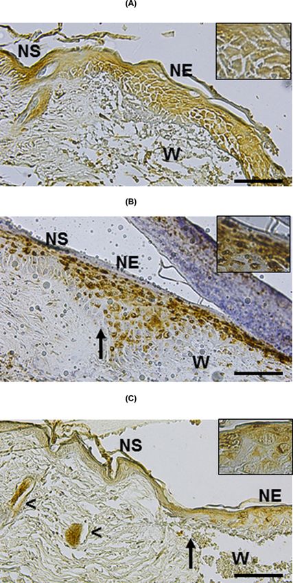
Bioscience Reports (2020) 40 BSR20200461
                                                                                                                                         https://doi.org/10.1042/BSR20200461

            migration, proliferation and differentiation of skin EPC [3,19]. Importantly, due to the low expression of MHC class
           I and II and no expression of costimulatory molecules, the possibility of rejection in allogeneic or xenogeneic models
           of transplantation is very low [20,21]. Based on this knowledge, the use of MSC as a potential therapeutic strategy to
           induce cutaneous wound healing is highly considered in regenerative medicine. In this work, we used an animal skin
           wound model to evaluate the capacity of human MSC to induce wound healing and its possible relationship with the
           presence of EPC and the expression of growth factors.

           Materials and methods
           Animals
           Male C57BL/6 mice (8-week-old) were obtained from the IVIC Laboratory Animal Center and maintained on a
           standard laboratory diet and housed in a controlled environment. All animal experimentation was performed at the

                                                                                                                                                                                           Downloaded from http://portlandpress.com/bioscirep/article-pdf/40/7/BSR20200461/888680/bsr-2020-0461.pdf by guest on 25 December 2020
           IVIC following institutional guidelines and the National Institutes of Health guide for the care and use of laboratory
           animals. The study protocol was approved by the Animal Committee of IVIC (COBIANIM2014-04).

           Reagents and culture medium
           FITC or phycoerythrin (PE)-conjugated monoclonal antibodies anti-human CD73 and CD90 were from BD Bio-
           sciences (Franklin Lakes, NJ, U.S.A.). Monoclonal antibodies anti-mouse CD34 were from Biolegend (San Diego,
           CA, U.S.A.). Polyclonal antibody anti-human Lgr6 was from Novus Biologicals (Littleton, CO, U.S.A.). Polyclonal
           antibodies anti-human KGF and bFGF) were from R&D Systems (Minneapolis, MN, U.S.A.). Atelocollagen mem-
           branes (CM) were purchased from Cosmo Bio (Tokyo, Japan). α-MEM medium was from Life Technologies (U.S.A.)
           and Chang medium was from Irvine Scientific (U.S.A.).

           Isolation and culture of bone marrow MSC
           MSC used in this work were from healthy patients treated for bone regeneration, due to pseudarthrosis secondary to
           a fracture [22,23], who authorized the use of these cells by signing informed consent. These cells were from bone mar-
           row and isolated from the posterior iliac crest of patients [22,23]. MSC were cultured, expanded and stored at −70◦ C
           until its use. For the present study, MSCs were thawed and expanded in αMEM-Chang medium until becoming near
           confluent.

           Phenotypic and functional analysis
           The expression of MSC markers (CD73 and CD90) were evaluated by flow cytometry. Data collection and analysis
           of the fluorescent intensities were made using a BD Acurri™ C6 (Beckton Dickinson, U.S.A.). The multipotential
           capacity of MSC was examined following previous methodology using osteogenic, chondrogenic and adipogenic
           differentiation media [10,22,23].

           Transplantation of MSC on cutaneous wounds
           MSC were seeded (3 × 10⁵ cells) on transwells containing CM for six-well culture plates (Atelo Cell, Cosmo Bio.,
           Tokyo, Japan). The cells were maintained in culture until becoming near confluent. A skin wound model was per-
           formed in C57BL/6 mice. For this purpose, mice were anesthetized with a cocktail of Ketamine/Xylazine (100 mg/kg
           and 7.5 mg/kg body weight, respectively) and hair was removed from the dorsal surface area (Figure 1A). A 4-mm
           full thickness excisional skin wound was made on the dorsal area using a sterile disposable biopsy punch (Healthlink,
           Jacksonville, FL, U.S.A.) (Figure 1B). Inserts were removed from the plates and each CM containing MSC (MSC/CM)
           were cut in circles of approximately 4 mm in diameter and immediately applied on to the wound beds. A sterile
           band-aid was placed and centered over the wound. A transparent film dressing (Tegaderm 3M) was placed over the
           wound and an immediate-bonding adhesive was used to better fix the dressing to the skin (Figure 1C). As controls,
           groups of mice with wounds were uncovered or covered with CM without cells. The animals were housed individu-
           ally. After 3 days of implantation, mice were killed by cervical dislocation and wounds were photographed. Wound
           area was determined before (d0) and after wounding (d3) by using ImageJ v1.48 (http://imagej.nih.gov/ij) program.
           It was expressed as the percentage of wound closure at day 3 as compared with d0 in each group (% of wound closure
           = [wound area d0 − wound area d3/area d0] × 100). Samples from the wounds were collected and included in paraf-
           fin for histological and immunohistochemical (IH) analysis. We evaluated early cellular changes occurring at day 3
           after MSC transplantation because re-epithelialization of a skin wound of this size occurred at approximately day 7.
           Infiltrating polymorphonuclear cells were examined by two independent observers at low powered field microscopy.
           Data were reiterated by using a semiquantitative cross-scoring system (low = +, moderate = ++, high = +++).

2   © 2020 The Author(s). This is an open access article published by Portland Press Limited on behalf of the Biochemical Society and distributed under the Creative Commons Attribution
    License 4.0 (CC BY).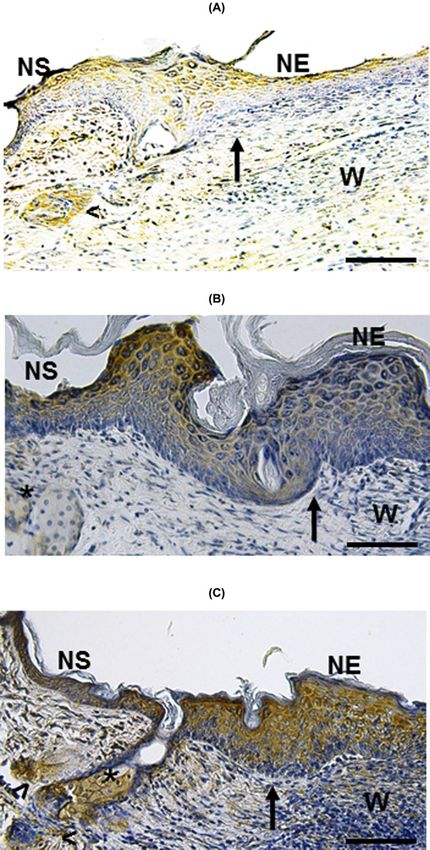
Bioscience Reports (2020) 40 BSR20200461
https://doi.org/10.1042/BSR20200461

                                                                                                                                                                                           Downloaded from http://portlandpress.com/bioscirep/article-pdf/40/7/BSR20200461/888680/bsr-2020-0461.pdf by guest on 25 December 2020
Figure 1. Schematic representation of the skin wound model
C57BL/6 mice were anesthetized and hair was removed from the dorsal surface area (A). A 4-mm full thickness excisional skin
wound was created on the dorsal area (B). A sterile band-aid was placed so that the wound was centered within it, and a transparent
film dressing (Tegaderm 3M) was placed over the wound using an immediate-bonding adhesive to better fix the dressing to the
skin (C).

Histological and IH analysis of skin wound
Samples of skin wounds, including normal skin (NS), were fixed in 10% formalin solution, embedded in paraffin
and sectioned at 4-μm-thick. Hematoxylin and Eosin (H&E) staining was performed. For immunostaining, paraffin
sections were dewaxed, rehydrated and incubated with antibodies against Lgr6, CD34, KGF and bFGF.

Wound analysis
Digital photographs from wounds were taken at days 0 and 3 post-wound. Percentage of re-epithelialization was
determined using an analysis program (ImageJ). Percentage re-epithelialization was defined as the distance traveled
by both epithelial tongues (long dn1 and long dn2 ) divided by the distance needed to travel to fully re-epithelialize
the wound (long dn0) × 100: % re-epithelialization = [long dn1 + long dn2 /long d0] × 100).

Statistical analysis
Results are reported as mean + − standard error. We tested the data from the experiments for statistical significance
using the Student’s t test for comparisons between groups. Differences were considered statistically significant at
P≤0.05.

Results
Culture, phenotypical and functional characterization of MSC
Cryopreserved MSC were thawed and cultured in α-MEM Chang medium. They showed fibroblast-like morphology
in culture (Figure 2A) and expressed the typical MSC markers CD73 and CD90 (Figure 2B). By culturing in differenti-
ation media, they showed their multipotential capacity of differentiation to adipogenic, osteogenic and chondrogenic
cells (Figure 2C–E, respectively).

Implant of MSC on cutaneous wounds
MSC were seeded on transwell inserts with CM (Figure 3A). After 72 h, cells grew reaching 100% confluence showing
a fibroblastoid-like morphology on the CM (Figure 3B). MSC/CM were removed from the inserts and cut to the
size of the wound (Figure 3C), and turned MSC side downward on to the wound bed (Figure 3D). The implanted

© 2020 The Author(s). This is an open access article published by Portland Press Limited on behalf of the Biochemical Society and distributed under the Creative Commons Attribution   3
License 4.0 (CC BY).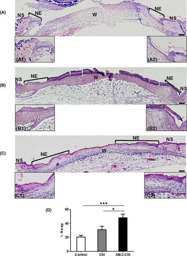
Bioscience Reports (2020) 40 BSR20200461
                                                                                                                                         https://doi.org/10.1042/BSR20200461

                                                                                                                                                                                           Downloaded from http://portlandpress.com/bioscirep/article-pdf/40/7/BSR20200461/888680/bsr-2020-0461.pdf by guest on 25 December 2020
           Figure 2. Phenotypical and functional characterization of MSC
           Microscopical observation shows the fibroblast-like morphology of MSC in culture (A). Flow cytometry analysis of MSC marker
           expression shows the expression of CD73 and CD90 (arrows). Negative controls were stained with the respective isotype (arrows)
           (B). Multipotent differentiation assays show the osteogenic (C), adipogenic (D) and chondrogenic (E) potential of MSC.

           MSC/CMs were in contact with the wound edges (Figure 3D). Finally, the wound was covered with a sterile band-aid
           and Tegaderm (Figure 3E).

           MSC promote early re-epithelialization of cutaneous wounds
           Because early cellular changes play a fundamental role in skin repair, we evaluated cutaneous wounds after 3 days
           of MSC implantation. For this purpose, animals were killed and wounds were evaluated. Macroscopic evaluation
           showed similar wound areas at day 0 and day 3 in each group (Figure 4A). Image analysis confirmed that there were
           not statistically significant difference in wound closure between day 3 and day 0 in all groups (Figure 4B). Signs
           of early re-epithelialization (whitish areas covering the wound surface) were observed in wounds from all groups
           (Figure 4C). However, they were more evident in the MSC/CM-treated group. The whole wound tissue, including
           NS, was collected and included in paraffin for histological analysis. Each sample was examined according to the
           presence of areas of NS, new epithelium (NE) and the wound area (W) (Figure 5). Histological studies showed small
           re-epithelialization areas (NE) in the periphery of wounds of control mice (non-treated) (Figure 5A). Similar results
           were observed in wounds implanted with CM alone (Figure 5B). In contrast, wounds treated with MSC/CM showed a
           larger re-epithelialization area from wound edge to the center of it (Figure 5C), as compared with those wounds treated
           with CM alone or without treatment (Figure 5A,B). These results were confirmed by using an image analysis software,
           which showed significant increases in re-epithelialization in wounds treated with MSC/CM, as compared with those
           treated with CM or control (Figure 5D). Epithelial thickening was observed in all groups, indicating the presence of
           hyperproliferative epidermis (Figure 5A–C). All wounds showed similar infiltration of PMN at day post-wounding
           (Figure 6).

4   © 2020 The Author(s). This is an open access article published by Portland Press Limited on behalf of the Biochemical Society and distributed under the Creative Commons Attribution
    License 4.0 (CC BY).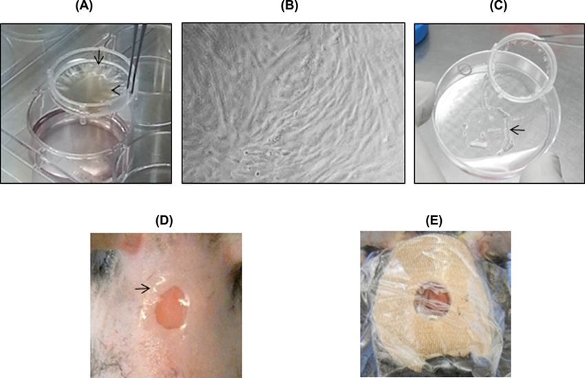
Bioscience Reports (2020) 40 BSR20200461
https://doi.org/10.1042/BSR20200461
Downloaded from http://portlandpress.com/bioscirep/article-pdf/40/7/BSR20200461/888680/bsr-2020-0461.pdf by guest on 25 December 2020
Figure 3. Implant of MSC on cutaneous wounds
Culture medium containing MSC (head arrow, A) was added on CM transwell (arrow, A). After 72 h, MSC reached 100% confluence
and exhibited fibroblast-like typical morphology on CM (B). MSC/CM were removed from the insert (arrow, C). CM (arrow) were cut
and implanted on the bed of cutaneous wounds (D). The wound was covered with a band-aid and Tegaderm (E).
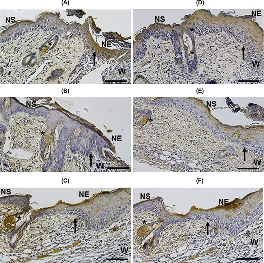
Detection of EPC expressing Lgr6 and CD34 in wounds treated with MSC
Based on evidence showing that EPC (Lgr6+ and CD34+ cells) are involved in skin wound repair [24], we evaluated
whether re-epithelialization of wounds induced by MSC transplantation was associated with the presence of these
cells. At day 3 post-MSC implant, IH evaluation showed Lgr6+ cells at the NS and NE of the control group and treated
with CM (Figure 7A,B, respectively). In the group treated with MSC/CM, most of the Lgr6+ cells were detected in
HFs and NE adjacent to the wound (Figure 7C). In contrast, few CD34+ cells were observed in all experimental
groups at day 3 of evolution of the wound (not shown). However, higher number of CD34 cells was observed at day 7
post-treatment in NS, HFs, sebaceous gland (SGs) and NE adjacent to the wound (Figure 8C), as compared with the
control and the group treated with CM (Figure 8A,B, respectively).
Increased expression of KGF and bFGF in wounds treated with MSC
Because KGF and bFGF are expressed during the skin repair process [25], we evaluated whether re-epithelialization
observed in wounds treated with MSC implantation was associated with increased expression of these factors. At
day 3 post-MSC implant, IH analysis showed that wounds treated with MSC/CM had an increased expression of
KGF (Figure 9C), as compared with untreated and CM-treated groups (Figure 9A,B, respectively). The increased
expression of KGF was mainly observed in NS, HF and SGs (Figure 9C). Expression of bFGF was also augmented
in wounds treated with MSC/CM (Figure 9F), as compared with untreated and CM-treated groups (Figure 9D,E,
respectively). Similar to KGF, the increased expression of bFGF was observed in NS, HF and SGs (Figure 9F).
Discussion
Numerous therapeutic procedures have been developed and applied to induce wound healing. However, many of
them have failed to re-epithelialize injured skin. Recently, several experimental protocols based on cellular therapy
© 2020 The Author(s). This is an open access article published by Portland Press Limited on behalf of the Biochemical Society and distributed under the Creative Commons Attribution 5
License 4.0 (CC BY).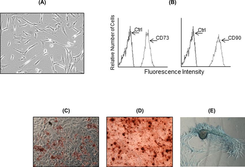
Bioscience Reports (2020) 40 BSR20200461
                                                                                                                                         https://doi.org/10.1042/BSR20200461

                                                                                                                                                                                           Downloaded from http://portlandpress.com/bioscirep/article-pdf/40/7/BSR20200461/888680/bsr-2020-0461.pdf by guest on 25 December 2020
           Figure 4. Evaluation of wound closure after MSC transplantation
           Wounds were evaluated before (d0) and after (d3) MSC transplantation. Wound closure was compared between the same experi-
           mental group (circle of the same size) (A). Wound closure was determined by using the ImageJ program. It was expressed as the
           percentage (means + − SE) of wound closure at day 3 as compared with d0 in each group (% of wound closure = [wound area
           d0 − wound area d3/area d0] × 100). There were not a statistically significant difference in wound closure between day 3 and
           d0 post-wounding in all groups (control, n=5; CM, n=5 and MSC/CM, n=4) (B). Signs of early re-epithelialization (whitish areas
           covering the wound surface) were observed in wounds (higher magnification) in all groups (C, arrows).

           have been used for inducing wound healing [26–28]. Here, we evaluate the capacity of MSC to induce cutaneous
           wound repair and its possible relationship with the presence of EPC and the expression of growth factors.
              It is well known that MSC are involved in the biological process of skin wound repair [29–31]. It has been sug-
           gested that MSC may induce skin repair through paracrine signals and anti-inflammatory and immune regula-
           tory effects [32–35]. Here, we used an experimental murine model to investigate the effect of human MSC on the
           re-epithelialization of full-thickness skin wound. Importantly, the possibility of rejection of human MSC, in this
           murine model, is very low because these cells have low immunogenicity [36–38]. In fact, we did not observe evi-
           dence of acute rejection and signs of an inflammatory reaction in wounds treated with human MSC.

6   © 2020 The Author(s). This is an open access article published by Portland Press Limited on behalf of the Biochemical Society and distributed under the Creative Commons Attribution
    License 4.0 (CC BY).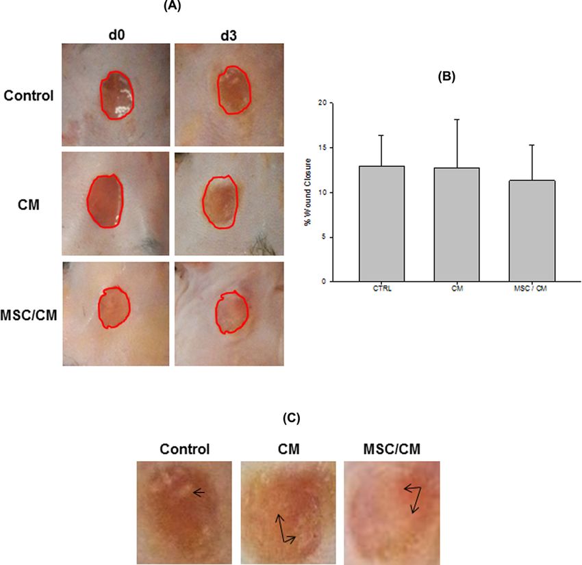
Bioscience Reports (2020) 40 BSR20200461
https://doi.org/10.1042/BSR20200461

                                                                                                                                                                                           Downloaded from http://portlandpress.com/bioscirep/article-pdf/40/7/BSR20200461/888680/bsr-2020-0461.pdf by guest on 25 December 2020

Figure 5. MSC transplantation enhances wound re-epithelialization
Histological studies of wounds were performed in untreated wounds (control, A), CM-treated wounds (B) and MSC/CM-treated
wounds (C), at day 3 post wounding. H&E-stained sections show NS and NE in the edges of wounds (W). Higher magnification of
the newly formed epidermis is shown in each section (control, A1–A2; CM, B1–B2; and MSC/CM C1–C2). Histologic sections of
wounds treated with MSC/CM show a larger area of re-epithelialization (C), as compared with those treated with CM alone (B) or
control (A). Image analysis from histological sections show a significant increase in the percentage of re-epithelialization in wound
treated with MSC/CM, as compared with control groups (D). Scale bar = 100 μm. Results are presented as means +         − SE (Control,
n=7; CM, n=7; MSC/CM, n=8). *P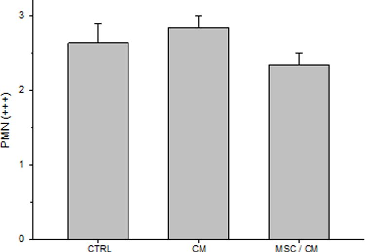
Bioscience Reports (2020) 40 BSR20200461
                                                                                                                                         https://doi.org/10.1042/BSR20200461

                                                                                                                                                                                           Downloaded from http://portlandpress.com/bioscirep/article-pdf/40/7/BSR20200461/888680/bsr-2020-0461.pdf by guest on 25 December 2020
           Figure 6. Evaluation of PMN in wounds
           PMN were evaluated before (d0) and after (d3) MSC implantation in control, MC- and MSC/MC-treated wounds. Analysis of PMN
           infiltrating wounds was performed by two independent observers (low powered field microscopy) by using a semiquantitative
           cross-scoring system (low = +, moderate = ++, high = +++). Results are presented as means +
                                                                                                     − SE. There was not a statistically
           significant difference in PMN infiltration between all experimental groups (control, n=8; CM, n=6; MSC/CM, n=9).

               Our results show histologically evidence that MSC enhance wound re-epithelialization as early as day 3 post-MSC
           implantation. Wounds treated with MSC showed larger re-epithelialization areas, from wound edge to the center of
           it, as compared with controls. It is known that the re-epithelialization process of skin wounds occurs by the prolifer-
           ation, migration and differentiation of EPC located at different sites of healthy peripheral skin (i.e., epidermis, HF)
           [24,39–41]. Likewise, it is known that different EPC participates upon wounding. Recently, it has been reported that
           Lgr6+ cells located in the follicular isthmus, SG, IFE and follicular bulge participate in the process of wound repair
           [42,43]. In this work, we show that Lgr6+ cells were present in the three experimental groups. However, there were
           differences in the location of these cells between MSC/CM-treated wound and the control groups. Histologic eval-
           uation showed Lgr6+ cells in the epidermis of NS and NE in control groups. In contrast, higher number of Lgr6+
           cells was observed in NE and in HFs of wounds treated with MSC. Because there is evidence showing that Lgr6+ cells
           are primed to respond to wound signals in unwounded healthy skin [44], it is possible that paracrine signals from
           MSC induce a rapid mobilization of these cells, which result in higher re-epithelialization rate of wounds treated with
           these cells. On the other hand, few CD34+ EPCs were present in all experimental groups at early stages of the wound
           evolution (3 days). However, after 7 days, higher numbers of CD34+ cells were observed in NS, NE, HFs and SGs of
           wound treated with MSC. These results suggest a delayed response of CD34+ EPCs to paracrine signals from MSC.
           Together, our results support previous works showing that different EPC population participate at different stages of
           wound repair [44]. Our results suggest that Lgr6+ cells are the ones to respond first, and CD34+ cells would be the
           last to respond to paracrine signals from MSC for producing re-epithelialization of full-thickness wounds.
               The wound re-epithelialization and repair process involves the expression of several growth factors [3]. Among
           them, it is known that KGF and bFGF participate in inducing skin wound re-epithelialization by regulating not only
           the proliferation and migration of EPC, but also of other cells (i.e., fibroblasts and endothelial cells) located in the
           periphery of the wounds [17,18,45]. In the present study we show that wounds treated with MSC had greater KGF and
           bFGF expression than those from the control groups. The high expression of KGF and bFGF may play an important
           role in inducing early re-epithelialization of wounds treated with MSC. The expression of these factors was mainly
           detected in NS, HF and SGs adjacent to the wound. The increased production of these growth factors could be due
           to paracrine signals from implanted MSC. However, other growth factors, produced as results of paracrine signals

8   © 2020 The Author(s). This is an open access article published by Portland Press Limited on behalf of the Biochemical Society and distributed under the Creative Commons Attribution
    License 4.0 (CC BY).
Bioscience Reports (2020) 40 BSR20200461
https://doi.org/10.1042/BSR20200461
Downloaded from http://portlandpress.com/bioscirep/article-pdf/40/7/BSR20200461/888680/bsr-2020-0461.pdf by guest on 25 December 2020
Figure 7. Detection of Lgr6+ progenitor cells in cutaneous wounds after 3 days of MSC implantation
IH studies to detect Lgr6+ cells were performed in untreated wounds (control, A), CM-treated wounds (B) and MSC/CM-treated
wounds (C). Tissue sections show NS and NE in the edges of wounds (W). Lgr6+ cells were present at the NS and NE of the control
group and treated with CM (A,B, respectively). Most of the Lgr6+ cells were detected at the NE adjacent to the wound treated with
MSC/CM (C). They were also detected in HFs close to the wound (head arrows). Each picture is representative of three different
experiments, all with similar results. Scale bar = 50 μm.
© 2020 The Author(s). This is an open access article published by Portland Press Limited on behalf of the Biochemical Society and distributed under the Creative Commons Attribution 9
License 4.0 (CC BY).Bioscience Reports (2020) 40 BSR20200461
https://doi.org/10.1042/BSR20200461
Downloaded from http://portlandpress.com/bioscirep/article-pdf/40/7/BSR20200461/888680/bsr-2020-0461.pdf by guest on 25 December 2020
Figure 8. Detection of CD34+ cells in cutaneous wounds after 7 days of MSC implantation
IH studies to detect CD34+ were performed in untreated wounds (control, A), CM-treated wounds (B) and MSC/CM-treated wounds
(C). Tissue sections show NS and NE in the edges of wounds (W). A larger number of CD34+ cells are observed in epidermis adjacent
to the wound, NE, HFs (head arrow) and SGs (asterisks) in the group treated MSC/CM (C), as compared with the control and the
group treated with CM (A,B, respectively). Scale bar = 50 μm.
10 © 2020 The Author(s). This is an open access article published by Portland Press Limited on behalf of the Biochemical Society and distributed under the Creative Commons Attribution
License 4.0 (CC BY).Bioscience Reports (2020) 40 BSR20200461
https://doi.org/10.1042/BSR20200461
Downloaded from http://portlandpress.com/bioscirep/article-pdf/40/7/BSR20200461/888680/bsr-2020-0461.pdf by guest on 25 December 2020
Figure 9. Increased expression of KGF and bFGF in cutaneous wounds after 3 days of MSC implantation
Wounds treated with MSC/CM show an increased expression of KGF and bFGF (C,F, respectively) in NS, HF and SGs (asterisks),
as compared with untreated (A,D, respectively) and CM-treated groups (B,E, respectively). Arrows indicate the edge of wound.
Each picture is representative of three different experiments, all with similar results. Scale bar = 50 μm.
from transplanted MSC, may also participate in wound repair [17–46]. Interestingly, recent evidence has shown that
paracrine signals from exosomes released by MSC may also contribute to the wound healing process [47–49].
In conclusion, our work shows that transplantation of MSC induces an early re-epithelialization of skin wounds,
which is associated with an increase in EPC and epithelial growth factors. MSC loaded on CM may constitute a
potential treatment for skin repair in cases of cutaneous wounds.
Competing Interests
The authors declare that there are no competing interests associated with the manuscript.
© 2020 The Author(s). This is an open access article published by Portland Press Limited on behalf of the Biochemical Society and distributed under the Creative Commons 11
Attribution License 4.0 (CC BY).Bioscience Reports (2020) 40 BSR20200461
https://doi.org/10.1042/BSR20200461
Funding
This work was supported by the Mppeuct (Ministerio del Poder Popular para la Educacion Universitaria, Ciencia y Tecnologia),
LOCTI [grant number 2011000906].
Author Contribution
Giselle Ramos-Gonzalez designed the research, analyzed the data, and wrote the paper. Olga Wittig, Dylana Diaz-Solano and
Carlos Ayala-Grosso designed research and analyzed data. Lianeth Salazar designed the research. Jose E. Cardier designed the
research, analyzed the data, and wrote the paper.
Downloaded from http://portlandpress.com/bioscirep/article-pdf/40/7/BSR20200461/888680/bsr-2020-0461.pdf by guest on 25 December 2020
Acknowledgements
The authors wish to thank the support of Esal and Tresta.
Abbreviations
bFGF, basic fibroblast growth factor; CM, collagen membrane; EPC, epithelial progenitor cell; HF, hair follicle; IFE, interfollicular
epidermis; IH, immunohistochemical; KGF, keratinocyte growth factor; MSC, mesenchymal stromal cell; MSC/CM, CM contain-
ing MSC; NE, new epithelium; NS, normal skin; PMN, polymorphonuclear; SG, sebaceous gland.
References
1 Gurtner, G.C., Werner, S., Barrandon, Y. and Longaker, M.T. (2008) Wound repair and regeneration. Nature 453, 314–321,
https://doi.org/10.1038/nature07039
2 Pastar, I., Stojadinovic, O., Yin, N.C., Ramirez, H., Nusbaum, A.G., Sawaya, A. et al. (2014) Epithelialization in wound healing: a comprehensive review.
Adv. Wound Care 3, 445–464, https://doi.org/10.1089/wound.2013.0473
3 Sivamani, R.K., Garcia, M.S. and Isseroff, R.R. (2007) Wound re-epithelialization: modulating keratinocyte migration in wound healing. Front. Biosci. 12,
2849–2868
4 Sorice, S., Rustad, K.C., Li, A.Y. and Gurtner, G.C. (2016) The rol of stem cell therapeutics in wound healing: Current understanding and future
directions. Plast. Reconstr. Surg. 138, 31S–41S, https://doi.org/10.1097/PRS.0000000000002646
5 Satoh, H., Kishi, K., Tanaka, T., Kubota, Y., Nakajima, T., Akasaka, Y. et al. (2004) Transplanted mesenchymal stem cells are effective for skin
regeneration in acute cutaneous wounds. Cell Transplant. 13, 405–412, https://doi.org/10.3727/000000004783983765
6 McFarlin, K., Gao, X., Lio, Y.B., Dulchavsky, D.S., Kwon, D., Arbab, A.S. et al. (2006) Bone marrow-derived mesenchymal stromal cells accelerate
wound healing in the rat. Wound Repair Regen. 14, 471–478, https://doi.org/10.1111/j.1743-6109.2006.00153.x
7 Lee, S.H., Lee, J.H. and Cho, K.H. (2011) Effects of human adipose-derived stem cells on cutaneous wound healing in nude mice. Ann. Dermatol. 23,
150–155, https://doi.org/10.5021/ad.2011.23.2.150
8 Boink, M.A., van den Broek, L.J., Roffel, S., Nazmi, K., Bolscher, J.G.M., Gefen, A. et al. (2016) Different wound healing properties of dermis, adipose,
and gingiva mesenchymal stromal cells. Wound Repair Regen. 24, 100–109, https://doi.org/10.1111/wrr.12380
9 Oswald, J., Boxberger, S., Jorgensen, B., Feldmann, S., Ehninger, G., Bornhäuser, M. et al. (2004) Mesenchymal stem cells can be differentiated into
endothelial cells in vitro. Stem Cells 22, 377–384, https://doi.org/10.1634/stemcells.22-3-377
10 Domini, M., Le Blanc, K., Mueller, I., Slaper-Cortenbach, I., Marini, F.C., Krause, D.S. et al. (2006) Minimal criteria for defining multipotent mesenchymal
stromal cells. Cytotherapy 8, 315–317, https://doi.org/10.1080/14653240600855905
11 Liu, J.W., Dunoyer-Geindre, S., Serre-Beinier, V., Mai, G., Lambert, J.-F., Fish, R.J. et al. (2007) Characterization of endothelial-like cells derived from
human mesenchymal stem cells. J. Thromb. Haemost. 5, 826–834, https://doi.org/10.1111/j.1538-7836.2007.02381.x
12 Fathle, C., Wilson, L., Hutter, J., Kapoor, V., Smith, A., Hocking, A. et al. (2004) Contribution of bone marrow-derived cells to skin: collagen deposition
and wound repair. Stem Cells 22, 812–822, https://doi.org/10.1634/stemcells.22-5-812
13 Luo, G., Cheng, W., He, W., Wang, X., Tan, J., Fitzgerald, M. et al. (2010) Promotion of cutaneous wound healing by local application of mesenchymal
stem cells derived from human umbilical cord blood. Wound Repair Regen. 18, 506–513, https://doi.org/10.1111/j.1524-475X.2010.00616.x
14 Huang, S., Wu, Y., Gao, D. and Fu, X. (2015) Paracrine action of mesenchymal stromal cells delivered by microspheres contributes to cutaneous wound
healing and prevents scar formation in mice. Cytotherapy 17, 922–931, https://doi.org/10.1016/j.jcyt.2015.03.690
15 Horton, J.A., Hudak, K.E., Chung, E.J., White, A.O., Scroggins, B.T., Burkeen, J.F. et al. (2013) Mesenchymal stem cells inhibit cutaneous
radiation-induced fibrosis by suppressing chronic inflammation. Stem Cells 31, 2231–2241, https://doi.org/10.1002/stem.1483
16 Bernardo, M.A. and Fibbe, W.E. (2013) Mesenchymal stromal cells: sensors and switchers of inflammation. Cell Stem Cell 13, 392–402,
https://doi.org/10.1016/j.stem.2013.09.006
17 Chen, L., Tredget, E.E., Wu, P.Y.G. and Wu, Y. (2008) Paracrine factors of mesenchymal stem cells recruit macrophages and endothelial lineage cells
and enhance wound healing. PLoS ONE 3, e1886, https://doi.org/10.1371/journal.pone.0001886
18 Chen, L., Xu, Y., Zhao, J., Zhang, Z., Yang, R., Xie, J. et al. (2014) Conditioned medium from hypoxic bone marrow-derived mesenchymal stem cells
enhances wound healing in mice. PLoS ONE 9, e96161, https://doi.org/10.1371/journal.pone.0096161
12 © 2020 The Author(s). This is an open access article published by Portland Press Limited on behalf of the Biochemical Society and distributed under the Creative Commons
Attribution License 4.0 (CC BY).Bioscience Reports (2020) 40 BSR20200461
https://doi.org/10.1042/BSR20200461
19 Javazon, E.H., Keswani, S.G., Badillo, A.T., Crombleholme, T.M., Zoltick, P.W., Radu, A.P. et al. (2007) Enhanced epithelial gap closure and increased
angiogenesis in wounds of diabetic mice treated with adult murine bone marrow stromal progenitor cells. Wound Repair Regen. 15, 350–359,
https://doi.org/10.1111/j.1524-475X.2007.00237.x
20 Chen, L., Tredget, E.E., Liu, C. and Wu, Y. (2009) Analysis of allogenicity of mesenchymal stem cells in engraftment and wound healing in mice. PLoS
ONE 4, e7119, https://doi.org/10.1371/journal.pone.0007119
21 Kota, D.J., Wiggins, L.L., Yoon, N. and Lee, R.H. (2013) TSG-6 produced by HMSCs delays the onset ofautoimmune diabetes by suppressing Th1
development and enhancing tolerogenicity. Diabetes 62, 2048–2058, https://doi.org/10.2337/db12-0931
22 Wittig, O., Romano, E., Gonzalez, C., Diaz-Solano, D., Marquez, M.E., Tovar, P. et al. (2016) A method of treatment for nonunion after fractures using
mesenchymal stromal cells loaded on collagen microspheres and incorporated into PRP clots. Int. Orthop. 40, 1033–1038,
https://doi.org/10.1007/s00264-016-3130-6
23 Wittig, O., Diaz-Solano, D. and Cardier, J.E. (2018) Viability and functionality of mesenchymal stromal cells loaded on collagen microspheres and
incorporated into plasma clots for orthopaedic application: effect of storage conditions. Injury 49, 1052–1057,
https://doi.org/10.1016/j.injury.2018.04.005
Downloaded from http://portlandpress.com/bioscirep/article-pdf/40/7/BSR20200461/888680/bsr-2020-0461.pdf by guest on 25 December 2020
24 Aragona, M., Dekoninck, S., Rulands, S., Lenglez, S., Mascré, G., Simons, B.D. et al. (2017) Defining stem cell dynamics and migration during wound
healing in mouse skin epidermis. Nat. Commun. 8, 14684, https://doi.org/10.1038/ncomms14684
25 Werner, S. and Grose, R. (2003) Regulation of wound healing by groth factors and cytokines. Physiol. Rev. 83, 835–870,
https://doi.org/10.1152/physrev.2003.83.3.835
26 Velander, P., Theopold, C., Bleiziffer, O., Bergmann, J., Svensson, H., Feng, Y. et al. (2009) Cell suspensions of autologous keratinocytes or autologous
fibroblasts accelerate the healing of full thickness skin wounds in a diabetic porcine wound healing model. J. Surg. Res. 157, 14–20,
https://doi.org/10.1016/j.jss.2008.10.001
27 Nakamura, Y., Ishikawa, H., Kawai, K., Tabata, Y. and Suzuki, S. (2013) Enhanced wound healing by topical administration of mesenchymal stem cells
transfected with stromal cell-derived factor-1. Biomaterials 34, 9393–9400, https://doi.org/10.1016/j.biomaterials.2013.08.053
28 Li, Y., Zheng, L., Xu, X., Song, L., Li, Y., Zhang, S. et al. (2013) Mesenchymal stem cells modified with angiopoietin-1 gene promote wound healing.
Stem Cell Res. Ther. 4, 113, https://doi.org/10.1186/scrt324
29 Liu, L., Yu, Y., Hou, Y., Chai, J., Duan, H., Chu, W. et al. (2014) Human umbilical cord mesenchymal stem cells transplantation promotes cutaneous
wound healing of severe burned rats. PLoS ONE 9, e88348, https://doi.org/10.1371/journal.pone.0088348
30 Rodriguez-Menocal, L., Shareef, S., Salgado, M., Shabbir, A. and Badivas, E. (2015) Role of whole bone marrow, cultured cells, and mesenchymal stem
cells in chronic wound healing. Stem Cell Res. Ther. 6, 24, https://doi.org/10.1186/s13287-015-0001-9
31 Doi, H., Kitajima, Y., Luo, L., Yan, C., Tateishi, S., Ono, Y. et al. (2016) Potency of umbilical cord blood- and Wharton’s jelly-derived mesenchymal stem
cells for scarless wound healing. Sci. Rep. 6, 18844, https://doi.org/10.1038/srep18844
32 Shohara, R., Yamamoto, A., Takikawa, S., Iwase, A., Hibi, H., Kikkawa, F. et al. (2012) Mesenchymal stromal cells of human umbilical cord Wharton’s
jelly accelerate wound healing by paracrine mechanisms. Cytotherapy 14, 1171–1181, https://doi.org/10.3109/14653249.2012.706705
33 Santos, J.M., Camoes, S.P., Filipe, E., Cipriano, M., Barcia, R.N., Filipe, M. et al. (2015) 3D spheroid cell culture of umbilical cord tissue-derived MSCs
(UCX® ) leads to enhanced paracrine induction of wound healing. Stem Cell Res. Ther. 6, 90, https://doi.org/10.1186/s13287-015-0082-5
34 Fang, F., Huang, R.L., Zheng, Y., Liu, M. and Huo, R. (2016) Bone marrow derived mesenchymal stem cells inhibit the proliferative and profibrotic
phenotype of hypertrophic scar fibroblast and keloid fibroblasts through paracrine signaling. J. Dermatol. Sci. 83, 95–105,
https://doi.org/10.1016/j.jdermsci.2016.03.003
35 Wang, S., Yang, H., Tang, Z., Long, G. and Huang, W. (2016) Wound dressing model of human umbilical cord mesenchymal stem cells-alginates
complex promotes skin wound healing by paracrine signaling. Stem Cells Int. 3269267, https://doi.org/10.1155/2016/3269267
36 Lee, R.H., Pulin, A.A., Seo, M.J., Kota, D.J., Ylostalo, J., Larson, B. et al. (2009) Intravenous hMSCs improve myocardial infarction in mice because cells
embolized in lung are activated to secrete the anti-inflammatory protein TSG-6. Cell Stem Cell 5, 54–63, https://doi.org/10.1016/j.stem.2009.05.003
37 Liu, S., Jiang, L., Li, H., Shi, H., Luo, H., Zhang, Y. et al. (2014) Mesenchymal stem cells prevent hypertrophic scar formation via inflammatory
regulation when undergoing apoptosis. J. Invest. Dermatol. 134, 2648–2657, https://doi.org/10.1038/jid.2014.169
38 Park, K.H., Mun, C.H., Kang, M.I., Lee, S.W., Lee, S.K. and Park, Y.B. (2016) Treatment of collagen-induced arthritis using immune modulatory
properties of human mesenchymal stem cells. Cell Transplant. 25, 1057–1072, https://doi.org/10.3727/096368915X687949
39 Ito, M., Liu, Y., Yang, Z., Nguyen, J., Liang, F., Morris, R. et al. (2005) Stem cells in the hair follicle bulge contribute to wound repair but not to
homeostasis of the epidermis. Nat. Med. 11, 1351–1354
40 Levy, V., Lindon, C., Zheng, Y., Harfe, B.D. and Morgan, B.A. (2007) Epidermal stem cells arise from the hair follicle after wounding. FASEB J. 21,
1358–1366, https://doi.org/10.1096/fj.06-6926com
41 Langton, A.K., Herrick, S.E. and Headon, D.J. (2008) An extended epidermal response heals cutaneous wounds in the absence of a hair follicle stem
cell contribution. J. Invest. Dermatol. 128, 1311–1318, https://doi.org/10.1038/sj.jid.5701178
42 Snippert, H.J., Haegebarth, A., Kasper, M., Jaks, V., van Es, J.H., Barker, N. et al. (2010) Lgr6 marks stem cells in the hair follicle that generate all cell
lineages of the skin. Science 327, 1385–1389, https://doi.org/10.1126/science.1184733
43 Lough, D.M., Wetter, N., Madsen, C., Reichensperger, J., Cosenza, N., Cox, L. et al. (2016) Transplantation of an LGR6+ epithelial stem cell-enriched
scaffold for repair of full-thickness soft-tissue defects. Plast. Reconstr. Surg. 137, 495–507, https://doi.org/10.1097/01.prs.0000475761.09451.00
44 Joost, S., Jacob, T., Sun, X., Annusver, K., La Manno, G., Sur, I. et al. (2018) Single-cell transcriptomics of traced epidermal and hair follicle stem cells
reveals rapid adaptations during wound healing. Cell Rep. 25, 585–597, https://doi.org/10.1016/j.celrep.2018.09.059
45 Werner, S., Smola, H., Liao, X., Longaker, M.T., Krieg, T., Hofschneider, P.H. et al. (1994) The function of KGF in morphogenesis of epithelium and
reepithelialization of wounds. Science 266, 819–822, https://doi.org/10.1126/science.7973639
© 2020 The Author(s). This is an open access article published by Portland Press Limited on behalf of the Biochemical Society and distributed under the Creative Commons 13
Attribution License 4.0 (CC BY).Bioscience Reports (2020) 40 BSR20200461
https://doi.org/10.1042/BSR20200461
46 da Silva Meirelles, L., Fontes, A.M., Covas, D.T. and Caplan, A. (2009) Mechanisms involved in the therapeutic properties of mesenchymal stem cells.
Cytokine Growth Factor Rev. 20, 419–427, https://doi.org/10.1016/j.cytogfr.2009.10.002
47 Liang, X., Ding, Y., Zhang, Y., Tse, H.F. and Lian, Q. (2014) Paracrine mechanisms of mesenchymal stem cell-based therapy: current status and
perspectives. Cell Transplant. 23, 1045–1059, https://doi.org/10.3727/096368913X667709
48 Shabbir, A., Cox, A., Rodriguez-Menocal, L., Salgado, M. and Badiavas, E.V. (2005) Mesenchymal stem cell exosomes induce proliferation and migration
of normal and chronic wound fibroblasts, and enhance angiogenesis in vitro. Stem Cells Dev. 24, 1635–1647, https://doi.org/10.1089/scd.2014.0316
49 Hu, L., Wang, J., Zhou, X., Xiong, Z., Zhao, J., Yu, R. et al. (2016) Exosomes derived from human adipose mesenchymal stem cells accelerates
cutaneous wound healing via optimizing the characteristics of fibroblasts. Sci. Rep. 6, 32993, https://doi.org/10.1038/srep32993
Downloaded from http://portlandpress.com/bioscirep/article-pdf/40/7/BSR20200461/888680/bsr-2020-0461.pdf by guest on 25 December 2020
14 © 2020 The Author(s). This is an open access article published by Portland Press Limited on behalf of the Biochemical Society and distributed under the Creative Commons Attribution
License 4.0 (CC BY).You can also read