Screening and development of monoclonal antibodies for identification of ferret T follicular helper cells - Nature
←
→
Page content transcription
If your browser does not render page correctly, please read the page content below
www.nature.com/scientificreports
OPEN Screening and development
of monoclonal antibodies
for identification of ferret T
follicular helper cells
Wenbo Jiang1, Julius Wong1, Hyon‑Xhi Tan1, Hannah G. Kelly1, Paul G. Whitney2, Ian Barr2,
Daniel S. Layton3, Stephen J. Kent1,4,5, Adam K. Wheatley1* & Jennifer A. Juno1*
The ferret is a key animal model for investigating the pathogenicity and transmissibility of important
human viruses, and for the pre‐clinical assessment of vaccines. However, relatively little is known
about the ferret immune system, due in part to a paucity of ferret‐reactive reagents. In particular,
T follicular helper (Tfh) cells are critical in the generation of effective humoral responses in humans,
mice and other animal models but to date it has not been possible to identify Tfh in ferrets. Here,
we describe the screening and development of ferret-reactive BCL6, CXCR5 and PD-1 monoclonal
antibodies. We found two commercial anti-BCL6 antibodies (clone K112-91 and clone IG191E/A8) had
cross-reactivity with lymph node cells from influenza-infected ferrets. We next developed two murine
monoclonal antibodies against ferret CXCR5 (clone feX5-C05) and PD-1 (clone fePD-CL1) using a single
B cell PCR-based method. We were able to clearly identify Tfh cells in lymph nodes from influenza
infected ferrets using these antibodies. The development of ferret Tfh marker antibodies and the
identification of ferret Tfh cells will assist the evaluation of vaccine-induced Tfh responses in the ferret
model and the design of novel vaccines against the infection of influenza and other viruses, including
SARS-CoV2.
Ferrets (Mustela putorius furo) are a well-established animal model for influenza research and are widely used
to investigate the pathogenesis and transmission of influenza viruses and pre‐clinically evaluate the efficacy
of influenza v accines1,2. In addition, ferrets serve as an animal model for the study of other viruses, including
human respiratory syncytial virus (HRSV)3, human metapneumovirus (HMPV)4, Hendra virus (HeV)5, Nipah
virus (NiV)5, different species of e bolavirus6 and severe acute respiratory syndrome coronavirus (SARS‐CoV)7.
Martina et al. demonstrated that ferrets are susceptible to experimental infection by SARS‐CoV, and that the
virus is efficiently transmitted to animals living with them8. It has now been shown that ferrets are similarly
susceptible to the pandemic virus severe acute respiratory syndrome coronavirus 2 (SARS-CoV-2), and that the
virus replicates efficiently in the upper respiratory tract of ferrets9,10. Ferrets are therefore a useful model in which
to assess neutralizing antibody responses to viral challenge and to test novel vaccine candidates11.
Characterization of the ferret immune response following infection and/or vaccination is informative for
developing vaccines and anti-viral therapies. However, a diverse range of ferret-specific immunological reagents
are not currently available. One major gap is the lack of reagents to study T follicular helper (Tfh) cells, which
are critical for the generation and maturation of the antibody response. Tfh cells are a subset of CD4 T cells that
provide help with B cells for high-affinity antibody production in germinal centres (GC)12–14. These specialised
cells are crucial for the formation of GC, affinity maturation and maintenance of B cell memory. BCL6 is the
master transcription factor for Tfh differentiation. Distinguishing phenotypic markers of Tfh cells include the
high expression of CXCR5, PD-1, and I COS12. High CXCR5 expression facilitates Tfh cells migration to B cell
follicles where its ligand, CXCL13 is produced abundantly by follicular stromal cells. PD-1 and ICOS are required
1
Department of Microbiology and Immunology, Peter Doherty Institute for Infection and Immunity, University of
Melbourne, Melbourne, VIC, Australia. 2WHO Collaborating Centre for Reference and Research On Influenza, Peter
Doherty Institute for Infection and Immunity, University of Melbourne, Melbourne, VIC, Australia. 3CSIRO Health
and Biosecurity, Australian Animal Health Laboratories, Geelong, VIC, Australia. 4Melbourne Sexual Health Clinic
and Infectious Diseases Department, Alfred Hospital, Monash University Central Clinical School, Carlton, VIC,
Australia. 5ARC Centre for Excellence in Convergent Bio‑Nano Science and Technology, University of Melbourne,
Melbourne, Australia. *email: a.wheatley@unimelb.edu.au; jennifer.juno@unimelb.edu.au
Scientific Reports | (2021) 11:1864 | https://doi.org/10.1038/s41598-021-81389-z 1
Vol.:(0123456789)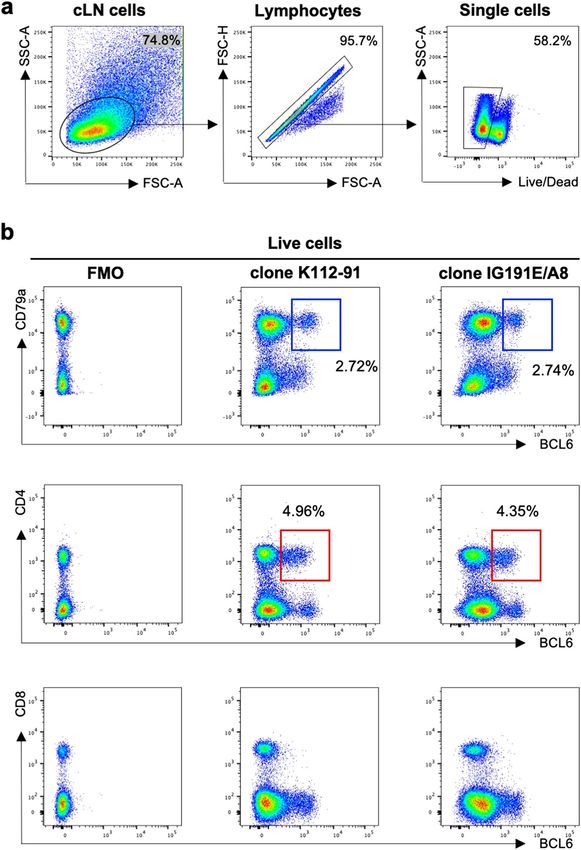
www.nature.com/scientificreports/
to engage with their respective ligands, PD-L1/PDL-2 and ICOSL, which are expressed by GC B cells to support
the development of Tfh c ells12.
Here, we describe the screening and development of ferret Tfh marker monoclonal antibodies and identi-
fication of ferret Tfh cells using these antibodies. We first identified two commercial anti-human/mouse BCL6
antibodies which had cross-reactivity with ferret lymph node (LN) cells. We next developed mouse anti-ferret
CXCR5 and PD-1 monoclonal antibodies using single cell PCR-based method. Finally, we detected Tfh cells in
lymph nodes from influenza infected ferrets using these antibodies.
Results
Screening of commercial anti‑human or mouse BCL6, CXCR5 and PD‑1 antibodies for
cross‑reactivity with ferret lymph node cells. BCL6 expression is the canonical transcription factor
that distinguishes Tfh cells from other CD4+ T cells in human and mouse15–17. However, high co-expression of
CXCR5 and PD-1 serves as surrogate or confirmatory surface markers for the Tfh population in human and
mouse lymphoid tissues18. Commercial anti-human/mouse BCL6 antibodies were screened for cross-reactivity
against ferrets by staining LN cell suspensions recovered from influenza infected ferrets. The gating strategy to
identify live lymphocytes in the ferret LN is shown in Fig. 1a. We found that clones K112-91 and IG191E/A8,
originally developed for human B CL615,16, showed cross-reactivity with ferret cLN cells (Fig. 1b). The BCL6+ B
cell (CD79a+) population represents a putative GC B cell population, while BCL6+ CD4+ cells are likely to mark
Tfh cells although additional markers are needed.
We next screened commercial antibodies raised against mouse or human CXCR5 (clones L138D7 and RF8B2)
and PD-1 (clones 29F.1A12 and EH12.2H7) for ferret cross-reactivity. Unfortunately, all screened murine and
human antibodies showed no cross-reactivity with ferret lymph node cells (data not shown) although they
showed good reactivity with mouse or human lymph node cells. Thus, we initiated the generation of ferret CXCR5
and PD-1-specific monoclonal antibodies.
Homology analysis of ferret, human and mouse BCL6, CXCR5 and PD‑1. Comparison of amino
acid homology of ferret, human and mouse BCL6, CXCR5 and PD-1 confirmed ferret BCL6 was a highly con-
served, with high homology to both human BCL6 (95.47%) and mouse BCL6 (93.78%) (Table 1). In contrast,
ferret CXCR5 had only moderate homology with human CXCR5 (84.22%) and mouse CXCR5 (87.43%) and
ferret PD-1 had low homology with both human (67.24%) and mouse PD-1 (54.48%), consistent with the lack of
ferret cross-reactivity of commercially available CXCR5 and PD-1-specific monoclonal antibodies.
Generation of mouse anti‑ferret CXCR5 and PD‑1 monoclonal antibodies. Due to the low
sequence conservation, we initiated the de novo development of anti-ferret PD1 and CXCR5 monoclonal anti-
bodies for flow cytometric use (workflow in Fig. 2). The cDNA sequence of the ectodomains of ferret CXCR5
and PD-1 were identified using a NGS dataset (Wong et al. in press). These genes were synthesized and cloned
into a mammalian expression vector containing human IgG1 Fc tag used for protein purification. Recombinant
proteins were expressed by Expi293 cells and purified by protein A agarose. Next, we immunized C57BL/6 mice
with recombinant ferret CXCR5 or PD-1 proteins. At day 21 post-immunization, we isolated draining lymph
nodes from the mice, stained lymph node cell suspensions with a panel of antibodies as well as immunogen
probes. The murine GC B cells binding to the fluorescent ferret CXCR5 or PD-1 probes were single-cell-sorted
into 96-well PCR plates. The BCR sequences of sorted B cells were then recovered by single cell PCR with mouse
IgG heavy or light chain primers19. The variable domains genes of heavy or kappa chains from clonally expanded
families of B cells were synthesized and cloned into mammalian expression vectors containing mouse IgG1 or
kappa chain constant domain gene. Antibodies were expressed in Expi293 and purified by protein G agarose.
Validation of anti‑ferret CXCR5 and PD‑1 monoclonal antibodies by ELISA. The binding speci-
ficity of the putative mouse anti-ferret CXCR5 and PD-1 antibodies was first assessed by ELISA. An irrelevant
antigen with the same Fc tag as recombinant ferret CXCR5 or PD-1 proteins was used as a control. Anti-ferret
CXCR5 clone A09 (feX5-A09), B04 (feX5-B04), C05 (feX5-C05) and E04 (feX5-E04) showed high binding activ-
ity with ferret CXCR5 proteins with an EC50 of 0.0109 μg/ml, 0.0036 μg/ml, 0.0027 μg/ml, 0.0033 μg/ml, respec-
tively (Fig. 3a). Anti-ferret PD-1 clone CL1 (fePD-CL1) similarly displayed high binding activity with ferret
PD-1 proteins with an EC50 of 0.0052 μg/ml while clone CL2 (fePD-CL2) showed no binding activity with ferret
PD-1 proteins by ELISA (Fig. 3b). All antibodies showed no binding with control proteins, demonstrating that
the antibodies were not targeted against Fc tag region of the recombinant proteins.
Identification of Tfh cells in lymph node cells from influenza infected ferrets. The ability of mAb
feX5-C05 (anti-CXCR5) and fePD-CL1 (anti-PD1) to stain ferret lymphocytes was examined using flow cytom-
etry. Single cell suspensions from the LN of influenza infected ferrets were stained with a panel consisting of anti-
BCL6, CD4 and CD79a antibodies and anti-CXCR5 (feX5-C05) and anti-PD1 (fePD-CL1) conjugated to biotin
and PE, respectively (gating in Fig. 4a). CXCR5++ PD-1++ CD4 T cells display elevated expression of BCL6
relative to non-Tfh cells (CD4+ CXCR5− PD−1−), consistent with a Tfh cell identity (Fig. 4b). Furthermore, the
CXCR5 and PD-1 expression pattern of ferret CD4 T cells is similar to that of mouse and macaque CD4 T cells
(Fig. 4c). In addition to CD4 T cells, CXCR5 is highly expressed by mouse and macaque B cells (Fig. 4d). Con-
sistent with mouse and macaque data, we found that ferret CD79a+ B cells were also predominately CXCR5+
(Fig. 4d). Taken together, these data show that ferret Tfh cells are detected by our in-house developed anti-ferret
CXCR5 and PD-1 antibodies or combination with a commercial cross-reactive anti-BCL6 antibody.
Scientific Reports | (2021) 11:1864 | https://doi.org/10.1038/s41598-021-81389-z 2
Vol:.(1234567890)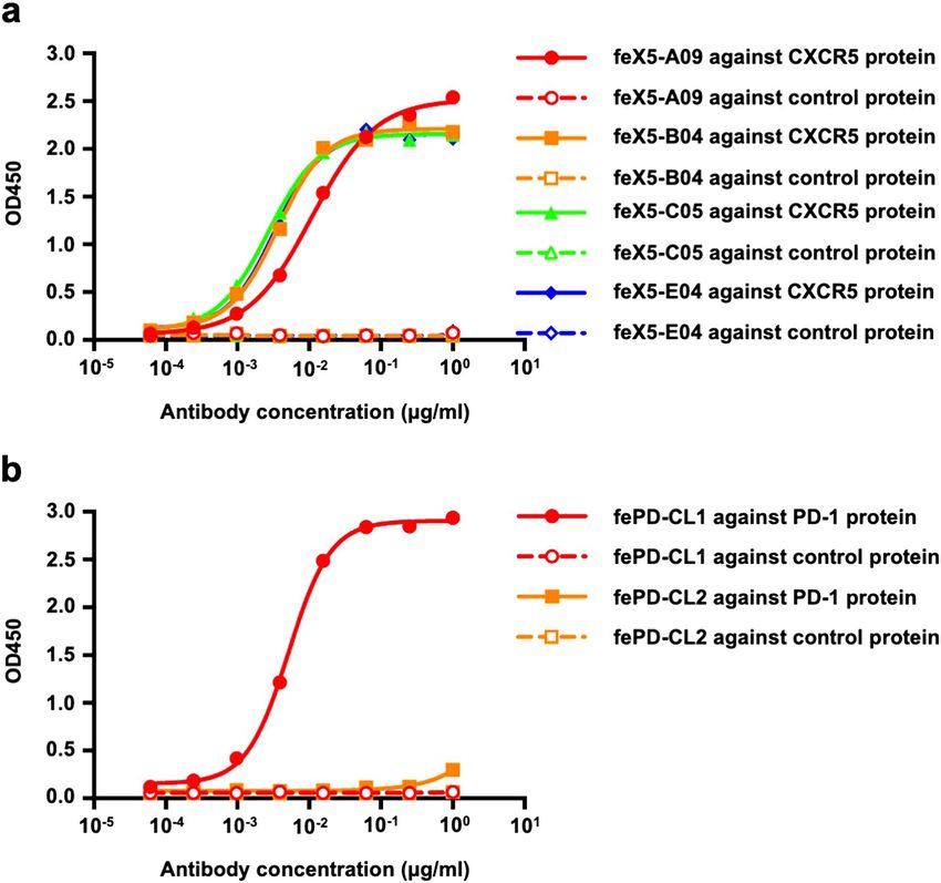
www.nature.com/scientificreports/

                                  Figure 1.  Cross-reactivity of commercial anti-human/mouse BCL6 antibodies with lymph node cells from
                                  influenza infected ferret. (a) Gating strategy to identify live lymphocytes in the ferret LN. Lymphocytes were
                                  identified by forward scatter area (FSC-A) and side-scatter area (SSC-A). Doublets were excluded by gating on
                                  single cells as determined by FSC-A versus FSC-H, and live cells were identified by viability dye exclusion. For
                                  each step, the parental population is indicated above the plot. (b) Representative plots of influenza-infected
                                  ferret lymph node cells stained with anti-BCL 6 (clone K112-91 and clone IG191E/A8), CD79a, CD4 and CD8
                                  antibodies. The parental population is indicated above the plot. Blue box indicates GC B cells (BCL6+CD79a+)
                                  and red box indicates Tfh cells (BCL6+CD4+).

Scientific Reports |   (2021) 11:1864 |               https://doi.org/10.1038/s41598-021-81389-z                                                     3

                                                                                                                                             Vol.:(0123456789)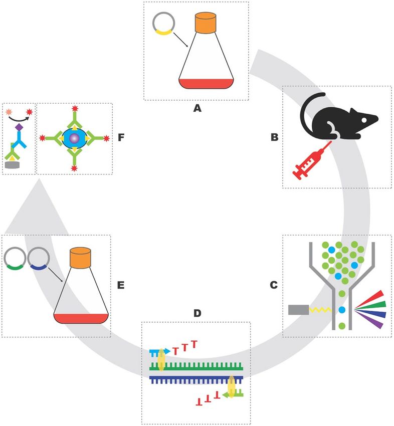
www.nature.com/scientificreports/
Antigen homology (%)
Antigen Ferret vs human Ferret vs mouse Human vs Mouse
BCL6 95.47 93.78 94.63
CXCR5 84.22 87.43 83.16
PD-1 67.24 54.48 59.31
Table 1. Antigen homology of ferret, human and mouse BCL6, CXCR5 and PD-1.
Figure 2. Flow chart to develop mouse anti-ferret CXCR5 and PD-1 monoclonal antibodies. Antigens were
expressed by transiently transfection of Expi293 cells (a), C57BL/6 mice were intramuscularly immunized with
purified antigens (b), antigen-specific B cells were single-cell-sorted by Aria III (c), the sequences of heavy
and light chain variable domains of BCR were recovered by single cell PCR (d), candidate antibody clones
were expressed by transiently transfection of Expi293 cells with heavy and light chain plasmids (e), purified
antibodies were validated by ELISA and FACS (f). Illustration was drawn using Adobe Illustrator 2020.
Discussion
Protective antibody responses induced by vaccination play a central role in defense against virus infection14.
Ferrets are a useful animal model for studying the pathogenicity and transmissibility of several human viruses
and for pre‐clinical evaluation of the in vivo protective efficacy of vaccines. The help signal provided by Tfh cells
is a critical factor which determines the magnitude and quality of antibody r esponses20. Identification of Tfh cells
in ferrets could greatly assist providing an immunological rationale for the design of novel vaccines against the
infection of influenza and other viruses, including SARS-CoV2. However, key immunological reagents, such as
ferret reactive Tfh marker monoclonal antibodies, were lacking until this report.
In the present study, we developed ferret-specific CXCR5 and PD-1 monoclonal antibodies and identified
Tfh cells in ferrets. Identification of Tfh cells by surface CXCR5 and PD-1 staining is of great utility if live Tfh
cell staining is needed (such as for antigen induced activation or RNA studies) since intranuclear BCL6 staining
involves cell fixation and permeabilization. In addition, CXCR5 could be a surrogate surface B cell marker if
intracellular staining is not warranted or wanted since the currently available marker for ferret B cells (a com-
mercial cross-reactive CD79a antibody) requires intracellular staining. Furthermore, anti-ferret PD-1 antibody
can be useful for studies of non-TFH T cell activation or exhaustion in this animal model (as demonstrated in
Fig. 4), thereby increasing the complexity and detail of immunophenotype studies.
Ferret Tfh responses remain largely unexplored, and basic questions, such as what the magnitude and quality
of ferret Tfh responses are in the context of virus infection or vaccination, and how Tfh responses correlate with
antibody responses following vaccination, remain to be answered. With these ferret-specific CXCR5 and PD-1
antibodies, these questions can be investigated, including longitudinal tracking of Tfh activity following virus
infection. Results from ferret Tfh response studies can provide useful insights regarding how Tfh cells influence
Scientific Reports | (2021) 11:1864 | https://doi.org/10.1038/s41598-021-81389-z 4
Vol:.(1234567890)www.nature.com/scientificreports/
Figure 3. Binding activity of anti-CXCR5 and PD-1 antibodies with autologous and control antigens. (a)
Binding activity of anti-ferret CXCR5 clone A09 (feX5-A09), B04 (feX5-B04), C05 (feX5-C05) and E04
(feX5-E04) against recombinant ferret CXCR5 protein or an irrelevant control protein with the same Fc tag.
(b) Binding activity of anti-ferret PD-1 clone CL1 (fePD-CL1) and CL2 (fePD-CL2) against recombinant ferret
PD-1 protein or an irrelevant control protein with the same Fc tag.
vaccine immunogenicity. These insights will assist the design and evaluation of novel Tfh-targeting vaccines
against the infection of influenza and other viruses in ferrets, as they have for other animal models. Considering
the current situation with coronavirus disease (COVID-19) pandemic, these reagents will be helpful in evalu-
ation of the Tfh responses induced by COVID-19 vaccines in ferret model and accelerate the development of
vaccines against SARS-CoV2 i nfection9,10.
In summary, we developed ferret-specific CXCR5 and PD-1 monoclonal antibodies which were next used
for detection of ferret Tfh cells. As these reagents identify surface-expressed antigens, they are compatible with
live cell sorting and downstream RNA sequencing analysis, which can be challenging with antibodies requiring
cell permeabilization and intracellular staining. The sequences of the heavy and light chain variable domains of
anti-ferret CXCR5 and PD-1 antibodies are provided (Fig. 5) so that the field can use these reagents to advance
the study of ferret Tfh. Recombinant antibodies can be expressed in a short time frame by transiently transfec-
tion of mammalian Expi293 cells. These ferret specific CXCR5 and PD-1 antibodies provide a starting point to
allow in-depth study of the Tfh responses to viral infections, such as influenza and SARS-CoV2. Further reagents
(such as anti-ferret CD154 monoclonal antibody) that are in critical need to identify antigen-specific Tfh are
under development and will be made publicly available in the future.
Materials and methods
Animals and ethics. Mouse studies and related experimental procedures were approved by the University
of Melbourne Animal Ethics Committee (#1914874) and were conducted in accordance with the Prevention of
Cruelty to Animals Act (1986), the Australian National Health and Medical Research Council Code of Practice
for the Care and Use of Animals for Scientific Purposes (1997) and the ARRIVE guidelines. Female C57BL/6
mice (6–8 weeks old) were immunized with 10 μg of recombinant ferret CXCR5 or PD-1 proteins with Addavax
Scientific Reports | (2021) 11:1864 | https://doi.org/10.1038/s41598-021-81389-z 5
Vol.:(0123456789)www.nature.com/scientificreports/
Scientific Reports | (2021) 11:1864 | https://doi.org/10.1038/s41598-021-81389-z 6
Vol:.(1234567890)www.nature.com/scientificreports/
◂Figure 4. Identification of Tfh cells in lymph node cells from influenza infected ferrets. (a) Gating strategy to
identify CD4 T cells in the ferret LN. Lymphocytes were identified by forward scatter area (FSC-A) and side-
scatter area (SSC-A). Doublets were excluded by gating on single cells as determined by FSC-A versus FSC-H, live
cells were identified by viability dye exclusion, CD4 T cells were identified as CD4+ CD79a−. For each step, the
parental population is indicated above the plot. (b) Representative plots of ferret CD4 T cells stained with anti-
BCL 6 (clone K112-91), anti-ferret CXCR5 (clone feX5-C05) and anti-ferret PD-1 (clone fePD-CL1) antibodies.
The BCL6 expression of CXCR5++ PD-1++ CD4 T cells (Tfh, blue oval) were compared with CXCR5− PD-1−
CD4 T cells (non-Tfh, red oval) cells. The parental population is indicated above the plot. (c) Representative plots
of CD4 T cells from influenza-infected ferret, mouse and macaque LNs stained with species-appropriate CXCR5
and PD-1 antibodies (mouse, clones L138D7 and 29F.1A12 respectively; macaque, MU5UBEE and EH12.2H7
respectively). The parental population is indicated above the plot. (d) Representative plots of live lymphocytes
from influenza-infected ferret, mouse and macaque LNs stained with their respective CXCR5 antibodies as well
as CD79, B220 and CD19 antibodies. The parental population is indicated above the plot.
(1:1 ratio; InvivoGen) at both hind quadriceps using 29G needles. 21 days post vaccination, draining lymph
nodes (inguinal lymph nodes (inLN) and iliac lymph nodes (ilLN)) were collected and mashed into single cell
suspension using 70 μm strainer (Miltenyi Biotec) for staining.
Ferret specimens were collected from culled animals that were involved in experiments approved by The
University of Melbourne Animal Ethics Committee (AEC 1714183) and were conducted in accordance with the
Prevention of Cruelty to Animals Act (1986) and the Australian National Health and Medical Research Council
Code of Practice for the Care and Use of Animals for Scientific Purposes (1997). Male ferrets were intranasally
infected with between 1 × 10⁴ to 1 × 10⁶ TCID50 in 0.5 ml of A/Perth/265/2009. At day 14 post-infection, para-
tracheal lymph nodes from influenza-infected ferrets were removed, cut into small pieces and passing through
a 70 μm strainer (Miltenyi Biotec). Single cell suspensions were then frozen in freezing media (90% fetal calf
serum with 10% DMSO) and stored in liquid nitrogen.
As a comparator for pan-species expression of CXCR5 on T and B cell population, we assessed macaque
Tfh and B cell phenotypes from lymph node samples. The macaque samples used in this were obtained from a
macaque influenza vaccination trial and were processed as described p reviously21.
Sequence analysis and recombinant protein generation. The nucleic acid sequences of ferret,
human and mouse BCL6, CXCR5 and PD-1 were extracted from Ensembl w ebsite22 and Centre for Biotechnol-
ogy Information (NCBI). Amino acid homology of ferret, human and mouse BCL6, CXCR5 and PD-1 were
compared using Geneious.
The ectodomain of ferret CXCR5 and PD-1 were identified using the NGS dataset (Wong et al. manuscript
submitted). The gene sequence of ectodomains of ferret CXCR5 and PD-1 were codon-optimized and synthe-
sized (GeneArt) and cloned into mammalian expression vector containing human IgG1 Fc tag. Plasmids were
extracted using NucleoBond Xtra Midi Plus plasmid DNA kit (MACHEREY-NAGEL). Recombinant ferret
CXCR5 and PD-1 proteins were expressed by transient transfection of Expi293 (Thermo) suspension cultures
with 2.7 μl ExpiFectamine (Thermo) and 1ug DNA/ml cell culture. At day 5 post-transfection, proteins in culture
supernatant were purified by protein A agarose affinity chromatography and gel filtration.
Sorting of ferret CXCR5 and PD‑1 specific B cells. Recombinant ferret CXCR5 and PD-1 proteins
were conjugated with APC or PE (Abcam) according to the manufacturer’s instructions. The resulting fluores-
cent proteins were designated as probes. Cell suspension of draining lymph nodes from ferret CXCR5 and PD-1
immunized mice were stained with probes and the following panel: live/dead Aqua (Thermo Fisher), CD45
APC-Cy7 (30-F11; BD), CD3 BV785 (145-2C11; BioLegend), F4/80 BV785 (BM8; BioLegend), Streptavidin
BV785 (BD), B220 BV650 (RA3-6B2; BD), IgD PerCP-Cy5.5 (11-26c.2a, BD), CD38 PE-Cy7 (90; BioLegend),
GL7 AF488 (GL7; BioLegend). Probe-binding GC B cells were single-cell-sorted to 96-well PCR plates a BD
Aria III.
RT‑PCR. The BCR sequences of sorted ferret CXCR5 or PD-1 specific B cells were recovered as previously
described19. Briefly, the mRNA of sorted single B cells was reversely transcribed into cDNA using SuperScript III
reverse transcriptase (Thermo) and random hexamer primers (Bioline). The sequences of heavy and light chain
variable domains were then amplified by nested PCR using HotStarTaq DNA polymerase (Qiagen) and mouse
immunoglobulin heavy and light chain primers19. PCR products were sequenced in Macrogen.
Antibody generation. The gene of heavy or kappa chain variable domain was synthesized (GeneArt) and
cloned into mammalian expression vector containing mouse IgG1 or kappa chain constant domain. Heavy
and kappa chain plasmids were extracted using NucleoBond Xtra Midi Plus plasmid DNA kit (MACHEREY-
NAGEL). Antibodies were expressed by transient transfection of Expi293 (Thermo) suspension cultures with
2.7 μl ExpiFectamine (Thermo) and 1ug DNA (heavy: kappa = 1:1)/ml cell culture. At day 5 post-transfection,
antibodies in culture supernatant were purified by protein G agarose affinity chromatography. For flow cytomet-
ric application, antibodies were conjugated to biotin or PE using biotin or PE conjugation kit (Abcam).
Scientific Reports | (2021) 11:1864 | https://doi.org/10.1038/s41598-021-81389-z 7
Vol.:(0123456789)www.nature.com/scientificreports/
Figure 5. The nucleic acid sequences of variable domains of anti-ferret CXCR5 and PD-1 antibodies. (a) The
nucleic acid sequences of variable domain of anti-ferret CXCR5 (clone feX5-C05) heavy chain. (b) The nucleic
acid sequences of variable domain of anti-ferret CXCR5 (clone feX5-C05) kappa chain. (c) The nucleic acid
sequences of variable domain of anti-ferret PD-1 (clone fePD-CL1) heavy chain. (d) The nucleic acid sequences
of variable domain of anti-ferret PD-1 (clone fePD-CL1) kappa chain.
ELISA. ELISAs were performed based on a modified protocol previously described23. 96-well MaxiSorp
plates (Thermo) were coated with recombinant ferret CXCR5 or PD-1 or control proteins (1 μg/ml at 100 μl/
well) overnight at 4 °C. After blocking with 2.5% BSA in PBS, anti-ferret CXCR5 or PD-1 antibodies at differ-
ent dilutions (starting at 1 μg/ml, four-fold serial dilutions) were added and incubated for two hours at room
temperature. Plates were washed with 0.05% Tween 20 in PBS prior to incubation with 1:5000 dilution of HRP-
conjugated goat anti-mouse IgG (Sera-Care) for 1 h at room temperature. Plates were washed again and devel-
oped using 3,3′,5,5′-Tetramethylbenzidine (TMB) substrate (Sigma) and read at 450 nm using CLARIOstar
microplate reader (BMG LABTECH).
FACS. Lymph node cell suspensions from influenza infected ferret were stained with the following panel: live/
dead Blue (Thermo Fisher), CD79a PerCP-Cy5.5 (HM47; BioLegend)24, CD8 AF700 (OKT8; Thermo)24, CD4
FITC (from CSIRO)25, BCL6 AF647 (K112-91; BD), BCL6 AF647 (IG191E/A8; BioLegend), CXCR5 BV421
(L138D7; BioLegend), CXCR5 BB515 (RF8B2; BD), CXCR5 PE (2G8, BD); CXCR5 Biotin (in-house), Strepta-
vidin BV421 (BD), PD-1 BV786 (29F.1A12; BioLegend), PD-1 BV421 (EH12.2H7; BioLegend), PD-1 PE (in-
house). For BCL6 staining, cells were fixed, permeabilized, and stained using the BD Transcription Factor Buffer
kit (BD) according to the manufacturer’s instructions. Macaque LN suspensions were stained with Live/dead
Aqua (Thermo Fisher), CD4 BV605 (L200; BD), CXCR5 PeCy7 (MU5UBEE, Thermo Fisher), and CD3 BUV737
(SP34-2, BD). All samples were acquired on a BD LSR Fortessa using BD FACS Diva and data was analyzed in
FlowJo v10.
Scientific Reports | (2021) 11:1864 | https://doi.org/10.1038/s41598-021-81389-z 8
Vol:.(1234567890)www.nature.com/scientificreports/
Received: 9 June 2020; Accepted: 21 December 2020
References
1. Albrecht, R. A. et al. Moving forward: Recent developments for the ferret biomedical research model. mbio https: //doi.org/10.1128/
mBio.01113-18 (2018).
2. Wong, J., Layton, D., Wheatley, A. K. & Kent, S. J. Improving immunological insights into the ferret model of human viral infec-
tious disease. Influenza Other Respir. Viruses 13, 535–546. https://doi.org/10.1111/irv.12687 (2019).
3. Stittelaar, K. J. et al. Ferrets as a novel animal model for studying human respiratory syncytial virus infections in immunocompetent
and immunocompromised hosts. Viruses https://doi.org/10.3390/v8060168 (2016).
4. MacPhail, M. et al. Identification of small-animal and primate models for evaluation of vaccine candidates for human metapneu-
movirus (hMPV) and implications for hMPV vaccine design. J. Gen. Virol. 85, 1655–1663. https://doi.org/10.1099/vir.0.79805-0
(2004).
5. Vigant, F. & Lee, B. Hendra and nipah infection: Pathology, models and potential therapies. Infect Disord. Drug Targets 11, 315–336.
https://doi.org/10.2174/187152611795768097 (2011).
6. Cross, R. W. et al. The domestic ferret (Mustela putorius furo) as a lethal infection model for 3 species of ebolavirus. J. Infect. Dis.
214, 565–569. https://doi.org/10.1093/infdis/jiw209 (2016).
7. Subbarao, K. & Roberts, A. Is there an ideal animal model for SARS?. Trends Microbiol. 14, 299–303. https://doi.org/10.1016/j.
tim.2006.05.007 (2006).
8. Martina, B. E. et al. Virology: SARS virus infection of cats and ferrets. Nature 425, 915. https://doi.org/10.1038/425915a (2003).
9. Shi, J. et al. Susceptibility of ferrets, cats, dogs, and other domesticated animals to SARS-coronavirus 2. Science https://doi.
org/10.1126/science.abb7015 (2020).
10. Kim, Y. I. et al. Infection and rapid transmission of SARS-CoV-2 in ferrets. Cell Host Microbe 27, 704–709.e702. https://doi.
org/10.1016/j.chom.2020.03.023 (2020).
11. See, R. H. et al. Severe acute respiratory syndrome vaccine efficacy in ferrets: Whole killed virus and adenovirus-vectored vaccines.
J. Gen. Virol. 89, 2136–2146. https://doi.org/10.1099/vir.0.2008/001891-0 (2008).
12. Vinuesa, C. G., Linterman, M. A., Yu, D. & MacLennan, I. C. Follicular helper T cells. Annu. Rev. Immunol. 34, 335–368. https://
doi.org/10.1146/annurev-immunol-041015-055605 (2016).
13. Crotty, S. Follicular helper CD4 T cells (TFH). Annu. Rev. Immunol. 29, 621–663. https: //doi.org/10.1146/annure v-immuno l-03121
0-101400 (2011).
14. Linterman, M. A. & Hill, D. L. Can follicular helper T cells be targeted to improve vaccine efficacy?. F1000Research 5, 1. https://
doi.org/10.12688/f1000research.7388.1 (2016).
15. Nurieva, R. I. et al. Bcl6 mediates the development of T follicular helper cells. Science 325, 1001–1005. https://doi.org/10.1126/
science.1176676 (2009).
16. Johnston, R. J. et al. Bcl6 and Blimp-1 are reciprocal and antagonistic regulators of T follicular helper cell differentiation. Science
325, 1006–1010. https://doi.org/10.1126/science.1175870 (2009).
17. Yu, D. et al. The transcriptional repressor Bcl-6 directs T follicular helper cell lineage commitment. Immunity 31, 457–468. https
://doi.org/10.1016/j.immuni.2009.07.002 (2009).
18. Meli, A. P. & King, I. L. Identification of mouse T follicular helper cells by flow cytometry. Methods Mol. Biol. 1291, 3–11. https://
doi.org/10.1007/978-1-4939-2498-1_1 (2015).
19. von Boehmer, L. et al. Sequencing and cloning of antigen-specific antibodies from mouse memory B cells. Nat. Protoc. 11, 1908–
1923. https://doi.org/10.1038/nprot.2016.102 (2016).
20. Victora, G. D. & Nussenzweig, M. C. Germinal centers. Annu. Rev. Immunol. 30, 429–457. https: //doi.org/10.1146/annure v-immun
ol-020711-075032 (2012).
21. Kelly, H.G. et al. Self-assembling influenza nanoparticle vaccines drive extended germinal center activity and memory B cell
maturation. JCI Insight. 5, e136653. https://doi.org/10.1172/jci.insight.136653 (2020).
22. Peng, X. et al. The draft genome sequence of the ferret (Mustela putorius furo) facilitates study of human respiratory disease. Nat.
Biotechnol. 32, 1250–1255. https://doi.org/10.1038/nbt.3079 (2014).
23. Liu, Y. et al. Cross-lineage protection by human antibodies binding the influenza B hemagglutinin. Nat. Commun. 10, 324. https
://doi.org/10.1038/s41467-018-08165-y (2019).
24. Liu, W. C. et al. Sequential immunization with live-attenuated chimeric hemagglutinin-based vaccines confers heterosubtypic
immunity against influenza A viruses in a preclinical ferret model. Front. Immunol. 10, 756. https://doi.org/10.3389/fimmu
.2019.00756(2019).
25. Layton, D. S. et al. Development of an anti-ferret CD4 monoclonal antibody for the characterisation of ferret T lymphocytes. J.
Immunol. Methods 444, 29–35. https://doi.org/10.1016/j.jim.2017.02.009 (2017).
Acknowledgements
This work was supported by NHMRC Grants APP1149990 and GNT112909. WJ is supported by a Melbourne
International Research Scholarship and Melbourne International Fee Remission Scholarship. JAJ, AKW and SJK
are supported by NHMRC fellowships.
Author contributions
W.J., A.K.W., J.A.J. and S.J.K. designed the study. W.J., J.W. and H.X.T. performed experiments. P.G.W. provided
ferret samples. D.S.L. provided ferret CD4 antibody. W.J., A.K.W., J.A.J., P.G.W. and S.J.K. wrote the manuscript.
All authors read and revised the manuscript.
Competing interests
The authors declare no competing interests.
Additional information
Correspondence and requests for materials should be addressed to A.K.W. or J.A.J.
Reprints and permissions information is available at www.nature.com/reprints.
Publisher’s note Springer Nature remains neutral with regard to jurisdictional claims in published maps and
institutional affiliations.
Scientific Reports | (2021) 11:1864 | https://doi.org/10.1038/s41598-021-81389-z 9
Vol.:(0123456789)www.nature.com/scientificreports/
Open Access This article is licensed under a Creative Commons Attribution 4.0 International
License, which permits use, sharing, adaptation, distribution and reproduction in any medium or
format, as long as you give appropriate credit to the original author(s) and the source, provide a link to the
Creative Commons licence, and indicate if changes were made. The images or other third party material in this
article are included in the article’s Creative Commons licence, unless indicated otherwise in a credit line to the
material. If material is not included in the article’s Creative Commons licence and your intended use is not
permitted by statutory regulation or exceeds the permitted use, you will need to obtain permission directly from
the copyright holder. To view a copy of this licence, visit http://creativecommons.org/licenses/by/4.0/.
© The Author(s) 2021
Scientific Reports | (2021) 11:1864 | https://doi.org/10.1038/s41598-021-81389-z 10
Vol:.(1234567890)You can also read