Phase Separation of YAP Reprograms Cells for Long-term YAP Target Gene Expression - bioRxiv
←
→
Page content transcription
If your browser does not render page correctly, please read the page content below

bioRxiv preprint first posted online Oct. 11, 2018; doi: http://dx.doi.org/10.1101/438416. The copyright holder for this preprint
(which was not peer-reviewed) is the author/funder, who has granted bioRxiv a license to display the preprint in perpetuity.
                              It is made available under a CC-BY-NC-ND 4.0 International license.

                       Phase Separation of YAP Reprograms Cells for Long-term
                                    YAP Target Gene Expression

               Danfeng Cai1, Shahar Sukenik2, †, Daniel Feliciano3, ††, Martin Gruebele2,
                                 Jennifer Lippincott-Schwartz1, 3, *
   1
    Eunice Kennedy Shriver National Institute for Child Health and Human Development,
   National Institute of Health, Bethesda, Maryland 20892
   2
    Department of Chemistry, University of Illinois at Urbana–Champaign, Urbana, Illinois
   61801
   3
    Janelia Research Campus, Howard Hughes Medical Institute, Ashburn, Virginia 20147

   *Correspondence to: lippincottschwartzj@janelia.hhmi.org
   †
    Current address: Department of Chemistry and Chemical Biology, University of
   California at Merced, 5200 N Lake Dr, Merced, CA 95340
   ††
      Current address: Thoracic and Malignancies Branch, National Cancer Institute,
   National Institute of Health, Bethesda, Maryland 20892

                                                                      1
bioRxiv preprint first posted online Oct. 11, 2018; doi: http://dx.doi.org/10.1101/438416. The copyright holder for this preprint
(which was not peer-reviewed) is the author/funder, who has granted bioRxiv a license to display the preprint in perpetuity.
                              It is made available under a CC-BY-NC-ND 4.0 International license.

   Abstract

   Yes-associated Protein (YAP) regulates cell proliferation and survival, and is over-

   expressed in most malignant tumors. As a transcriptional co-activator, its nuclear

   localization is the key determinant of its function. Recent work has revealed that

   hyperosmotic stress leads to YAP nuclear translocation and target gene expression, but

   how this overrides canonical YAP regulation by the Hippo pathway is unclear. Here we

   showed that YAP has an intrinsic ability to form liquid-like condensates when

   macromolecular crowding inside a cell increases, and this caused YAP to translocate to

   the nucleus, initiating downstream YAP signaling. Within seconds of inducing

   macromolecular crowding by hyperosmotic stress, YAP partitioned into cytoplasmic and

   nuclear droplets. Cytoplasmic YAP droplets concentrated YAP into an environment

   where it could be phosphorylated by NLK for nuclear translocation, whereas YAP

   nuclear droplets (enriched in TEAD1 and TAZ) functioned to re-organize gene enhancer

   elements, readying transcription of proliferation genes once the cell had either adapted

   to or been relieved of hyperosmotic stress. Thus, principles of liquid phase partitioning

   are used for reprogramming cells into a YAP-dependent, proliferative state in response

   to macromolecular crowding.

                                                                      2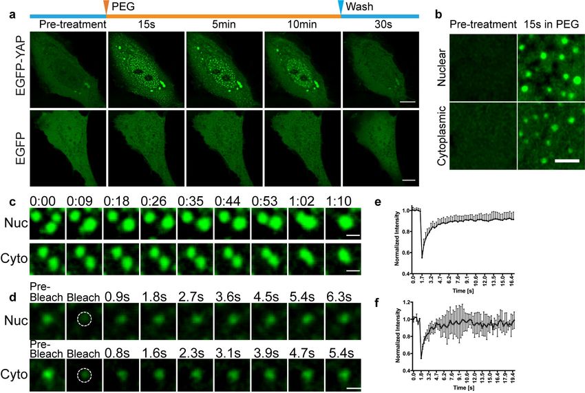
bioRxiv preprint first posted online Oct. 11, 2018; doi: http://dx.doi.org/10.1101/438416. The copyright holder for this preprint
(which was not peer-reviewed) is the author/funder, who has granted bioRxiv a license to display the preprint in perpetuity.
                              It is made available under a CC-BY-NC-ND 4.0 International license.

   Macromolecular crowding is a key feature of all eukaryotic cells, helping to drive

   polypeptide chains to fold into functional proteins, and impacting the diffusion and

   reaction kinetics of molecules. When molecular crowding inside a cell reaches a

   threshold, it can induce a process called liquid-liquid phase separation (LLPS)1, 2, in

   which labile, multivalent interactions among proteins and/or RNAs leads to their

   demixing from the surrounding milieu into liquid-like or gel-like condensates3-5. While the

   effects of macromolecular crowding on diffusion/reaction kinetics and protein phase

   separation have been studied in vitro, very little is known about how molecular crowding

   regulates general cell physiological processes, where changes in macromolecular

   crowding are known to occur. For example, changes in macromolecular crowding occur

   during starvation6, cell division7, and when cells experience differences in substrate

   stiffness8. Changes in macromolecular crowding also occur in cells of kidney9, gut10, 11

   and blood vessels12, when they undergo changes in osmotic pressure. What specific

   signaling pathways are triggered by these changes in macromolecular crowding to help

   the cell adapt to and survive in its transformed environment are unknown.

   Recently, hyperosmotic stress, which increases macromolecular crowding13, 14, has

   been shown to increase the nuclear localization of YAP, a key controller of organ size in

   animals. As a transcriptional co-activator, YAP is normally controlled by Hippo pathway.

   When the Hippo pathway is active, YAP binds to 14-3-3 proteins and is retained in the

   cytoplasm. When the Hippo pathway is inactive, YAP is freed from binding to 14-3-3

   proteins and redistributes into the nucleus, where it triggers transcription of proliferation-

   specific genes with the TEA domain family member (TEAD) transcription factors15, 18, 19.

                                                                      3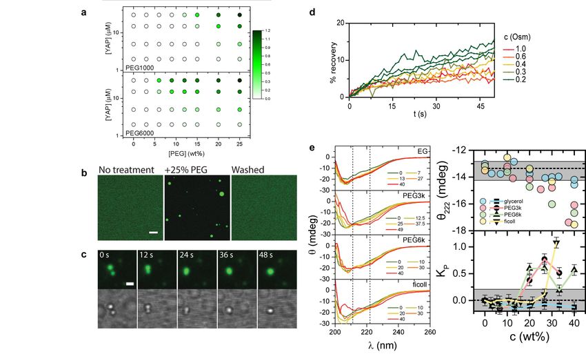
bioRxiv preprint first posted online Oct. 11, 2018; doi: http://dx.doi.org/10.1101/438416. The copyright holder for this preprint
(which was not peer-reviewed) is the author/funder, who has granted bioRxiv a license to display the preprint in perpetuity.
                              It is made available under a CC-BY-NC-ND 4.0 International license.

   However, hyperosmotic stress was found to cause YAP nuclear localization and target

   gene expression, overriding canonical Hippo pathway regulation17. Given YAP’s

   significance in controlling multiple aspects of cell behavior, including proliferation, cell

   stemness and malignancy15, 20, 21, we sought to investigate how increase in

   macromolecular crowding mediated by hyperosmotic stress leads to YAP activation.

   To study the mechanism by which macromolecular crowding regulates YAP activity, we

   treated U2-OS cells transiently expressing EGFP-YAP with 10% w/v PEG-300 (PEG),

   an osmotic stressor. Cytoplasmic and nuclear volume decreased by 22 ± 7 % and 32 ±

   9 % respectively (Fig. 1a, b), resulting in the cells having a more crowded intracellular

   environment (Fig. 1c) and higher dry mass density22 (Extended Data Fig. 1a). In

   response to this treatment, EGFP-YAP signal redistributed into the nucleus, peaking at

   10 min before attaining a steady state nuclear concentration that, while lower than at 10

   min, was higher than that seen before PEG treatment (Fig. 1d). Similar results were

   seen for endogenous YAP (Extended Data Fig. 1b). Washout of PEG resulted in EGFP-

   YAP’s signal quickly returning to its predominantly cytoplasmic, pre-treatment

   localization (Fig. 1d), indicating that the process is highly reversible. Within 3 h of PEG

   treatment, transcription of YAP target genes was increased by 2-fold (Fig. 1e),

   indicating that YAP’s nuclear redistribution led to downstream target gene expression,

   as previously reported17.

   Visualizing EGFP-YAP dynamics under hyperosmotic stress more closely, we found

   that YAP changed from being diffusely distributed in the cytoplasm to being localized in

                                                                      4
bioRxiv preprint first posted online Oct. 11, 2018; doi: http://dx.doi.org/10.1101/438416. The copyright holder for this preprint
(which was not peer-reviewed) is the author/funder, who has granted bioRxiv a license to display the preprint in perpetuity.
                              It is made available under a CC-BY-NC-ND 4.0 International license.

   discrete cytoplasmic and nuclear puncta within 15-30 s of adding PEG (Fig. 2a, upper

   panel and Movie S1). In zoomed-in images, these appeared spherical (Fig. 2b). This

   contrasted with the behavior of EGFP (Fig. 2a, lower panel) or EGFP-RhoA (Extended

   Data Fig. 2a) expressed in cells, which showed no change in subcellular distribution

   upon hyperosmotic stress. The EGFP-YAP puncta persisted for 10-15 min before slowly

   dissipating. Washing in isotonic medium when the puncta were still present caused the

   puncta to disappear in 30 seconds (Fig. 2a, upper panel, Movie S1). The foci were not

   artifacts of EGFP-YAP overexpression, since endogenous YAP-positive foci labeled by

   YAP antibody staining could be seen after PEG treatment in cells not expressing EGFP-

   YAP (Extended Data Fig. 2b). The formation of YAP foci was cell type-independent

   (Extended Data Fig. 2c), YAP isoform-independent (Extended Data Fig. 2d), and not

   limited to specific osmotic agent used (Extended Data Fig. 2e).

   Protein and RNA can partition into discrete fluid condensates in the process called

   liquid-liquid phase separation when they reach a local concentration threshold mediated

   by weak multivalent interactions2, 5, 23. These so-called ‘liquid droplets’ have spherical

   shapes, can coalesce into larger structures, and can exchange their contents with the

   rest of the cytosol or within the structure3. We found that YAP foci exhibited similar

   properties. They showed high sphericity in high resolution images (Extended Data Fig.

   2f), could fuse with each other to form larger spheres in both the nucleus and the

   cytoplasm (Fig. 2c, Movie S2-S4), and exhibited rapid fluorescence recovery upon

   photobleaching either the entire foci (t1/2 = 0.9 ± 0.4 sec, Fig. 2d, e) or part of one (Fig.

                                                                      5
bioRxiv preprint first posted online Oct. 11, 2018; doi: http://dx.doi.org/10.1101/438416. The copyright holder for this preprint
(which was not peer-reviewed) is the author/funder, who has granted bioRxiv a license to display the preprint in perpetuity.
It is made available under a CC-BY-NC-ND 4.0 International license.
2f). Therefore, YAP foci formed under hyperosmotic stress are genuine phase-
separated liquid droplets.
To determine whether YAP’s ability to condense into liquid droplets is an intrinsic
property or requires other co-factors, we expressed and purified EGFP-YAP from E. coli
and examined EGFP-YAP’s behavior under different conditions. Non-ionic crowders
larger than 300Da produced a turbid EGFP-YAP solution and this was dependent on
both the concentration of EGFP-YAP and concentration and size of PEG (Fig. 3a).
Confocal imaging of the turbid EGFP-YAP solution revealed micron-sized spheres that
displayed liquid-like characteristics (Fig. 3b), including droplet coalescence (Fig. 3c),
wetting the coverslip (Movie S5) and concentration-dependent recovery after
photobleaching (Fig. 3d), as reported for proteins capable of phase partitioning24. The
purified EGFP-YAP droplets retained their fluidity over 30 min and did not undergo
irreversible aggregation (Extended Data Fig. 3b). EGFP-YAP droplets disappeared
when the solution was re-suspended in 20mM Tris buffer, indicating their formation was
reversible (Fig. 3b, washed). Other conditions known to affect protein phase separation
(i.e., salts, inert proteins, and nucleic acids)24, 25 did not trigger YAP droplet formation
(Extended Data Fig. 3a). Indeed, varying salt concentration failed to change the turbidity
of EGFP-YAP solution, suggesting that strong electrostatic interactions don’t play an
important role in YAP phase separation (Extended Data Fig. 3a). UV circular dichroism
(CD) analysis revealed the presence of possible α-helical structure in YAP condensates
(Fig. 3e), indicating that weak multivalent interactions among coiled-coil structures might
help drive YAP’s co-acervation into droplets under crowded conditions. These data
6bioRxiv preprint first posted online Oct. 11, 2018; doi: http://dx.doi.org/10.1101/438416. The copyright holder for this preprint
(which was not peer-reviewed) is the author/funder, who has granted bioRxiv a license to display the preprint in perpetuity.
It is made available under a CC-BY-NC-ND 4.0 International license.
indicate that under macromolecular crowding YAP has an intrinsic ability to partition into
liquid condensates through a mechanism independent of salts or nucleic acids.
Phase-separated condensates provide cells with an additional way to organize their
internal space, functioning as hubs to facilitate protein-protein interactions or to
sequester proteins away from their normal partners3. With this in mind, we examined
what other proteins co-segregate with YAP in YAP-enriched droplets seen under
hyperosmotic stress. Focusing first on cytoplasmic YAP droplets, we concluded they
were not stress granules because they neither contained the stress granule component
G3BP (Extended Data Fig. 4a) nor co-segregated with G3BP-containing stress granules
under arsenite treatment (Extended Data Fig. 4b). YAP droplets also were not
processing bodies (P-bodies) involved in RNA processing as they lacked critical P-body
components, including GW182 and Ago2 (Extended Data Fig. 4c, d). However, the P-
body component Dcp1a did co-segregate with YAP droplets (Fig. 4a, d), suggesting
some unknown link to RNA processing. Intriguingly, YAP droplets contained proteins
involved in YAP-specific post-translational modifications, including Nemo-like kinase
(NLK) (Fig. 4b, d) and Hippo pathway kinase large tumor suppressor 1 (LATS1) (Fig. 4c,
e). NLK phosphorylates YAP on Serine 128, releasing YAP from 14-3-3 protein
association thereby triggering its nuclear re-localization17, 26. Co-segregation of NLK and
YAP in droplets thus could facilitate this reaction, leading to YAP nuclear redistribution.
Co-segregation of LATS1 with YAP in droplets seemed at odds with NLK’s role because
LATS1 phosphorylates YAP at Serine 127, an event that leads to tighter association of
YAP with 14-3-3 proteins18, 27. However, MST2, one of the kinases that activates LATS1
7bioRxiv preprint first posted online Oct. 11, 2018; doi: http://dx.doi.org/10.1101/438416. The copyright holder for this preprint
(which was not peer-reviewed) is the author/funder, who has granted bioRxiv a license to display the preprint in perpetuity.
It is made available under a CC-BY-NC-ND 4.0 International license.
to enable its phosphorylation of YAP, did not localize to YAP droplets (Extended Data
Fig. 4e). YAP droplet association of LATS1 thus might sequester LATS1 away from
MST2, leading to decreased LATS1 activity. These findings show that YAP
condensates are neither stress granules nor P-bodies, but are a novel type of
cytoplasmic droplet that sequesters kinases that target YAP for possible nuclear
translocation.
We next investigated what molecules co-segregate with YAP condensates in the
nucleus, hoping to gain clues as to the roles of YAP in this environment. YAP nuclear
condensates did not co-localize with known nuclear body markers such as SC35
(nuclear speckle, Extended Data Fig. 4f), PML (promyelocytic leukemia bodies,
Extended Data Fig. 4g), or Coilin (Cajal bodies, Extended Data Fig. 4h), suggesting
they perform functions different from these well-known nuclear bodies. Notably, YAP
condensates were enriched in the transcription factor TEAD1 (Fig. 4f, j) and another
coactivator of the YAP/TAZ signaling pathway, TAZ (Extended Data Fig. 4i). They also
were enriched in β-catenin, the transcription coactivator of the Wnt signaling pathway
that is often co-regulated with YAP28-30 (Fig. 4g, j). These properties of nuclear YAP
droplets were similar to phase-separated structures in the nucleus called super
enhancers (SEs)31, 32, which contain gene enhancer elements in clusters that are close
with each other and the promoters of genes they regulate33-35.
To gain more insight into the function of nuclear YAP droplets, we probed them with
antibodies to RNA Pol II, discovering that RNA Pol II separated away from YAP nuclear
8bioRxiv preprint first posted online Oct. 11, 2018; doi: http://dx.doi.org/10.1101/438416. The copyright holder for this preprint
(which was not peer-reviewed) is the author/funder, who has granted bioRxiv a license to display the preprint in perpetuity.
It is made available under a CC-BY-NC-ND 4.0 International license.
droplets upon hyperosmotic shock (Fig. 4h, j). YAP nuclear droplets also did not overlap
with nascent connective tissue growth factor (Ctgf) RNA as shown by RNA-FISH (Fig.
4i). This suggests that YAP nuclear droplets, while potentially driving clustering of gene
enhancer elements to form SEs, do not recruit RNA Pol II. This could be important for
preventing transcriptional activity at these super enhancer sites in the initial phase of
hyperosmotic shock. Our RT-PCR results showing an initial decrease at 20 min in YAP
signaling upon hyperosmotic shock (see Fig. 1e) is consistent with this possibility. We
thus hypothesized that only later, when YAP droplets have disappeared as visible
structures, would newly formed, YAP-induced SEs impact cell reprograming by
recruiting RNA Pol II, leading to rapid transcription of cell proliferation genes. Supporting
this possibility, we observed that treating cells with PEG for just 10 min was enough to
confer cells with a proliferation advantage 2 hours later compared with cells in normal
medium, as shown by EdU labeling (Fig. 4k). A recent paper reported that hyperosmotic
shock depletes RNA Pol II from promoter regions of genes, and after relieving cells of
hyperosmotic stress, cells accumulate more Pol II at promoter regions than before
treatments36. Our results confirmed this finding, and further suggested that it is YAP
nuclear droplets that re-organize gene enhancer elements to facilitate rapid transcription
of cell proliferation genes once cells have either adapted to or been relieved of
hyperosmotic stress.
We next sought to identify and remove regions in YAP responsible for its phase
separation under hyperosmotic stress to test whether YAP droplet formation is
necessary for the above effects on transcription and DNA synthesis. The YAP protein
9bioRxiv preprint first posted online Oct. 11, 2018; doi: http://dx.doi.org/10.1101/438416. The copyright holder for this preprint
(which was not peer-reviewed) is the author/funder, who has granted bioRxiv a license to display the preprint in perpetuity.
It is made available under a CC-BY-NC-ND 4.0 International license.
mainly consists of disordered sequences except for the central WW domain and C-
terminal PDZ binding motif (Fig. 5a). To identify regions responsible for YAP phase
separation, we first deleted the C-terminal transcriptional activation domain (TAD) of
YAP and made an EGFP-YAPΔTAD fusion protein (Fig. 5b). We found that EGFP-
YAPΔTAD was unable to phase separate under hyperosmotic shock (Fig. 5c, d),
indicating that the TAD sequence is indispensable for YAP phase separation. We
further found that phase separation-deficient EGFP-YAPΔTAD disrupts endogenous
YAP droplet formation (Extended Data Fig. 5), and has impaired nuclear localization
compared with wildtype (WT) EGFP-YAP at 15 min after PEG treatment (Fig. 5e). To
test whether YAP phase separation seen immediately after PEG addition is necessary
for the long-term promotion of YAP transcriptional activity, we expressed EGFP-YAP-
ΔTAD in U2-OS cells and studied the localization of endogenous TEAD1 transcription
factor. In the absence of YAP liquid droplets, TEAD1 did not enrich in any nuclear foci
(Fig. 5f). Correspondingly, the transcriptional activity of YAP under hyperosmotic stress
was much decreased in cells expressing ΔTAD compared to those expressing WT-YAP
as assessed by Ctgf mRNA expression 3 hrs after PEG treatment (Fig. 5g), indicating
that formation of liquid droplets enhances YAP target gene transcription in the long run.
These results indicated that the C-terminal TAD domain was necessary for YAP
condensate formation, and that the formation of YAP condensates was required for
downstream YAP signaling.
The N-terminal 51 amino acid region of YAP is a low complexity proline-rich domain
(comprised of 35% proline) (Fig. 5b), which might enhance the solubility of YAP
10bioRxiv preprint first posted online Oct. 11, 2018; doi: http://dx.doi.org/10.1101/438416. The copyright holder for this preprint
(which was not peer-reviewed) is the author/funder, who has granted bioRxiv a license to display the preprint in perpetuity.
It is made available under a CC-BY-NC-ND 4.0 International license.
condensates (i.e., preventing their gelation) by acting as a hydrophilic agent37, 38.
Consistent with this possibility, deletion of this region did not inhibit formation of YAP
liquid droplets. Rather, the EGFP-YAPΔP condensates persisted longer in the cell,
being visible 15 min after PEG treatment when the EGFP-YAP full length condensates
had already disappeared (Fig. 5c, d). These results indicate that only the C-terminal
TAD was responsible for YAP condensate formation, with other disordered sequences
in the protein (i.e., proline-rich domain) acting to maintain fluidity of the condensates.
In conclusion, we describe a mechanism for how hyperosmotic stress causes YAP to
translocate to the nucleus and initiate downstream YAP signaling that is based on
YAP’s intrinsic ability to phase partition into liquid droplets under macromolecular
crowding. In this mechanism, YAP’s low complexity TAD domain causes YAP to
partition into cytoplasmic and nuclear droplets within seconds of increased
macromolecular crowding. Whereas cytoplasmic YAP droplets concentrate YAP into an
environment where it becomes modified for nuclear translocation, YAP nuclear droplets
(enriched in TEAD1, TAZ and β-catenin) function to re-organize gene enhancer
elements, readying transcription of proliferation genes once the cell has either adapted
to or been relieved of hyperosmotic stress. Transient nuclear YAP droplet formation
under increased macromolecular crowding thus represents a novel way that cells
reprogram their long-term gene expression patterns. Its operation may be relevant for
understanding cell state changes under various physiological conditions where
macromolecular crowding increases.
11bioRxiv preprint first posted online Oct. 11, 2018; doi: http://dx.doi.org/10.1101/438416. The copyright holder for this preprint
(which was not peer-reviewed) is the author/funder, who has granted bioRxiv a license to display the preprint in perpetuity.
It is made available under a CC-BY-NC-ND 4.0 International license.
References and Notes:
1. Delarue, M. et al. mTORC1 Controls Phase Separation and the Biophysical Properties of
the Cytoplasm by Tuning Crowding. Cell 174, 338-+ (2018).
2. Woodruff, J.B. et al. The Centrosome Is a Selective Condensate that Nucleates
Microtubules by Concentrating Tubulin. Cell 169, 1066-+ (2017).
3. Shin, Y. & Brangwynne, C.P. Liquid phase condensation in cell physiology and disease.
Science 357 (2017).
4. Banani, S.F., Lee, H.O., Hyman, A.A. & Rosen, M.K. Biomolecular condensates: organizers
of cellular biochemistry. Nat Rev Mol Cell Biol 18, 285-298 (2017).
5. Jain, A. & Vale, R.D. RNA phase transitions in repeat expansion disorders. Nature 546,
243-+ (2017).
6. Joyner, R.P. et al. A glucose-starvation response regulates the diffusion of
macromolecules. Elife 5 (2016).
7. Son, S. et al. Resonant microchannel volume and mass measurements show that
suspended cells swell during mitosis. Journal of Cell Biology 211, 757-763 (2015).
8. Guo, M. et al. Cell volume change through water efflux impacts cell stiffness and stem
cell fate. P Natl Acad Sci USA 114, E8618-E8627 (2017).
9. Sheikh-Hamad, D. & Gustin, M.C. MAP kinases and the adaptive response to
hypertonicity: functional preservation from yeast to mammals. Am J Physiol Renal
Physiol 287, F1102-1110 (2004).
10. Shiau, Y.F., Feldman, G.M., Resnick, M.A. & Coff, P.M. Stool electrolyte and osmolality
measurements in the evaluation of diarrheal disorders. Ann Intern Med 102, 773-775
(1985).
11. Klaschik, E., Nauck, F. & Ostgathe, C. Constipation--modern laxative therapy. Support
Care Cancer 11, 679-685 (2003).
12. Jacob, M., Chappell, D. & Becker, B.F. Regulation of blood flow and volume exchange
across the microcirculation. Crit Care 20, 319 (2016).
13. Miermont, A. et al. Severe osmotic compression triggers a slowdown of intracellular
signaling, which can be explained by molecular crowding. P Natl Acad Sci USA 110, 5725-
5730 (2013).
14. Finan, J.D. & Guilak, F. The effects of osmotic stress on the structure and function of the
cell nucleus. J Cell Biochem 109, 460-467 (2010).
15. Pan, D. The hippo signaling pathway in development and cancer. Dev Cell 19, 491-505
(2010).
16. Totaro, A., Panciera, T. & Piccolo, S. YAP/TAZ upstream signals and downstream
responses. Nat Cell Biol 20, 888-899 (2018).
17. Hong, A.W. et al. Osmotic stress-induced phosphorylation by NLK at Ser128 activates
YAP. Embo Reports 18, 72-86 (2017).
18. Dong, J.X. et al. Elucidation of a universal size-control mechanism in Drosophila and
mammals. Cell 130, 1120-1133 (2007).
19. Meng, Z., Moroishi, T. & Guan, K.L. Mechanisms of Hippo pathway regulation. Genes
Dev 30, 1-17 (2016).
20. Zanconato, F., Cordenonsi, M. & Piccolo, S. YAP/TAZ at the Roots of Cancer. Cancer Cell
29, 783-803 (2016).
12bioRxiv preprint first posted online Oct. 11, 2018; doi: http://dx.doi.org/10.1101/438416. The copyright holder for this preprint
(which was not peer-reviewed) is the author/funder, who has granted bioRxiv a license to display the preprint in perpetuity.
It is made available under a CC-BY-NC-ND 4.0 International license.
21. Lian, I. et al. The role of YAP transcription coactivator in regulating stem cell self-renewal
and differentiation. Gene Dev 24, 1106-1118 (2010).
22. Wang, Z. et al. Spatial light interference microscopy (SLIM). Opt Express 19, 1016-1026
(2011).
23. Shin, Y. et al. Spatiotemporal Control of Intracellular Phase Transitions Using Light-
Activated optoDroplets. Cell 168, 159-+ (2017).
24. Lin, Y., Protter, D.S.W., Rosen, M.K. & Parker, R. Formation and Maturation of Phase-
Separated Liquid Droplets by RNA-Binding Proteins. Mol Cell 60, 208-219 (2015).
25. Smith, J. et al. Spatial patterning of P granules by RNA-induced phase separation of the
intrinsically-disordered protein MEG-3. Elife 5 (2016).
26. Moon, S. et al. Phosphorylation by NLK inhibits YAP-14-3-3-interactions and induces its
nuclear localization. EMBO Rep 18, 61-71 (2017).
27. Hao, Y.W., Chun, A., Cheung, K., Rashidi, B. & Yang, X.L. Tumor suppressor LATS1 is a
negative regulator of oncogene YAP. J Biol Chem 283, 5496-5509 (2008).
28. Benham-Pyle, B.W., Pruitt, B.L. & Nelson, W.J. Cell adhesion. Mechanical strain induces
E-cadherin-dependent Yap1 and beta-catenin activation to drive cell cycle entry. Science
348, 1024-1027 (2015).
29. Azzolin, L. et al. YAP/TAZ incorporation in the beta-catenin destruction complex
orchestrates the Wnt response. Cell 158, 157-170 (2014).
30. Konsavage, W.M., Jr. & Yochum, G.S. Intersection of Hippo/YAP and Wnt/beta-catenin
signaling pathways. Acta Biochim Biophys Sin (Shanghai) 45, 71-79 (2013).
31. Cho, W.K. et al. Mediator and RNA polymerase II clusters associate in transcription-
dependent condensates. Science (2018).
32. Sabari, B.R. et al. Coactivator condensation at super-enhancers links phase separation
and gene control. Science (2018).
33. Chong, S.S. et al. Imaging dynamic and selective low-complexity domain interactions
that control gene transcription. Science 361, 378-+ (2018).
34. Hnisz, D., Shrinivas, K., Young, R.A., Chakraborty, A.K. & Sharp, P.A. A Phase Separation
Model for Transcriptional Control. Cell 169, 13-23 (2017).
35. Hnisz, D. et al. Super-Enhancers in the Control of Cell Identity and Disease. Cell 155, 934-
947 (2013).
36. Erickson, B., Sheridan, R.M., Cortazar, M. & Bentley, D.L. Dynamic turnover of paused
Pol II complexes at human promoters. Genes Dev 32, 1215-1225 (2018).
37. Crick, S.L., Ruff, K.M., Garai, K., Frieden, C. & Pappu, R.V. Unmasking the roles of N- and
C-terminal flanking sequences from exon 1 of huntingtin as modulators of
polyglutamine aggregation. P Natl Acad Sci USA 110, 20075-20080 (2013).
38. Bergeron-Sandoval, L.P., Safaee, N. & Michnick, S.W. Mechanisms and Consequences of
Macromolecular Phase Separation. Cell 165, 1067-1079 (2016).
13bioRxiv preprint first posted online Oct. 11, 2018; doi: http://dx.doi.org/10.1101/438416. The copyright holder for this preprint
(which was not peer-reviewed) is the author/funder, who has granted bioRxiv a license to display the preprint in perpetuity.
It is made available under a CC-BY-NC-ND 4.0 International license.
Acknowledgements:
We are grateful to the thoughtful discussions with Duojia Pan (UTSW), Ming Guo (MIT),
and members of the Lippincott-Schwartz laboratory.
Funding:
This work was supported by the Intramural Research Program of the National Institutes
of Health (J.L.-S., D.C.), the Howard Hughes Medical Institute (J.L.-S.), and Damon
Runyon Cancer Research Fellowship (D.C. DRG2233-15), as well as NSF MCB
1803786 (S.S. and M.G.).
Authors Contributions:
D.C. and J.L.-S. conceived the project and designed the study. D.C. performed all the
experiments in cell. S.S. performed all the experiments in vitro. D. F. constructed
mutant constructs for experiments. J.L.-S. and M.G. supervised research, interpreted
data, and participated in project planning. D.C. and J.L.-S. wrote the manuscript with
inputs from all the authors.
Competing Interests:
The authors have no competing interests.
Data and materials availability:
All data is available in the main text or the supplementary materials.
14bioRxiv preprint first posted online Oct. 11, 2018; doi: http://dx.doi.org/10.1101/438416. The copyright holder for this preprint
(which was not peer-reviewed) is the author/funder, who has granted bioRxiv a license to display the preprint in perpetuity.
It is made available under a CC-BY-NC-ND 4.0 International license.
Figure Legends:
Fig. 1. Cells change volume and form YAP-positive condensates during hyperosmotic
stress. (a) Side-view of a cell before, undergoing, and after hyperosmotic stress, 3-D
rendered by Imaris. Color bar: volume (µm3). (b) Quantification of normalized U2-OS
nuclear (cyan), cytoplasmic (pink) and total (blue) volume. Unpaired t test. Comparing to
volume before treatment. EGFP-YAP cellular localization in a U2-OS cell at indicated
time. (c) Ratiometric images and quantification of crowding sensor FRET expressed in
U2-OS cells. Rainbow-RGB showing changes in FRET indices. Color bar: FRET index
(a.u.). (d) Localization of EGFP-YAP in U2-OS cells and quantification of normalized
nuclear to cytoplasmic EGFP-YAP ratio. Unpaired t test. Comparing to ratio pre-
treatment. (e) Relative Ctgf and Cyr61 mRNA levels in control and PEG-treated HEK
293T cells expressing EGFP-YAP. Unpaired t test. Error bars are SEM. *pbioRxiv preprint first posted online Oct. 11, 2018; doi: http://dx.doi.org/10.1101/438416. The copyright holder for this preprint
(which was not peer-reviewed) is the author/funder, who has granted bioRxiv a license to display the preprint in perpetuity.
It is made available under a CC-BY-NC-ND 4.0 International license.
FISH against Ctgf RNA, in control or in PEG medium. (j) Quantification of (f-h).
Unpaired t tests comparing PEG with control-treated cells. (k) Quantification of EdU
incorporation within 2hrs, after treating cells first with control medium for 10min or
10%PEG for 10min.
Fig. 5. Phase separation of YAP is important for its nuclear localization and signaling. (a)
Disorder analysis of YAP 454aa isoform. Algorithms used: IUPred (blue), VLXT (cyan)
and VSL2 (magenta). (b) Schematic of wild-type (WT) and mutant YAP structures. (c)
Phase separation of EGFP-YAP variants in cell. (d) Percentage of cells expressing
EGFP-YAP variants with liquid droplets at different time after PEG treatment. (e)
Nuclear localization of different EGFP-YAP variants. (f) Relative localization of EGFP-
YAP-ΔTAD with TEAD1 before and after PEG treatment. Immunofluorescence (g)
Relative Ctgf mRNA expression in EGFP-YAP and EGFP-YAP-ΔTAD mutant after
control or PEG treatment. Error bars show SEM.
16You can also read