4d inner shell ionization of Xe+ ion and subsequent Auger decay - Refubium
←
→
Page content transcription
If your browser does not render page correctly, please read the page content below
4d inner shell ionization of Xe+ ion
                           and subsequent Auger decay

    M. A. Khalal1, P. Lablanquie1, L. Andric1,2, J. Palaudoux1, F. Penent1, K. Bučar3, M. Žitnik3,
            R. Püttner4, K. Jänkälä5, D. Cubaynes6,7, S. Guilbaud6,7 and J.-M. Bizau6,7

      1
       Sorbonne Université, UPMC Université Paris 06, CNRS, LCP-MR (UMR 7614), 4 place Jussieu,
                                           75252 Paris Cedex 05, France
          2
            Université Paris-Est, 5 boulevard Descartes, F-77454 Marne-la Vallée Cedex 2, France
                      3
                        Jozef Stefan Institute, Jamova cesta 39, SI-1001 Ljubljana, Slovenia
           4
             Fachbereich Physik, Freie Universität Berlin, Arnimallee 14, D-14195 Berlin, Germany
    5
     Nano and Molecular Systems Research Unit, University of Oulu, P.O. Box 3000, 90014 Oulu, Finland
               6
                 ISMO, CNRS UMR 8214, Univ. Paris-Sud, Université Paris-Saclay, 91405 Orsay, France
7
  Synchrotron SOLEIL, l’Orme des Merisiers, Saint-Aubin, Boîte Postale 48, 91192 Gif-sur-Yvette Cedex, France

Abstract
       We have studied Xe+ 4d inner shell photoionization in a direct experiment on Xe+ ions, merging
an ion and a photon beam and detecting the ejected electrons with a cylindrical mirror analyzer. The
measured 4d photoelectron spectrum is compared to the 4d core valence double ionization spectrum
of the neutral Xe atom, obtained with a magnetic bottle spectrometer. This multi-coincidence
experiment gives access to the spectroscopy of the individual Xe2+ 4d-15p-1 states and to their respective
Auger decays, which are found to present a strong selectivity. The experimental results are interpreted
with the help of ab-initio calculations.

                                                                                                        1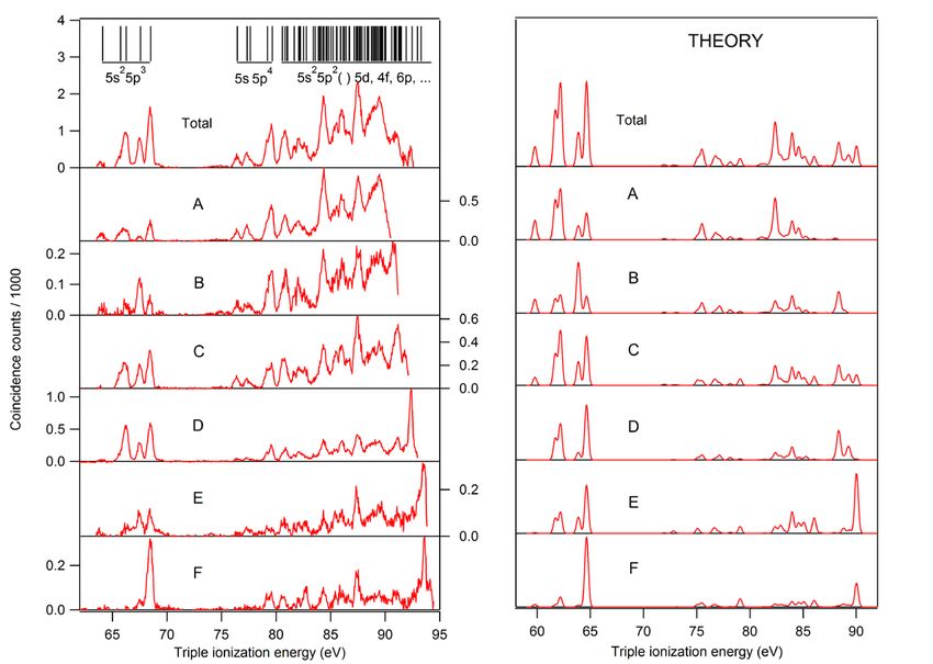
I. INTRODUCTION
          Direct photoelectron spectroscopy experiments on ions are extremely difficult due to the
  relatively low target ion density in the source volume of an electron spectrometer, and also because of
  the high cross section for ionizing collision of ions on the background gas : photoionization of this
  background gas (even in ultra-high vacuum conditions) produce a high background electron signal.
  Therefore direct electron spectroscopic experiments on ions have been reported only in a few cases in
  the 90’s, such as the resonant Auger decay of in Ca+ ions [1] [2] [3] or the 4d photoionization of Xe+
  ions [4]. Recently, the field has regained interest thanks to the development of powerful synchrotron
  radiation sources and to the use of new dedicated experiments such as the MAIA setup installed on the
  PLEIADES beam line at SOLEIL [5]. As an example, this setup enabled the detailed study of the
  Auger Decay of the 4d → nf (n= 4, 5) resonances in the Xe5+ ion [6]. Here we present another example
  of these new experimental studies of ions and revisit the 4d inner shell ionization of the Xe+ ions first
  reported by Gottwald et al in 1999 [4].
         In addition, we use a complementary experimental approach for this study, namely: core-
  valence double ionization of the neutral xenon atoms. The high efficiency of multiple coincidence
  experiments with a magnetic bottle allows one to observe in detail these core valence double ionization
  paths as demonstrated by Hikosaka et al in 2006 on Ne and N2 targets [7]. It was later shown that this
  approach can be applied to infer properties of the inner shell ionization of atomic ions, with Ar+ as an
  example [8]. The targets Hg+ [9] [10] and Kr+ [11] were recently addressed. We show here that the
  present core valence results on Xe atoms validate the new experimental results obtained using the
  MAIA setup on 4d shell ionization of Xe+ ions. Furthermore coincidence filtering of data collected by
  the magnetic bottle gives access to the Auger decay of each selected Xe2+ 4d-15p-1 state.
          Below we report spectroscopic study of the Xe2+* states resulting from photoionization of a 4d
  shell in Xe+ ion, and present the corresponding state resolved Auger decay. Calculations of transition
  energies and decay rates have been performed to guide and support the interpretation of the
  experimental results.

II. EXPERIMENTS

  II. A MAIA Experiment
        The experiment on Xe+ ion was performed with the MAIA merged-beam setup on the
  PLEIADES beam line of the synchrotron radiation facility SOLEIL [6] [5]. The ions are produced in
  a 12.4 GHz permanent magnet electron cyclotron resonance ion source (ECRIS) by heating 129Xe
  isotopic gas. They are accelerated to 4 keV and selected using a dipole magnet. The Xe+ ions are then
  focused and merged with the photon beam into the source volume of a cylindrical mirror electron
  analyzer (CMA) [12]. Both ion and photon beams are collinear to the CMA axis. Typical currents of
  focused ions were 2 µA. A second dipole magnet separates the incident Xe+ ions from the Xeq+ (q =
  2, 3) ions produced in the photoionization processes, which are counted by the microchannel plate
  detector. The photoelectrons emitted at the magic angle in the laboratory frame are counted as a
  function of their kinetic energy by 8 channeltron detectors placed at the CMA focal plane. In order to
  reduce the strong background in the electron signal produced by collisional processes between the fast

                                                                                                         2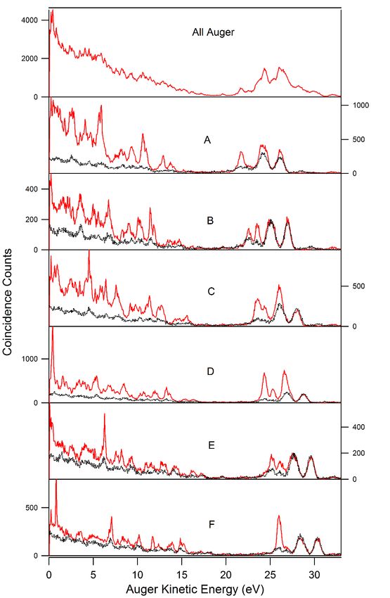
Xe+ beam and the residual gas in the CMA chamber or the metallic surfaces, and to suppress the signal
resulting from photoionization of neutral Xe coming from the ECR source, the photoelectrons are
detected in coincidence with the photoions (PEPICO) [6] [4]. To improve the ratio of true-to-false
coincidences further, a 150 V negative bias was applied to a 20 cm long region centered at the source
volume of the CMA. The photoions produced inside this region therefore have different velocity from
the ions produced outside and can be discriminated by the second dipole magnet.
         Because the plasma in the ECRIS is quite hot, the Xe+ ions are produced not only in the ground
state Xe+ 4d10 5s2 5p5 (2P3/2), but also in the metastable state Xe+ 4d10 5s2 5p5 (2P1/2). The latter state
does not decay before the ions arrive in the interaction region (lifetime 49 ms [13]) and thus contributes
to our photoelectron spectra. To estimate relative populations of the ions in the two levels, we have
measured the yield of Xe2+ photoions in the energy region of the Xe+ 4d10 5s2 5p5 (2PJi) → Xe+ 4d9 5s2
5p6 (2DJf) photoexcitations. The result is shown in Figure 1. The strongest resonance at 55.4 eV
corresponds to the transition Ji = 3/2 → Jf = 5/2 [14] of ions in the ground state, and the one at 56.1
eV to the transition Ji = 1/2 → Jf = 3/2 of ions in the metastable state. Figure 1 also displays the
calculated results for the corresponding transitions (see section III). For the resonance lower in energy
we obtain an oscillator strength of 0.1336 and for that higher in energy a value of 0.1660. The
calculated position and natural linewidth are 55.28 eV and 132 meV for the lower and 55.92 eV and
125 meV for the upper resonance, in quite good agreement with experimental values from the literature
reporting 55.39 eV and 56.08 eV for the energy positions [14], and 111±3 meV and 104±3 meV for
the widths [15], respectively. A comparison of the measured intensities of these two well-separated
resonances with the calculated oscillator strengths allows us to estimate the initial populations: by
assuming that the calculated oscillator strengths are correct one obtains that the observed intensity ratio
equals the ratio of calculated oscillator strengths multiplied by the population ratio. The best
description of the experimental spectrum is obtained taking 74% of the ions in the ground level and
26% in the excited level, which is close to the statistical population ratio (66% for 2P3/2 and 33% for
2
  P1/2).
       Figure 2 shows the electron spectra recorded at 120.7 eV photon energy. This energy was chosen
to avoid any overlap between the 4d-1 photoelectron and Auger lines. The top panel displays the total
electron spectrum as a function of electron kinetic energy. The spectrum is dominated by a high and
decreasing background produced by the collisions of the beam of Xe+ ions with residual gas (mainly
Xe and H2). The most intense lines can be identified as resulting from 4d photoionization of neutral
xenon. The strongest line at 50 eV corresponds to the Xe 4d3/2-1 photoelectrons and the less intense
lines below 35 eV to the N4.5OO Auger electrons. One possible reason for the presence of these lines
is the drifting of neutral Xe from the ion source. The pressure in the CMA chamber was 8·10-9 mbar.
These spurious lines are, however, useful for determination of the precise photon energy and for the
calibration of the CMA energy scale. They allow us to quantify electron energy shifts induced by the
contact and plasma potentials and to extract the electron kinetic energy in the laboratory frame. A
further correction due to the Doppler effect is applied to obtain electron kinetic energy in the frame of
the Xe+ ions which is used to present the spectra in Figure 2.
      The weak photoelectron lines produced in the photoionization in 4d subshell of the Xe+ ions are
separated out by the application of electron / ion coincidence technique. These lines are clearly
unveiled in the electron spectrum shown in Figure 2 middle (black points with statistical error bars)
which represents the electron signal recorded in coincidence with the Xe3+ photoions. The background
and the neutral Xe lines are still present because of the false coincidence events (true/false ratio ~1:10),
but their intensity is reduced by two orders of magnitude. The Xe3+ ions generated by double Auger
decay subsequent to the 4d ionization of neutral Xe are not detected by the present experimental setup
since they are not moving with the ion beam. The false coincidences signal can be estimated (red

                                                                                                          3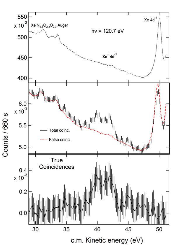
curve) by normalization to the electron signal, because the photoion signal remained constant during
  the recording of the spectra. Upon subtraction, only the signal due to the Xe+ ions remains visible
  (Figure 2, bottom).

  II. B HERMES Experiment
          The core + valence double ionization experiment on neutral Xe atoms was performed with the
  son of MAIA, the HERMES (High Energy Resolution Multi Electron coincidence Spectroscopy)
  experiment, on SEXTANTS beam line of the synchrotron radiation facility Soleil [16]. The HERMES
  set up has been described previously (see [17] and references included). Briefly, multi electron
  coincidences were recorded using a magnetic bottle electron time-of-flight spectrometer of the type
  developed by Eland et al [18]. The single-bunch operation mode of the synchrotron was used and the
  electron flight times were measured by a Time to Digital Converter (TDC) with 120 ps discretization
  step (‘TDC-V4’ developed at the LUMAT federation in Orsay, France). A mechanical chopper was
  applied to reduce the number of light bunches by a factor of ten, in order to effectively extend the 1184
  ns single bunch repetition time to ~12.5µs [19]. Calibration and conversion from electron time of flight
  to the electron kinetic energy was based on the Xe 4d Auger electrons whose energies are precisely
  known [20]. The energy resolution ∆E for electrons of energy E is found to obey the relation ∆E / E =
  1.6 % for E above 1 eV and is limited to ~20meV for smaller energies. The core-valence double
  ionization of Xe atoms was investigated at two photon energies (135 and 120 eV). The results
  presented here were obtained at the lower photon energy after accumulating the spectra for ~5 hours.
  The photon energy resolution was set to 15 meV. In order to diminish false coincidences, the electron
  count rate was limited to 2 kHz.

III. THEORY:
          Calculations were performed within the well-established configuration interaction Dirac-Fock
  framework using the Grasp2k code [21]. In the method one first solves relativistic one-electron wave
  functions from the Dirac-Fock equations in an average level scheme. Then the final atomic state
  functions are solved by diagonalizing the Dirac-Coulomb Hamiltonian in a basis of jj-coupled
  configuration state functions. The final states are thus linear combinations of configuration state
  functions of the same parity P, total angular momentum J and its projection M. For further details, see
  for example Refs. [21] [22].
          The atomic and singly charged ionic states were constructed from Xe [4d105s25p6] and Xe+
  [4d95s25p6] configurations, respectively. The calculation of Xe2+ and Xe3+ states included also some
  excitations to 4f, 5d, 6s and 6p orbitals. The Xe2+ states were constructed from odd parity Xe2+
  [4d95s25p5, 4d95s25p44f, 4d95s25p46p] configurations and Xe3+ states from Xe3+ [5s05p5, 5s5p4, 5s25p3]
  and Xe3+ [5p4nl, 5s5p3nl, 5s25p2nl], where nl is 4f, 5d, 6s or 6p. From the Xe2+ states only the lowest
  12 states in energy were calculated as they correspond to the measured Xe2+ [4d95s25p5] states. For
  Xe3+ the above given configurations yield 411 states from which the lowest 129 are energetically
  accessible from the 12 Xe2+ initial states by the Auger decay. The lifetimes of Xe+ [4d9] states were
  obtained from calculated Auger rates to states constructed from the main Xe2+[5s-15p-1] configurations
  and the correlating configurations Xe2+[4f25s5p3, 4f25s05p4, 4f5s5p4, 4f5s25p3, 4f5s05p5, 5s25p35d,
  5s25p25d2, 5s5p45d, 5s05p45d2].

                                                                                                         4
The fluorescence, one-electron photoionization and Auger decay transition matrix elements
   were obtained using the reos, photo and Auger components of the RATIP utility package of GRASP
   [23], respectively. All dipole matrix elements were calculated in the length gauge. The relative
   Xe→Xe2+ double ionization cross-sections were estimated simply by assuming them to be directly
   proportional to the statistical weights (2J+1) of the final LS states of the doubly charged ion. The LS
   coupling coefficients were obtained by transforming the calculated jj-coupled configuration state
   function coefficients using the LSJ program [24].

IV. RESULTS and discussion
   IVa) Results with MAIA experiment on the 4d innershell
   ionization of Xe+ ions.
          Figure 3 (B) shows our measurement of the photoelectron spectrum resulting from 4d
   photoionization of Xe+ ions:
          Xe+ + hν →       Xe2+* (4d-15p-1) + e−ph              (1)

            The data are identical to those in Fig. 2 bottom with the difference that they are here presented
   on a binding energy scale relative to the ground state of Xe+ ion. Note that for the experiments
   performed on the neutral species, see below, a binding energy scale relative to the ground state of the
   Xe atom is used; these two scales differ by 12.13 eV equal to the 5p ionization potential in Xe I. The
   data are compared with the pioneering measurements published in 1999 by Gottwald et al (A) [4].
   Large error bars clearly testify the difficulty of such an experiment. However, we clearly observe a
   broad unresolved photoelectron peak located in the 78-82 eV binding energy region. This is in
   disagreement with Gottwald et al who instead report on three broad structures at 72.2, 74.9 and 76.2
   eV, that is in a region where we do not detect any photoelectron signal within our error bars. The
   expected energy positions of the Xe2+ 4d-15p-1 states which are populated upon 4d shell ionization of
   Xe+ (5p-1) states can be deduced from the literature, namely from the measurement by Kivimäki et al
   [25] of the ‘4p’ photoelectron spectrum of the Xe atom and of the associated ‘N3’ N4,5O2,3 Coster-
   Kronig decay to these Xe2+ 4d-15p-1 states. They are summarized in Table 1 using the binding energy
   scale relative to the ground state of the Xe atom. Moreover, they are indicated by vertical bars in Figure
   3, where the broad blue and thin red bars show the expected energy positions for an ionization of Xe+
   ion in the ground state 5p-1 (2P3/2) and the metastable state 5p-1 (2P1/2), respectively. Clearly, these
   estimates match our experimental results and not those of Gottwald et al.
           To confirm this interpretation further, we have calculated the cross section for 4d inner shell
   photoionization of Xe+ ions, both from the 2P3/2 ground state and from the 2P1/2 excited state. The
   results of the calculation performed at a photon energy of 120.7eV are given in Table 1. The calculated
   cross sections are displayed in Figure 3 (C) using the same color convention as for the expected
   positions, see above: blue shows photoionization from the Xe+ ground state 5p-1 (2P3/2), while red
   displays photoionization from the Xe+ excited state 5p-1 (2P1/2). The two contributions have been
   weighted by taking into account the estimated initial populations of ground state and metastable Xe+
   ions (see § II.A). The shape of the peaks is a pure Lorentzian curve, reflecting the calculated lifetime
   of the individual Xe2+ 4d-15p-1 states (see Table 1). A convolution with a 700 meV FWHM Gaussian
   function (in black) is used to simulate experimental resolution; the comparison fully confirms that the

                                                                                                           5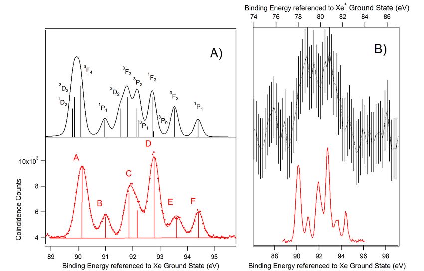
broad unresolved structure that we observe at 78-82eV corresponds to 4d photoelectron peaks in Xe+
ion. A closer look at the intensity of the calculated photoelectron peaks (see Figure 3 and Table 1)
shows that the photoionization of the Xe+ ion in the ground and in the metastable state results in
completely different populations of the Xe2+ 4d-15p-1 final states. For instance, the population of the
Xe2+ states at lower binding energy is suppressed when starting from the Xe+ metastable state. This
effect originates from the selection and propensity rules of the single photoionization process. For
instance, if we consider 4d photoionization from the metastable Xe+ state 5p-1 (2P1/2), and assume that
5p1/2-1 hole remains a spectator, then the resulting Xe2+ final states will be in major part composed of
4d3/2-15p1/2-1 or 4d5/2-15p1/2-1 configuration. The SLj terms generated from these configurations do not
include the low-lying 3F4 and 3P0 terms, which explains why these levels are found to have zero
intensity in our calculation (see Table 1).
A further argument demonstrating the validity of our present measurement of the 4d
photoelectron spectrum of Xe+ ions is given by a comparison with the results of the core valence double
ionization of the neutral Xe atom that we present next.
IVb) Results with HERMES experiment on the core valence
double ionization of neutral Xe atoms.
IV b1) Spectroscopy of the Xe2+ 4d-15p-1states.
The HERMES experiment was used to study the core-valence double photoionization of neutral
Xe atoms, and the subsequent Auger decay of these Xe2+ intermediate states:
Xe + hν → Xe2+* (4d-15p-1) + e−ph1 + e−ph2 (2a)
→ Xe2+* (4d-15p-1) →Xe3+ + e−Auger (2b)
This study is motivated by the fact that the same Xe2+* (4d-15p-1) intermediate states are
populated here as in the photoionization of Xe+ ions, see equation (1). Although they are formed
presumably with different relative populations because of the different formation mechanisms, it is
possible to compare directly the binding energies obtained by the two methods. The experiment
consists in searching for the three-electron coincidence events (e−ph1, e−ph2, e−Auger) associated with
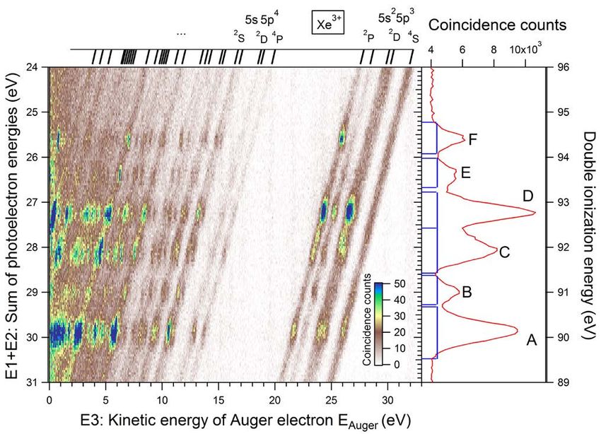
reaction (2) from the multi coincidence data set obtained by HERMES. In Figure 4 we plot energy
correlations for such three electrons events, presenting them as a function of the sum E1+E2 of the
kinetic energies of two electrons from the triplet -y axis- and of the kinetic energy E3 of the third
electron -x axis-. The energy scale ranges in Figure 4 are selected so that E1+E2 matches the sum of
kinetic energies of the two photoelectrons and E3 the energy of the Auger electron. The two-
dimensional plot shows diagonal lines on which intense dots are superimposed, plus a weak
background continuum of false coincidences which increases in intensity at low E3 values.
Along each diagonal line, the sum of the kinetic of the three electrons (E1 + E2 + E3) is
constant, indicating that these lines correspond to true coincidences associated with the formation of
different Xe3+ final states, which are labelled above the triple coincidence map in Figure 4. Apart from
the core valence double ionization followed by an Auger decay (2), two other processes can contribute
to the intensity of diagonal lines: direct triple ionization which is expected to be weak and unstructured,
6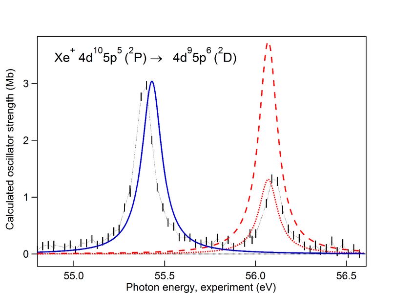
and double Auger decay of Xe+ 4d-1 states and of their satellites. Double Auger decay of Xe+ 4d-1 holes
has been studied by Penent et al [26]; that of the 4d-1 satellites has not been reported in the literature
to the best of our knowledge, but it is expected that they behave similar to those of the Kr+ 3d-1 satellites
studied by Anderssen et al [27]. From these studies and from energy considerations, we can deduce
that Double Auger decay of Xe+ 4d-1 and of the corresponding satellite states will not contribute in the
energy range of Figure 4. The core valence double ionization contribution that we are looking for, will
increase the intensities at specific ‘’spots’’ along the diagonal lines when the third electron e−3 is the
Auger electron. These spots are elongated in the vertical direction because the absolute energy
resolution for the low energy E3 electrons is better than for the sum of (E1+E2). Since in the present
case all Auger electron energies lie within the range covered by the possible energies of the two
photoelectrons, the core valence double ionization itself will also contribute to the background
intensity along the diagonal lines of Figure 4. These contributions are caused by the combinations (e−1,
e−2, e−3) of the type (e−ph1, e−Auger, e−ph2), i.e. where e−3 is a photoelectron and e−1 or e−2 is the Auger
electron; they show for a given Auger energy EAuger the energy sharing between the two photoelectrons.
This type of energy sharing is found to be mainly flat and unstructured due to the dominant direct core
valence double photoionization process. However, the sharing contains also minor structured
contributions of weak cascade processes caused by the formation of highly excited Xe+ 4d-1 5p-1 nl
satellite states and their subsequent spin-flip Auger decay to Xe2+ 4d-15p-1 states with the release of a
low energy Auger electron. Such type of energy sharing is not shown here, but it is similar to those
observed in the core valence double photoionization paths1s-1 2p-1 in Ne [7], 2p-1 3p-1 in Ar [28],
4f-16s-1 and 4f-15d-1 in Hg [9] or 3d-14p-1 in Kr [11].
         The spots along the lines in Figure 4 thus give access to process (2). The horizontal bands
formed by the spots trace the different Xe2+* (4d-15p-1) intermediate states, while the abscissa of the
spots along the lines give the kinetic energies for their Auger decays. Integration along the x-axis gives
the spectrum of the Xe2+* (4d-15p-1) states which is presented on the right hand side of Figure 4. This
spectrum is presented by dots in the bottom of Figure 5 (A) and shows six broad peaks. In order to
reproduce also the asymmetric shape of peak C it is fitted by seven Voight profiles with a fixed
Lorentzian contribution and widths of 70 ± 30 meV, which agrees well with the averaged calculated
lifetime broadening reported in Table 1. The Gaussian widths for the two components of peak C, as
well as for peaks B, E and F which are predicted to correspond to a single Xe2+* (4d-15p-1) state amount
to 350 ± 30meV and are mainly caused by the experimental resolution. Larger Gaussian widths were
necessary to fit peaks A and D, reflecting the presence of the unresolved components predicted by
theory. The resulting experimental binding energies are reported in Table 1. They match very well the
results deduced from the ‘4p’ Auger decay of Kivimäki et al [25], but are found ~200-500 meV lower
than the values obtained by Bolognesi et al [29] in their threshold-photoelectron coincidence spectra.
Our calculated results, shown in the top panel of Figure 5 (A), reproduce well the photoelectron
spectrum, enabling the identification of the observed Xe2+* (4d-15p-1) states. The calculated binding
energies are found to lie ~2eV below the observed values, which is typical of this kind of ab-initio
calculations. In the present approach the intensities are obtained by simply assuming that individual
Xe2+* (SLJ) states are populated according to their (2J+1) statistical weight. In this way a good overall
agreement with the measured spectrum is obtain which possibly reflects the nature of the observed
dominant direct double photoionization process. Small differences remain, such as a too low predicted
intensity for the D band; it is expected that they can be explained with more accurate ab-initio
calculations, such as those developed by S. Fritzsche for Kr [11] and Hg [9] core-valence double
ionization. Note that the predicted lifetimes of the Xe2+* (4d-15p-1) states vary by a factor 2 and are on
average longer than the lifetimes of a single 4d-1 hole: Jurvansuu et al [15] measured a lifetime
broadening of 104±3 meV for the Xe+ (4d-1) 2D3/2 state and of 111±3meV for the (4d-1) 2D3/2 one. This
on average longer lifetime can be explained by the main decay processes of the 4d-1 hole, which is the

                                                                                                           7
N4,5OO Auger decay. In case of a 4d-1 hole in Xe2+* (4d-15p-1) there is one 5p electron less in the O
shell as compared to the Xe+ (4d-1) state. This reduces the number of possible N4,5OO Auger channels
and increases the lifetime. The resolution in the present experiment is unfortunately not sufficient to
probe lifetime predictions.
Finally we compare in Figure 5 (B) our measurement of the Xe2+* (4d-15p-1) states reached by
core valence double ionization of the Xe atom, to that of Figure 3 where they are reached by 4d
ionization of the Xe+ ion. This comparison demonstrates the difficulty of performing photoelectron
spectroscopy on ionic targets, but also fully confirms that our determination of the 4d photoelectron
spectrum from Xe+ ions is correct.
IV b2) Auger decay of the Xe2+ 4d-15p-1states.
From Figure 4 one can extract the Auger spectrum for each of the 6 resolved components A to
F of Xe (4d-15p-1) initial states. These spectra are given by the intensity along each the corresponding
2+*
horizontal lines in the three-electron correlation map, and are represented in Figure 6. The background
signal due to the unstructured intensity along the diagonal lines is represented by black curves. It has
been estimated from the signal at E1+E2 energies in the 24-25eV range where no Xe2+* (4d-15p-1) states
are present. The Auger spectra of the six components A to F are obviously very different. Note that
the fastest Auger electron has a kinetic energy around 28 eV, an energy well below the fastest Auger
electron energy (36.422eV) associated with the decay of Xe+ (4d-1) states [20].
In order to understand better the Auger decay path (2b), we have calculated these Auger spectra
following the method described in chapter III. They are represented in the right panel of Figure 7 and
are plotted, contrary to Figure 6, as a function of the triple ionization energy, i.e. the binding energy
of the final Xe3+ states. In this way only 5 peaks are obtained for the Auger decay to Xe3+ (5s25p3)
states, even for Xe2+* bands with unresolved states, such as A. These predictions are compared to the
experiment results shown in the left panel of Figure 7, which have been obtained by integration along
the diagonal lines of the two dimensional plot of Figure 4 (instead of a simple projection on the x-axis,
as was done for Figure 6). Background has been subtracted from the experimental curves in Figure 7.
One observes an excellent agreement between theory and experiment, especially for the Auger decays
ending in the lower Xe3+ (5s25p3) states. The strong selectivity of the Auger decay is nicely reproduced,
see for instance the decay of the Xe2+* (1P1) state (F peak) which is found both experimentally and by
theory to populate selectively the 2P3/2 level of the Xe3+ (5s25p3) states. Figure 7 shows that agreement
is less good for the slower Auger electrons associated with the population of the higher energy Xe3+
final states. Two reasons can be invoked: some background may remain in our experimental spectrum
and more configurations should be included in the calculations.
V CONCLUSION
We have measured directly the 4d photoelectron spectrum of Xe+ ions of both, the 2P3/2 ground
state and the 2P1/2 metastable state. Such a measurement is extremely difficult due to the low density
of ion beams and was possible only thanks to the availability of an intense synchrotron source such as
the PLEIADES beam line, and thanks to the use of electron / ion coincidence techniques. Our results
are at variance with the earlier result from Gottwald et al [4], but we validate them by calculations and
8by a complementary experimental method, namely the core-valence double ionization of the neutral
Xe atom. This approach gives detailed access to the spectroscopy of the Xe2+* (4d-15p-1) levels which
are also populated by inner shell ionization of Xe+ ions. Moreover, we studied the Auger decay of the
Xe2+* (4d-15p-1) levels, which is found to be extremely selective. These results are another example of
the power of multi-electron coincidence experiments studies of the multi-photoionization of neutral
atoms, to retrieve information on the photoionization process of ions, that we illustrated in the case of
a 2p inner shell ionization of an Ar+ ion [30].
Acknowledgments:
The experiment was performed at SOLEIL Synchrotron (France) at the PLEIADES and
SEXTANTS beam line, with the approval of the SOLEIL Peer Review Committee (Projects No.
20150198 and 20150359). We are grateful to J. Bozek, A. R. Milosavljevic, C. Nicolas, E. Robert, N.
Jaouen and PLEIADES and SEXTANTS teams for help during the measurements, and to SOLEIL
staff for stable operation of the storage ring. We are indebted to P. Selles for fruitful discussions. M.
A. K. acknowledges the support of the Labex Plas@Par managed by the Agence Nationale de la
Recherche, as part of the “Programme d’Investissements d’Avenir” under Reference No. ANR-11-
IDEX-0004-02. This work has been financially supported by the Research Council for Natural
Sciences and Engineering of the Academy of Finland.
9________________________________________________________________________________________________________________
4d-15p-1 Binding Energy 4d-15p-1 Assignment Lifetime Calculated cross section for
(relative to Xe Ground State) JJ coupling LS coupling Broadening 4d photoionization (hv 120.7eV) of
Experiment [25] [29] Theory (j1, j2) J c2 2S+1
LJ c12 2S+1LJ c22 Xe+ 2P3/2 Xe+* 2P1/2
- eV - - % % % meV Mb Mb
1
90.39 1) 87.99 (5/2,3/2) 2 91 D2 69 48 0.335 0.063
A) 90.14 90.05 90.67 2) 88.07 (5/2,3/2) 3 94 3
D3 90 79 0.586 0.067
3
90.20 90.86 3) 88.28 (5/2,3/2) 4 100 F4 100 35 2.095 0
B) 91.00 90.97 3D1 91.19 3D1 4) 89.18 (5/2,3/2) 1 92 1
P1 43 3
D1 32 51 0.213 0.020
3
91.57 91.93 5) 89.74 (3/2,3/2) 2 67 D2 87 81 0.256 0.298
C1) 91.86 91.88 92.30 6) 90.00 (5/2,1/2) 3 71 3
F3 93 37 0.179 2.506
C2) 92.17 92.74 7) 90.36 (5/2,1/2) 2 61 3
P2 78 43 0.130 0.769
3 3
8) 90.40 (3/2,3/2) 1 89 P1 50 D1 49 70 0.207 0.030
D) 92.78 92.70 93.47 9) 90.91 (3/2,3/2) 3 70 1
F3 87 47 0.820 0.430
3
93.12 10) 90.96 (3/2,3/2) 0 100 P0 100 98 0.069 0
E) 93.60 93.45 94.11 11) 91.73 (3/2,1/2) 2 99 3
F2 82 37 0.006 2.759
F) 94.41 94.32 94.98 12) 92.59 (3/2,1/2) 1 89 1
P1 56 3
P1 25 44 0.026 0.743
______________________________________________________________________________________________________________
Table 1 : Binding Energies (with respect to the Xe ground state) and assignments of the Xe2+ (4d-15p-1) core-valence states in both, (j,j) J and LSJ
coupling (j1 = 4d-1j1 j2=5p-1j2 ). The present experimental values (1st column) are compared to the ‘4p’ Auger experiment by Kivimaki et al [25]
and to the threshold electron coincidence experiment by Bolognesi et al [29]. The table gives also the calculated lifetime broadening and the cross
section for formation by photoionization of a Xe+ ion.
10Figure 1: Experiment with MAIA setup on Xe+ ions. Xe2+ ion yield recorded in the region of the 4d
→ 5p resonances with 20meV band pass. The vertical error bars represent the statistical uncertainty.
The resonance at lower energy corresponds to the 2P3/2 → 2D5/2 transition and the higher one to the
2
P1/2 → 2D3/2 transition. The photon energy scale has been calibrated using data from ref [14].
Experimental results are scaled to the calculated cross section of the lower lying resonance (blue solid
line); adjustment of the calculated cross section for the second resonance (red dashed curve) with the
experiment values (red dotted curve) gives the proportion of Xe+ ions in the ground (2P3/2) and
metastable (2P1/2) states, which is found to be 74 % and 26 %. See text for details. Note that the
theoretical curves have been shifted by +150 meV to match better the positions of the resonances.
11Figure 2: Experiment with MAIA setup on Xe+ ions. Top: Raw photoelectron spectrum, represented
as a function of the Center of Mass kinetic energy. Middle: Photoelectron spectrum in coincidence
with Xe3+ ions (points with error bars). The red (dotted) curve gives the estimated false-coincidence
spectrum (see text for details). The bottom curve represents the resulting true coincidence spectrum
(line with error bars). The photon energy was 120.7eV and the spectral band pass was 110 meV. The
error bars give the statistical uncertainty.
12Figure 3: (B, middle) Experiment with MAIA setup: 4d photoelectron spectrum from photoionization
of Xe+ ions with 120.7eV photons (middle). It is compared to the previous experiment by Gottwald et
al [4] at hv = 103.3eV (A, top). The broad blue and thin red vertical bars indicate the expected energy
positions for the Xe+ 5p-1 (2P3/2) → Xe2+ 4d-15p-1 and the Xe+ 5p-1 (2P1/2) → Xe2+ 4d-15p-1 transitions,
respectively; the transition energies are estimated using the 4d-15p-1 binding energies of Kivimäki et al
[25]. The blue (solid) and red (dotted) curves in the bottom graph C show the calculated Xe+ (2P3/2) →
Xe2+ 4d-15p-1 and Xe+ (2P1/2) → Xe2+ 4d-15p-1 photoelectron spectra, respectively. The spectra are
weighted according to the estimated Xe+ (2P3/2) to Xe+ (2P1/2) ratio in the ion beam. The lines are
broadened by the Lorentzian lifetime contribution only. The black line shows the calculated spectrum
with additional 700 meV Gaussian broadening to simulate the effect of the experimental resolution.
Note that theoretical curves have been shifted by +2eV to match the positions expected from reference
[25] -vertical bars-.
13Figure 4: Experiment with HERMES setup. Core valence double photoionization of neutral Xe atoms
at 120eV photon energy. Left: Coincidence map for events where three electrons have been detected
in coincidence. The coincidence map shows the energy correlations between the sum of the kinetic
energies of two of them (E1+E2) -y axis- versus the kinetic energy of the third one E3 -x axis-. The right
panel shows the projection of the coincidence map on the y axis, revealing the Xe2+ 4d-15p-1 states.
The rectangles indicate the peak boundaries selected for the analysis of the Auger spectra in Figure 6
and 7.
14Figure 5. A) Bottom (red): Xe2+* (4d-15p-1) states observed by core valence double ionization, from
Figure 4 (right). The vertical bars represent the energy positions and intensities obtained from the fit
analysis, see text. Top: our calculations, see Table 1 for assignment of peaks; intensity is given by
statistical populations. Note that the theoretical curves have been shifted by +2eV to match the
positions of the experimental peaks. B): Comparison between MAIA and HERMES experiments.
15Figure 6: Experiment with HERMES setup. Auger spectra for the decay of each group of Xe2+ 4d-15p-
1
core-valence states defined in Figure 4 and 5. These Auger spectra are deduced from the experiment
in Figure 4, as the intensity along each line associated to the corresponding group of core-valence
states. The black dotted lines give the estimated background, obtained out of the core-valence peak
region (see text for further explanation).
16Figure 7: The Xe3+ final state energy spectra for the Auger decay of the Xe2+ 4d-15p-1 core-valence
states, represented as a function of the binding energy of the final Xe3+ states: experiment with
HERMES setup (left) and theory (right). The top panels present the total final state energy spectra
while the panels A to F show the spectra of the different groups of 4d-15p-1 states defined in Figs. 4
and 5. The experimental results have been obtained by integration along the diagonal lines in Fig. 4.
Contrary to Fig. 6, the background has been subtracted here. The spectra are calibrated using a triple
ionization energy of 64.09 eV [26] for the ground state of Xe3+. The vertical bars above the uppermost
panels indicate the energy positions and assignments of the Xe3+ levels as reported in the NIST tables.
17REFERENCES:
[1] J.M. Bizau, D. Cubaynes, M. Richter, F.J. Wuilleumier, J. Obert, J.C. Putaux, T.J. Morgan, E.
Källne, S. Sorensen, A. Damany, First observation of photoelectron spectra emitted in the
photoionization of a singly charged-ion beam with synchrotron radiation, Phys. Rev. Lett. 67
(1991) 576–579. doi:10.1103/PhysRevLett.67.576.
[2] S. Al Moussalami, J.M. Bizau, B. Rouvellou, D. Cubaynes, L. Journel, F.J. Wuilleumier, J.
Obert, J.C. Putaux, T.J. Morgan, M. Richter, First Angle-Resolved Photoelectron Measurements
following Inner-Shell Resonant Excitation in a Singly Charged Ion, Phys. Rev. Lett. 76 (1996)
4496–4499. doi:10.1103/PhysRevLett.76.4496.
[3] A. Gottwald, S. Anger, J.-M. Bizau, D. Rosenthal, M. Richter, Inner-shell resonances in
metastable Ca + ions, Phys. Rev. A. 55 (1997) 3941–3944. doi:10.1103/PhysRevA.55.3941. and
A. Gottwald, PhD thesis, Technischen Universität Berlin (1999).
[4] A. Gottwald, C. Gerth, M. Richter, 4 d Photoionization of Free Singly Charged Xenon Ions,
Phys. Rev. Lett. 82 (1999) 2068–2070. doi:10.1103/PhysRevLett.82.2068.
[5] J.M. Bizau, D. Cubaynes, S. Guilbaud, N. El Eassan, M.M. Al Shorman, E. Bouisset, J. Guigand,
O. Moustier, A. Marié, E. Nadal, E. Robert, C. Nicolas, C. Miron, A merged-beam setup at
SOLEIL dedicated to photoelectron–photoion coincidence studies on ionic species, J. Electron
Spectrosc. Relat. Phenom. 210 (2016) 5–12. doi:10.1016/j.elspec.2016.03.006.
[6] J.-M. Bizau, D. Cubaynes, S. Guilbaud, F. Penent, P. Lablanquie, L. Andric, J. Palaudoux, M.M.
Al Shorman, C. Blancard, Photoelectron Spectroscopy of Ions: Study of the Auger Decay of the
4 d → n f ( n = 4 , 5 ) Resonances in Xe 5 + Ion, Phys. Rev. Lett. 116 (2016).
doi:10.1103/PhysRevLett.116.103001.
[7] Y. Hikosaka, T. Aoto, P. Lablanquie, F. Penent, E. Shigemasa, K. Ito, Experimental
Investigation of Core-Valence Double Photoionization, Phys. Rev. Lett. 97 (2006).
doi:10.1103/PhysRevLett.97.053003.
[8] S.-M. Huttula, P. Lablanquie, L. Andric, J. Palaudoux, M. Huttula, S. Sheinerman, E. Shigemasa,
Y. Hikosaka, K. Ito, F. Penent, Decay of a 2 p Inner-Shell Hole in an Ar + Ion, Phys. Rev. Lett.
110 (2013). doi:10.1103/PhysRevLett.110.113002.
[9] M. Huttula, S.-M. Huttula, S. Fritzsche, P. Lablanquie, F. Penent, J. Palaudoux, L. Andric, Core-
valence double photoionization of atomic mercury, Phys. Rev. A. 89 (2014).
doi:10.1103/PhysRevA.89.013411.
[10] S.-M. Huttula, J. Soronen, M. Huttula, F. Penent, J. Palaudoux, L. Andric, P. Lablanquie,
Auger decay of core valence double photoionized states in atomic mercury, J. Phys. B At. Mol.
Opt. Phys. 48 (2015) 115001. doi:10.1088/0953-4075/48/11/115001.
[11] E. Andersson, P. Linusson, S. Fritzsche, L. Hedin, J.H.D. Eland, L. Karlsson, J.-E.
Rubensson, R. Feifel, Formation of Kr 3 + via core-valence doubly ionized intermediate states,
Phys. Rev. A. 85 (2012). doi:10.1103/PhysRevA.85.032502.
[12] B. Rouvellou, J.M. Bizau, D. Cubaynes, L. Journel, S. Al Moussalami, F.J. Wuilleumier, A
dedicated electron spectrometer for photoionization studies of atomic ions with synchrotron
radiation, J. Electron Spectrosc. Relat. Phenom. 76 (1995) 237–243. doi:10.1016/0368-
2048(95)02536-7.
[13] S. Jullien, J. Lemaire, S. Fenistein, M. Heninger, G. Mauclaire, R. Marx, Radiative lifetimes
of Xe+ and Kr+ in their 2P12 spin—orbit states, Chem. Phys. Lett. 212 (1993) 340–346.
doi:10.1016/0009-2614(93)89335-F.
[14] P. Andersen, T. Andersen, F. Folkmann, V.K. Ivanov, H. Kjeldsen, J.B. West, Absolute cross
sections for the photoionization of 4d electrons in Xe + and Xe 2+ ions, J. Phys. B At. Mol. Opt.
Phys. 34 (2001) 2009–2019. doi:10.1088/0953-4075/34/10/314.
[15] M. Jurvansuu, A. Kivimäki, S. Aksela, Inherent lifetime widths of Ar 2 p − 1 , Kr 3 d − 1 , Xe
3 d − 1 , and Xe 4 d − 1 states, Phys. Rev. A. 64 (2001). doi:10.1103/PhysRevA.64.012502.
18[16] M. Sacchi, N. Jaouen, H. Popescu, R. Gaudemer, J.M. Tonnerre, S.G. Chiuzbaian, C.F.
Hague, A. Delmotte, J.M. Dubuisson, G. Cauchon, B. Lagarde, F. Polack, The SEXTANTS
beamline at SOLEIL: a new facility for elastic, inelastic and coherent scattering of soft X-rays, J.
Phys. Conf. Ser. 425 (2013) 72018. doi:10.1088/1742-6596/425/7/072018.
[17] J. Palaudoux, S. Sheinerman, J. Soronen, S.-M. Huttula, M. Huttula, K. Jänkälä, L. Andric,
K. Ito, P. Lablanquie, F. Penent, J.-M. Bizau, S. Guilbaud, D. Cubaynes, Valence Auger decay
following 3 s photoionization in potassium, Phys. Rev. A. 92 (2015).
doi:10.1103/PhysRevA.92.012510.
[18] J.H.D. Eland, O. Vieuxmaire, T. Kinugawa, P. Lablanquie, R.I. Hall, F. Penent, Complete
Two-Electron Spectra in Double Photoionization: The Rare Gases Ar, Kr, and Xe, Phys. Rev.
Lett. 90 (2003). doi:10.1103/PhysRevLett.90.053003.
[19] K. Ito, F. Penent, Y. Hikosaka, E. Shigemasa, I.H. Suzuki, J.H.D. Eland, P. Lablanquie,
Application of a simple asynchronous mechanical light chopper to multielectron coincidence
spectroscopy, Rev. Sci. Instrum. 80 (2009) 123101. doi:10.1063/1.3258200.
[20] T.. Carroll, J.. Bozek, E. Kukk, V. Myrseth, L.. Sæthre, T.. Thomas, K. Wiesner, Xenon
N4,5OO Auger spectrum—a useful calibration source, J. Electron Spectrosc. Relat. Phenom. 125
(2002) 127–132. doi:10.1016/S0368-2048(02)00134-2.
[21] P. Jönsson, X. He, C. Froese Fischer, I.P. Grant, The grasp2K relativistic atomic structure
package, Comput. Phys. Commun. 177 (2007) 597–622. doi:10.1016/j.cpc.2007.06.002.
[22] K.G. Dyall, I.P. Grant, C.T. Johnson, F.A. Parpia, E.P. Plummer, GRASP: A general-purpose
relativistic atomic structure program, Comput. Phys. Commun. 55 (1989) 425–456.
doi:10.1016/0010-4655(89)90136-7.
[23] S. Fritzsche, The Ratip program for relativistic calculations of atomic transition, ionization
and recombination properties, Comput. Phys. Commun. 183 (2012) 1525–1559.
doi:10.1016/j.cpc.2012.02.016.
[24] G. Gaigalas, T. Zalandauskas, S. Fritzsche, Spectroscopic LSJ notation for atomic levels
obtained from relativistic calculations, Comput. Phys. Commun. 157 (2004) 239–253.
doi:10.1016/S0010-4655(03)00518-6.
[25] A. Kivimäki, H. Aksela, J. Jauhiainen, M. Kivilompolo, E. Nõmmiste, S. Aksela,
Interpretation of the N2,3N4,5O2,3 Coster–Kronig spectrum of xenon, J. Electron Spectrosc.
Relat. Phenom. 93 (1998) 89–94. doi:10.1016/S0368-2048(98)00160-1.
[26] F. Penent, J. Palaudoux, P. Lablanquie, L. Andric, R. Feifel, J.H.D. Eland, Multielectron
Spectroscopy: The Xenon 4 d Hole Double Auger Decay, Phys. Rev. Lett. 95 (2005).
doi:10.1103/PhysRevLett.95.083002.
[27] E. Andersson, S. Fritzsche, P. Linusson, L. Hedin, J.H.D. Eland, J.-E. Rubensson, L.
Karlsson, R. Feifel, Multielectron coincidence study of the double Auger decay of 3 d -ionized
krypton, Phys. Rev. A. 82 (2010). doi:10.1103/PhysRevA.82.043418.
[28] M. Nakano, Y. Hikosaka, P. Lablanquie, F. Penent, S.-M. Huttula, I.H. Suzuki, K. Soejima,
N. Kouchi, K. Ito, Auger decay of Ar 2 p satellite states studied with a multielectron coincidence
method, Phys. Rev. A. 85 (2012). doi:10.1103/PhysRevA.85.043405.
[29] P. Bolognesi, L. Avaldi, M.C.A. Lopes, G. Dawber, G.C. King, M.A. MacDonald, C. Villani,
F. Tarantelli, Direct observation of the Kr ( 3 d − 1 4 p − 1 ) and Xe ( 4 d − 1 5 p − 1 ) doubly
charged ion states by threshold-photoelectron coincidence spectroscopy, Phys. Rev. A. 64
(2001). doi:10.1103/PhysRevA.64.012701.
[30] S.-M. Huttula, P. Lablanquie, L. Andric, J. Palaudoux, M. Huttula, S. Sheinerman, E.
Shigemasa, Y. Hikosaka, K. Ito, F. Penent, Decay of a 2 p Inner-Shell Hole in an Ar + Ion, Phys.
Rev. Lett. 110 (2013). doi:10.1103/PhysRevLett.110.113002.
1920
You can also read


