Case Report: Reconstruction After Anterior Pubic Hemipelvectomy - Frontiers
←
→
Page content transcription
If your browser does not render page correctly, please read the page content below
CASE REPORT
                                                                                                                                                 published: 21 May 2021
                                                                                                                                        doi: 10.3389/fsurg.2021.585600

                                             Case Report: Reconstruction After
                                             Anterior Pubic Hemipelvectomy
                                             Benjamin Murphy 1*, Tharsa Thillainadesan 1 , Kerian Robinson 1 , Anita Clarke 2 and
                                             Peter Choong 1,3
                                             1
                                               Department of Orthopaedic Surgery, St Vincent’s Hospital, Melbourne, VIC, Australia, 2 Department of Urology, St Vincent’s
                                             Hospital, Melbourne, VIC, Australia, 3 Department of Surgery, The University of Melbourne, Melbourne, VIC, Australia

                                             We report on a case of a large atypical cartilaginous tumor of the pelvis and its novel
                                             surgical resection with an anterior hemipelvectomy and reconstruction with an iliac
                                             crest graft. Surgical intervention is the mainstay treatment of pelvic chondrosarcomas.
                                             However, there have been reports of concern regarding preventing pelvic visceral
                                             herniation and adequately reconstructing the pelvis. This report is unique within the
                                             literature and has yielded good functional outcomes whilst achieving satisfactory surgical
                                             margins and minimizing morbidity.
                                             Keywords: reconstruction of anterior pelvis, anterior hemipelvectomy, type III hemipelvectomy, pubic resection,
                                             chondrosarcoma

                                             INTRODUCTION
                            Edited by:
                        Marco Scarpa,
                                             Chondrosarcomas are malignant tumor of mesenchymal origin whose cells produce osteoid-free
     University Hospital of Padua, Italy
                                             chondroid matrix and can arise de novo or secondary to a pre-existing benign cartilaginous
                     Reviewed by:
                                             neoplasm (1–4). They are the second most common primary bone tumor, after osteosarcoma, and
               Jonathan Stevenson,
                                             are most commonly found within the pelvis and proximal femur (1, 5, 6). Chondrosarcomas exist
         Royal Orthopaedic Hospital,
                    United Kingdom           along a continuum of severity with low grade lesions exhibiting an indolent growth pattern, whilst
                  Tommaso Stecca,            high grade lesions readily metastasize and have a poor prognosis (2, 6). Patients almost always
        ULSS2 Marca Trevigiana, Italy        present with insidious and progressive pain, usually for months to years and especially at night
                *Correspondence:             (1, 2). Other common presentations include a palpable lump, pathological fracture and mass-effect
                  Benjamin Murphy            symptoms including nerve impingement (1, 2).
         benpdsmurphy@outlook.com                Surgical resection has been the mainstay of treatment for these tumors as their abundant
                                             extracellular matrix, poor vascularity and low percentage of actively dividing cells render the
                    Specialty section:       tumors chemotherapy and radiotherapy resistant (4, 7). The surgical management of pelvic
          This article was submitted to      chondrosarcomas needs to involve en-bloc resection, especially for high grade lesions, as
                     Surgical Oncology,      intralesional resection has been demonstrated to be ineffective (8–10). Therefore, operating
                a section of the journal
                                             surgeons must balance the risk of significant morbidity against the outcomes of definitive resection
                    Frontiers in Surgery
                                             with options including hip disarticulation vs. more conservative limb sparing operations including
         Received: 28 October 2020           internal hemipelvectomy (2). Internal hemipelvectomy can be classified according to resection site
           Accepted: 26 April 2021
                                             into type 1 (ilium), type 2 (periacetabular) and type 3 (pubis) with this case involving a type 3
           Published: 21 May 2021
                                             resection of both left and right pubic bones (11).
                              Citation:
                                                 Herein, we report on a novel technique in the surgical management of a large pelvic atypical
            Murphy B, Thillainadesan T,
 Robinson K, Clarke A and Choong P
                                             cartilaginous tumor via anterior hemipelvectomy with iliac crest graft reconstruction. The tumor
  (2021) Case Report: Reconstruction         was classified as an atypical cartilaginous tumor emerging from the posterior aspect of the pubis
 After Anterior Pubic Hemipelvectomy.        causing significant displacement and obstruction of the pelvic viscera without any local infiltration.
                Front. Surg. 8:585600.       Due to the large mass resected, there was a need for reconstruction to prevent herniation which
      doi: 10.3389/fsurg.2021.585600         was achieved via computer-navigated resection followed by computer guided iliac crest autograft

Frontiers in Surgery | www.frontiersin.org                                          1                                              May 2021 | Volume 8 | Article 585600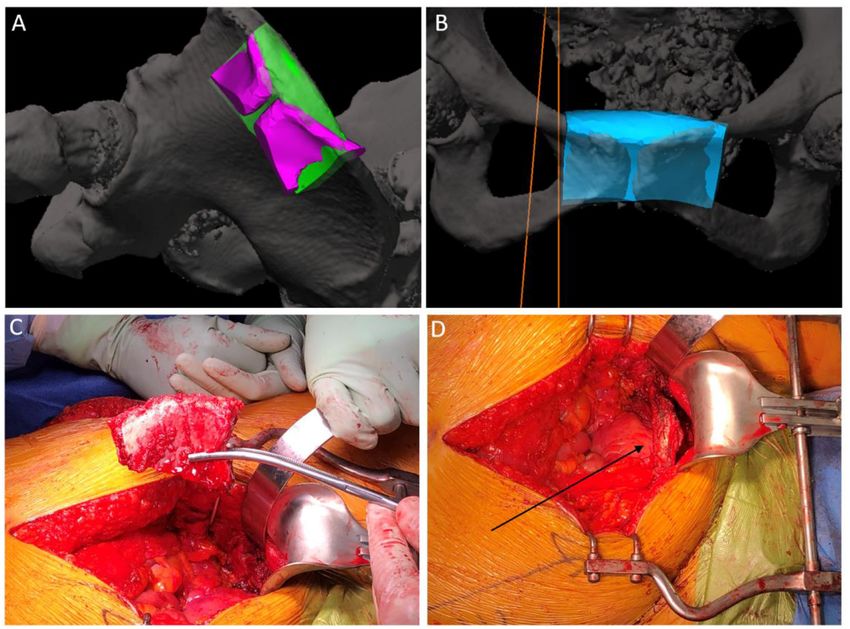
Murphy et al.                                                                                       Reconstruction After Anterior Pubic Hemipelvectomy

harvest. This case highlights key principles in orthopedic               Diagnostic Assessment
oncology as well as providing a unique approach to the                   In addition to an abdominal and pelvic CT scan, the
management of a complex case.                                            patient underwent further staging studies. These included a
                                                                         magnetic resonance imaging (MRI) which demonstrated a large
                                                                         heterogenous T2 bright lesion measuring 9 × 6 cm emerging
CASE DESCRIPTION                                                         from the posterior aspect of the right body of the pubis.
                                                                         The mass projected postero-superiorly with a large, 1 cm thick
A 54-year-old nulligravida female had a 10-year history of               cartliagenous cap and an underlying stalk that communicated
nocturia, urinary frequency and recurrent urinary tract infections       with the medullary cavity of the pelvis. There was also significant
which had been managed with short courses of antibiotics. On             effacement of the bladder with compression of the right ureter
presentation, physical examination revealed a firm hard mass             and resulting hydronephrosis. There was no infiltration of any
along the anterior vaginal wall. Her examination was otherwise           surrounding structures.
unremarkable and specifically no evidence of bony dysplasia                  Functional imaging including both thallium-201 and
of her limbs. Despite the length of symptoms, she had not                technetium-99 pentavalaent dimercaptsouccinic acid (DMSA V)
undergone any investigations for her long-standing urinary               scintigraphy were utilized and demonstrated minimal metabolic
symptoms until a computed tomography (CT) scan of her pelvis             activity within the tumor. A CT scan of the chest was performed
was organized and a large pelvic mass emerging from her pubis            which excluded pulmonary metastatic disease. Following these
was noted, prompting specialist sarcoma service referral. The            investigations, a CT-guided tissue biopsy targeting an area of
patient consented to the production of this report and it was            increased metabolic activity demonstrated a cartilaginous lesion
approved by our ethics committee.                                        with mild atypia and some focal calcification. This case was
                                                                         discussed at a multidisciplinary meeting involving sarcoma
                                                                         specialist surgeons, radiologists and pathologists. Given the size
Timeline                                                                 and histological nature of the lesion, the decision was made to
See Figure 1.                                                            manage this lesion as an atypical cartilaginous tumor.

                                                                         Therapeutic Intervention—Surgery
                                                                         This surgical intervention represents a unique approach to
                                                                         the management of a complex chondrosarcoma of the pelvis.
                                                                         The patient was given a general anesthetic and positioned in
                                                                         supine. The procedure commenced with a cystoscopy, bilateral
                                                                         retrograde pyelography and insertion of a left ureteral stent to
                                                                         alleviate obstruction. Significant distortion of the bladder and
                                                                         urethra were noted secondary to the external compression from
                                                                         the tumor. There was no local infiltration of the tumor into
                                                                         surrounding structures. The tumor was then approached via

                                                                          FIGURE 2 | 3D-CT reconstruction images of the pelvis demonstrating the
 FIGURE 1 | Timeline of case report.                                      tumor and computer navigation-system utilized intra-operatively.

Frontiers in Surgery | www.frontiersin.org                           2                                            May 2021 | Volume 8 | Article 585600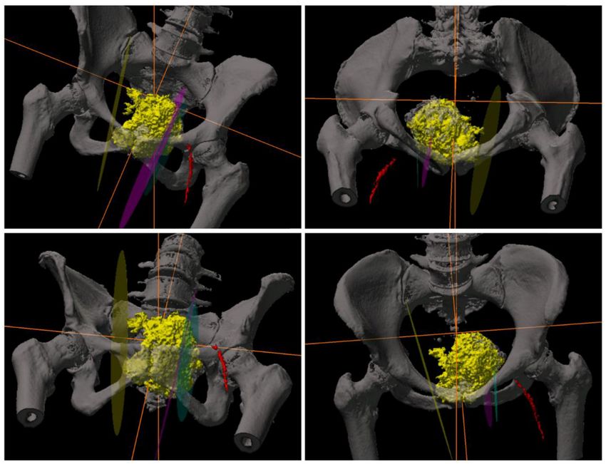
Murphy et al. Reconstruction After Anterior Pubic Hemipelvectomy
pelvic viscera from around the tumor within the lesser pelvis,
and detachment of the origins of the adductor musculature from
the anterior external pelvic wall. At this stage, navigation pins
were inserted into the iliac crest and registration of important
bony pelvic landmarks with the computer navigation system
was confirmed (Stryker Navigation System with Spinemap 3D
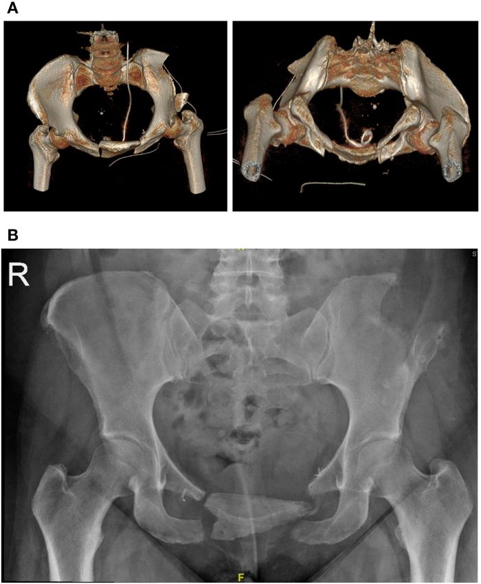
3.1 software R ) (12) (Figure 2). Using navigation, the planned
osteotomies were created through bilateral superior and inferior
pubic rami to mobilize the tumor (Figures 3A,B). The tumor was
carefully dissected from the bladder neck by rolling it forward
away from the female pelvic viscera and surrounding internal
pelvic neurovasculature and ultimately resected en-bloc from the
patient without complication. Using the same navigation system,
a preplanned segment of iliac bone was marked out on the ilium.
This segment was matched in size and curve to the resected pubic
bone using a pre-operative computer model. Osteotomy of the
FIGURE 3 | Intra-operative photos. (A) demonstration of the iliac graft was then computer-navigated (Figure 3C). The iliac
computer-navigated iliac crest resection with pubic cut overlay. (B) crest graft was prepared with drill holes and secured with Arthrex
demonstration of iliac crest craft positioning. (C) iliac crest graft post resection. fiber wire sutures (Arthrex FiberTape 2 mm R ) to the adjacent
(D) Arrow demonstrating positioning of iliac crest graft in the resected pubis.
pubic rami (Figure 3D) (13). The right and left rectus abdominis
musculature were sutured to the released ends of the right and left
gracilis muscles. Two surgical suction drains were placed, one at
the pubic resection site and the other at the iliac bone resection
site. During the operation, the patient was transfused 4 units of
packed red blood cells and 1 unit of fresh frozen plasma.
Follow-Up and Outcomes
Post-operatively, the patient was allowed to fully weight bear
and had a post-operative course consistent with a grade II
Clavien-Dindo surgical complication (i.e., minor alteration to
management) (14). She was successfully treated for a post-
operative urinary tract infection, presumably related to her Foley
catheter, with culture-specific oral antibiotics. Post-operative
scans demonstrated adequate positioning of the graft with no
immediate complications (Figure 4). The resected retropubic
mass was confirmed to be a grade one chondrosarcoma/atypical
cartilaginous tumor on histopathological analysis. There was
a >1 mm clear margin circumferentially around the tumor
which was completely intact with a thick fibrous capsule.
She was discharged from the acute surgical ward to a local
rehabilitation hospital on day 7. At the time of discharge home
from the rehabilitation ward, the patient was able to ambulate
independently with a frame for 30 meters and rise from supine to
standing independently At the 2-week post-operative mark her
surgical wounds were healing well with no signs of infection. At
the 6-week post-operative mark, the patient was ambulating well
with two crutches and had normal bowel and urinary continence.
She still described some pain and discomfort at her pubis with
a sense of decreasing movement and discomfort of the grafted
FIGURE 4 | (A) day 1 post surgery. 3D reconstructions of a CT pelvis
demonstrating the iliac crest graft in-situ. (B) 6-month follow up X-Ray
area. At 6-month follow-up there was no clinical or radiological
demonstrating stable positioning of the graft. evidence of tumor recurrence and she was able to ambulate
independently (Figure 4).
DISCUSSION
a longitudinal lower midline abdominal incision. Meticulous
haemostasis was undertaken whilst dissection was performed to This case report provides an insight into the diagnosis and
the level of the pubic body. This allowed the displacement of the management of pelvic chondrosarcoma as well as a novel
Frontiers in Surgery | www.frontiersin.org 3 May 2021 | Volume 8 | Article 585600Murphy et al. Reconstruction After Anterior Pubic Hemipelvectomy
approach in its surgical resection and reconstruction. It has been Computer navigation and preoperative 3D planning facilitated
well-established within the literature that pelvic chondrosarcoma not only the planning and execution of the pubic osteotomies
is difficult to diagnose pre-operatively (15). Firstly, whilst there but also the accurate and safe harvest of iliac bone to reconstruct
are characteristic radiological appearances, it is difficult to the defect.
distinguish chondrosarcomas from benign chondroid lesions
(16–18). Crim et al. demonstrated only a 21 and 58% sensitivity CONCLUSION
of plain radiographs and MRI for accurately diagnosing grade
1 chondrosarcomas (19). Additionally histological diagnosis, We report on a case of a large atypical cartilaginous tumor
especially in regards to pelvic chondrosarcoma, can be error- of the pelvis and its novel surgical resection with an anterior
prone due to sampling misrepresentation and lack of consensus pubic hemipelvectomy and reconstruction with an iliac crest
amongst experts (2, 15, 20). Tsuda et al. estimated this error graft. This report is unique within the literature and has
margin to be as high as 63% (21). In our case, the initial histology yielded good early functional outcomes whilst safely treating
and radiological investigations tended to favor a more benign the malignancy.
cause such as osteochondroma. However, given the size and
location of the tumor there was still a high index of suspicion Patient Experience
that this tumor represented an atypical cartilaginous tumor and Over many years I sought treatment from several different
thus the treatment was tailored to this diagnosis. general practitioners for recurring urinary infections
Pelvic chondrosarcomas also represent a surgical challenge, and urinary incontinence. It seemed to me that many
with local recurrences rates being higher and the tumors courses of antibiotics were having only short-term
conveying a poorer prognosis than peripheral bone effects. My urinary frequency had reached the point
chondrosarcomas (4, 5, 19, 20). There are few anatomical where urgent hourly visits to the toilet made work and
barriers to extension within the pelvis and therefore the sleep difficult.
tumors are often exceptionally large and with higher grade Since my surgery I have observed reduced urinary frequency
behavior (5, 6, 19). With our resection we achieved a wide to only 2–3 times nightly and therefore much improved sleep.
surgical margin (i.e., >1 mm in all directions) for this grade Also, I have much improved ease of bowel movements. My pain
1 chrondrosarcoma/atypical cartilaginous tumor, whilst post-surgery was minimal and the use of strong pain medication
minimizing morbidity. In a multicenter study by Tsuda only lasted 8 days. Additionally, after only 4 weeks, even with
et al. this margin was considered the desired outcome for the extent of the surgery, I was able to move around the house
all pelvic chondrosarcomas as they can often be prone unaided and walk for about 600 m in 20 min with the aid of
to inaccuracies in diagnosis and high local recurrence my wheelie walker. Now, after 8 weeks I am walking over 1 km
rates (21). in 30 min also with the aid of my walker with current physio
Type 3 hemipelvectomies are relatively uncommon and advice to start walking this distance unaided which I see as
account for ∼10% of internal hemipelvectomies (22, 23). quite achievable.
Traditionally, it was considered unnecessary to reconstruct
following type III hemipelvectomies given the preserved DATA AVAILABILITY STATEMENT
continuity of the weight bearing axis and the potential
complications associated with reconstruction (24). However, The original contributions presented in the study are included
Imanshi et al. in 2015 reported two case reports of type 3 in the article/supplementary material, further inquiries can be
internal hemipelvectomies in which bladder herniation was directed to the corresponding author/s.
noted to be a significant concern following pubic resection
and recommended reconstruction with a non-bony material ETHICS STATEMENT
(25). Similarly, von Rundstedt et al. and Arkoulis et al. both
reported on cases of bladder herniation following type 3 internal The studies involving human participants were reviewed and
hemipelvectomies (26, 27). The concern rising from these case approved by St Vincent’s Hospital (Melbourne) Human Research
reports is that previous reconstruction methods which relied on Ethics Committee (HREC). The patients/participants provided
soft tissue have not provided enough rigidity or durability to their written informed consent to participate in this study.
prevent herniation. Whilst bone graft with internal fixation could Written informed consent was obtained from the individual(s)
be considered an option, the natural human pubic symphysis for the publication of any potentially identifiable images or data
permits motion up to 2 mm of shift and 1 degree of rotation included in this article.
which would promote metal stress and potentially implant failure
(28, 29). Therefore, we opted to use the iliac crest as bone AUTHOR CONTRIBUTIONS
graft and fix it via thick fiber wire sutures to allow sufficient
post-operative movement to achieve a fibrous union much in All authors listed have made a substantial, direct and intellectual
the same way that the symphysis pubis is a fibrous joint. contribution to the work, and approved it for publication.
Frontiers in Surgery | www.frontiersin.org 4 May 2021 | Volume 8 | Article 585600Murphy et al. Reconstruction After Anterior Pubic Hemipelvectomy
REFERENCES Tomogr. (2011) 35:504–11. doi: 10.1097/RCT.0b013e318220
48ff
1. Flemming DJ, Murphey MD, editors. Enchondroma and Chondrosarcoma. 18. Kaya GC, Demir Y, Ozkal S, Sengoz T, Manisali M, Baran O, et
Seminars in Musculoskeletal Radiology. New York, NY: Thieme Medical al. Tumor grade-related thallium-201 uptake in chondrosarcomas.
Publishers Inc. (2000). doi: 10.1055/s-2000-6855 Ann Nucl Med. (2010) 24:279–86. doi: 10.1007/s12149-010-0
2. Leddy LR, Holmes RE. Chondrosarcoma of Bone. Orthopaedic Oncology. 361-2
Springer (2014). p. 117-30. doi: 10.1007/978-3-319-07323-1_6 19. Crim J, Schmidt R, Layfield L, Hanrahan C, Manaster BJ. Can imaging criteria
3. Katonis P, Alpantaki K, Michail K, Lianoudakis S, Christoforakis Z, distinguish enchondroma from grade 1 chondrosarcoma? Eur J Radiol. (2015)
Tzanakakis G, et al. Spinal chondrosarcoma: a review. Sarcoma. (2011) 84:2222–30. doi: 10.1016/j.ejrad.2015.06.033
2011:378957. doi: 10.1155/2011/378957 20. Eefting D, Schrage YM, Geirnaerdt MJ, Le Cessie S, Taminiau AH, Bovée
4. Gelderblom H, Hogendoorn PC, Dijkstra SD, van Rijswijk CS, Krol JV, et al. Assessment of interobserver variability and histologic parameters
AD, Taminiau AH, et al. The clinical approach towards chondrosarcoma. to improve reliability in classification and grading of central cartilaginous
Oncologist. (2008) 13:320–9. doi: 10.1634/theoncologist.2007-0237erratum tumors. Am J Surg Pathol. (2009) 33:50–7. doi: 10.1097/PAS.0b013e31817e
5. Wirbel RJ, Schulte M, Maier B, Koschnik M, Mutschler WE. Chondrosarcoma ec2b
of the pelvis: oncologic and functional outcome. Sarcoma. (2000) 4:161– 21. Tsuda Y, Evans S, Stevenson JD, Parry M, Fujiwara T, Laitinen M, et al. Is the
8. doi: 10.1155/2000/635246 width of a surgical margin associated with the outcome of disease in patients
6. Riedel RF, Larrier N, Dodd L, Kirsch D, Martinez S, Brigman BE. The with peripheral chondrosarcoma of the pelvis? A Multicenter Study. Clin
clinical management of chondrosarcoma. Curr Treatment Opt Oncol. (2009) Orthopaed Relat Res. (2019) 477:2432–40. doi: 10.1097/CORR.00000000000
10:94–106. doi: 10.1007/s11864-009-0088-2 00926
7. Lee FY, Mankin HJ, Fondren G, Gebhardt MC, Springfield DS, Rosenberg AE, 22. Angelini A, Drago G, Trovarelli G, Calabrò T, Ruggieri P. Infection
et al. Chondrosarcoma of bone: an assessment of outcome. J Bone Joint Surg after surgical resection for pelvic bone tumors: an analysis of 270
Am. (1999) 81:326–38. doi: 10.2106/00004623-199903000-00004 patients from one institution. Clin Orthopaed Relat Res. (2014) 472:349–
8. Deloin X, Dumaine V, Biau D, Karoubi M, Babinet A, Tomeno B, et al. Pelvic 59. doi: 10.1007/s11999-013-3250-x
chondrosarcomas: surgical treatment options. Orthop Traumatol Surg Res. 23. Chao AH, Neimanis SA, Chang DW, Lewis VO, Hanasono
(2009) 95:393–401. doi: 10.1016/j.otsr.2009.05.004 MM. Reconstruction after internal hemipelvectomy: outcomes
9. Bergh P, Gunterberg B, Meis-Kindblom JM, Kindblom LG. Prognostic and reconstructive algorithm. Ann Plastic Surg. (2015) 74:342–
factors and outcome of pelvic, sacral, and spinal chondrosarcomas: 9. doi: 10.1097/SAP.0b013e31829778e1
a center-based study of 69 cases. Cancer. (2001) 91:1201– 24. Karim SM, Colman MW, Lozano-Calderón SA, Raskin KA, Schwab JH,
12. doi: 10.1002/1097-0142(20010401)91:73.0. Hornicek FJ. What are the functional results and complications from allograft
CO;2-W reconstruction after partial hemipelvectomy of the pubis? Clin Orthopaed
10. Ozaki T, Lindner N, Hillmann A, Rödl R, Blasius S, Relat Res. (2015) 473:1442–8. doi: 10.1007/s11999-014-4009-8
Winkelmann W. Influence of intralesional surgery on 25. Imanishi J, Yazawa Y, Oda H, Okubo T. Type 3 internal
treatment outcome of chondrosarcoma. Cancer. (1996) 77:1292– hemipelvectomy: a report of two cases. J Orthopaed Surg. (2015)
7. doi: 10.1002/(SICI)1097-0142(19960401)77:7 23:255–8. doi: 10.1177/230949901502300231
3.0.CO;2-X 26. von Rundstedt FC, Waldner M, Mathers M, Brandt A, Lazica D,
11. Eilber FR, Grant TT, Sakai D, Morton DL. Internal hemipelvectomy–excision Roth SJU. “Scrotal Pouch” —scrotal herniation of bladder secondary
of the hemipelvis with limb preservation. An alternative to hemipelvectomy. to extensive bone resection due to chondrosarcoma: a simple and
Cancer. (1979) 43:806–9. doi: 10.1002/1097-0142(197903)43:33.0.CO;2-Y 8. doi: 10.1016/j.urology.2009.02.007
12. Stryker. SpineMap R 3D Navigation Software Stryker NAV3i R Platform 27. Arkoulis N, Savanis G, Simatos G, Zerbinis H, Nisiotis A. Incisional hernia of
United States: Stryker. (2019). Available online at: https://www. the urinary bladder following internal hemipelvectomy. Int J Surg Case Rep.
strykerneurotechnology.com/spinemap-spinemask-strykernav3i (accessed (2012) 3:316–8. doi: 10.1016/j.ijscr.2012.04.002
April 09, 2021). 28. Becker I, Woodley SJ, Stringer MD. The adult human pubic
13. Arthrex. FiberTape, 2 mm, 7“ (Blue) Tape with each end Tapered to #2 symphysis: a systematic review. J Anat. (2010) 217:475–
FiberWire, 30” - AR-7237-7. Available online at: https://www.arthrex.com/ 87. doi: 10.1111/j.1469-7580.2010.01300.x
products/AR-7237-7 29. Hoeppner D, Chandrasekaran VJW. Fretting in orthopaedic implants:
14. Clavien PA, Barkun J, de Oliveira ML, Vauthey JN, Dindo D, a review. Wear. (1994) 173:189–97. doi: 10.1016/0043-1648(94)9
Schulick RD, et al. The clavien-dindo classification of surgical 0272-0
complications: five-year experience. Ann Surg. (2009) 250:187–
96. doi: 10.1097/SLA.0b013e3181b13ca2 Conflict of Interest: The authors declare that the research was conducted in the
15. Roitman PD, Farfalli GL, Ayerza MA, Múscolo DL, Milano FE, Aponte- absence of any commercial or financial relationships that could be construed as a
Tinao LA. Is needle biopsy clinically useful in preoperative grading of central potential conflict of interest.
chondrosarcoma of the pelvis and long bones? Clin Orthopaed Relat Res.
(2017) 475:808–14. doi: 10.1007/s11999-016-4738-y Copyright © 2021 Murphy, Thillainadesan, Robinson, Clarke and Choong. This is an
16. Jo O, Schlicht S, Slavin J, Di Bella C, Pang G, Powell G, et al. The role open-access article distributed under the terms of the Creative Commons Attribution
of Thallium-201 scintigraphy and Tc-99m pentavalent dimercaptosuccinic License (CC BY). The use, distribution or reproduction in other forums is permitted,
acid in diagnosis and grading of chondrosarcoma. Eur J Radiol. (2020). provided the original author(s) and the copyright owner(s) are credited and that the
125:108846. doi: 10.1016/j.ejrad.2020.108846 original publication in this journal is cited, in accordance with accepted academic
17. Soldatos T, McCarthy EF, Attar S, Carrino JA, Fayad practice. No use, distribution or reproduction is permitted which does not comply
LM. Imaging features of chondrosarcoma. J Comput Assist with these terms.
Frontiers in Surgery | www.frontiersin.org 5 May 2021 | Volume 8 | Article 585600You can also read