Bcl xL mutant promotes cartilage differentiation of BMSCs by upregulating TGF β/BMP expression levels
←
→
Page content transcription
If your browser does not render page correctly, please read the page content below
EXPERIMENTAL AND THERAPEUTIC MEDICINE 22: 736, 2021

            Bcl‑xL mutant promotes cartilage differentiation of
           BMSCs by upregulating TGF‑β/BMP expression levels
             KAI XIAO1, LIN YANG2, WEI XIE1, XINFENG GAO1, RUOKUN HUANG1 and MING XIE1

                   1
                   Foot and Ankle Surgery, Wuhan Fourth Hospital, Puai Hospital, Tongji Medical College,
                 Huazhong University of Science and Technology; 2Department of Allergy, Tongji Hospital of
                            Tongji Medical College of HUST, Wuhan, Hubei 430033, P.R. China

                                      Received April 30, 2020; Accepted March 19, 2021

                                                  DOI: 10.3892/etm.2021.10168

Abstract. Bcl‑xL is a transmembrane molecule in the mito‑          This enhancement of chondrogenic differentiation was not
chondria, with apoptosis‑related and pro‑metabolic functions,      related to the expression of Bax and Bak. Taken together, these
that also plays a role in chondrogenesis and differentiation.      findings provided for improved application of bone tissue
A Bcl‑xL mutant, in which the GRI sequence is replaced             engineering technology in the treatment of articular cartilage
by ELN, has no anti‑apoptotic effect, while other biological       defects.
functions of this mutant remain unchanged. The present study
investigated the impact of this Bcl‑xL mutant on cartilage         Introduction
differentiation and the expression levels of TGF‑β and bone
morphogenetic protein (BMP). Human bone marrow mesen‑              Articular cartilage has complex biomechanical characteristics
chymal stem cells (BMSCs) were transfected with Bcl‑xL             and high durability (1). However, due to its limited repair
and Bcl‑xL mutant (∆ Bcl‑xL) overexpression vectors. The           activity, irreversible damage to its structure and function can
cells were divided into four groups: Control (not subjected to     result from external injuries or natural degeneration. Currently,
any transfection), EV (empty pcDNA3.1‑Bcl‑xL vector), OV           clinical treatment methods for articular cartilage injury include
(Bcl‑xL overexpression) and ∆OV (∆ Bcl‑xL overexpression).         micro‑fracture, autologous chondrocyte transplantation and
Saffron and toluidine blue staining was performed to observe       cartilage transplantation (2). However, long‑term therapeutic
cartilage tissue formation. Flow cytometry was conducted to        effects are not ideal due to challenges with the application,
measure BMSC apoptosis. The expression levels of TGF‑β and         such as difficulty in obtaining materials and etc. (3). Bone
BMP were evaluated using reverse transcription‑quantitative        marrow mesenchymal stem cells (BMSCs) have multi‑direc‑
PCR (RT‑qPCR) and western blotting. Compared with that             tional differentiation potential and have been widely used as
in the control group, the expression levels of Bcl‑xL in the       ideal seed cells in bone tissue engineering (4,5). Currently,
OV group increased significantly (P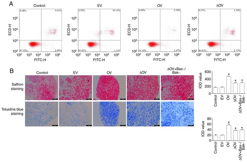
2 XIAO et al: Bcl‑xL MUTANT PROMOTES BMSC CARTILAGE DIFFERENTIATION
BMSCs transfected with Bcl‑xL mutant expression vectors with XhoI and NheI restriction sites. The digested PCR gene
was significantly increased (13,14). TGF‑β3 is a member of the fragments and linearized vector were ligated at 16˚C over‑
TGF‑β superfamily and plays a significant role in promoting night and the resulting Bcl‑xL overexpression vectors were
chondrogenic differentiation (13,14). Members of the bone transformed into competent DH5α cells. Target plasmids
morphogenetic protein (BMP) family also promote cartilage were extracted from the bacterial liquid according to the
repair (15). BMP‑2 enables for the migration and aggregation manufacturer's instructions. The transfection efficiency was
of mesenchymal stem cells into clusters, maintains them in a determined using reverse transcription‑quantitative PCR
tight state, stimulates Smad phosphorylation, enhances Sox9 (RT‑qPCR) as subsequently described.
expression and promotes mesenchymal stem cell differentia‑ The cDNA sequence corresponding to the GRI amino acid
tion into chondrocytes (16). BMP‑7 phosphorylates SMAD1 sequence of Bcl‑xL at positions 138‑140 was replaced with
and SMAD5, and induces the transcription of a variety of a sequence encoding ELN using the QuickChange Lighting
osteoblastic and chondrogenic genes (17). Site‑Directed Mutagenesis kit (Agilent Technologies, Inc.).
The present study investigated whether Bcl‑xL promotes The pcDNA3.1‑Δ Bcl‑xL plasmid was constructed using
chondrogenic differentiation of BMSCs in the microenviron‑ pcDNA3.1‑Bcl‑xL as a template and following the experi‑
ment of articular cartilage damage through its dual roles in mental procedures used in the construction of the parent vector.
anti‑apoptosis and TGF‑β/BMP upregulation. The aim was to The following Bax and Bak primer sequences were used:
provide a potential improvement to the application of bone tissue Bax forwards, 5'‑GGATGCTCTGAGCAGATCATGAAGATT
engineering for the treatment of articular cartilage defects. CAAGAGATCTTCATGATCTGCTCAGAGCTTTTTTCC‑3'
and reverse, 5'‑GAATAAAAAAGCTCTGAGCAGATCATG
Materials and methods AAGATCTCTTGAATCTTCATGATCTGCTCAGAGCTC‑3';
Bak forwards, 5'‑GGATGGCAGAGAATGCCTATGAGTATT
Isolation and identification of BMSCs. From January 1st 2018 CAAGAGATACTCATAGGCATTCTCTGCCTTTTTTCC‑3'
to September 30th 2018, bone marrow samples from 20 patients and reverse, 5'‑GAATAAAAAAGGCAGAGAATGCCTATG
(healthy volunteers with lower limb fractures; age, 18‑60 years; AGTATCTCTTGAATACTCATAGGCATTCTCTGCCTC‑3'.
10 males and 10 females) were collected from Pu Ai Hospital The cDNA for the human Bax and Bak genes were inserted
affiliated to Tongji Medical College, Huazhong University of into the pSuper‑Bax and pSuper‑Bak vectors (Addgene, Inc.),
Science and Technology. All operations were approved by the respectively, using XhoI and BamHI restriction sites. The target
Ethics Review Committee of Pu'ai Hospital affiliated with plasmids were extracted from the bacterial liquid and trans‑
Tongji Medical College of Huazhong University of Science fected using the Lipofectamine 2000 reagent according to the
and Technology, and all patients signed written informed manufacturer's instructions.
consent to participate in this study and for their samples to The BMSCs were divided into four groups: Control (not
be used for subsequent experiments. After skin preparation, subjected to transfection), EV (empty pcDNA3.1‑Bcl‑xL
disinfection and toweling in the patient's crotch area, 15 ml of vector), OV (Bcl‑xL overexpression) and ∆ OV (∆ Bcl‑xL
bone marrow solution was obtained and treated with 15 ml of overexpression).
lymphocyte separation solution (cat. no. XY08001T; Shanghai
Xinyu Biological Technology Co., Ltd.). The solution was Saffron and toluidine blue staining. The cells (7.5x10⁵ per
centrifuged at 400 x g for 15 min at 4˚C, resuspended in group) were resuspended in a 15‑ml centrifuge tube and
PBS and centrifuged again using the same conditions. After centrifuged at 150 x g for 5 min at 4˚C. The supernatant was
the supernatant was discarded, the cells were suspended aspirated and the cells were resuspended in the complete carti‑
in a complete medium (DMEM/F12 (cat. no. SH30023.01; lage differentiation induction medium (RASMX‑9004; Cyagen
HyClone; GE Healthcare Life Sciences) containing 10% FBS Biosciences, Inc.). The cells were centrifuged at 150 x g for
(cat. no. 10270‑106; Gibco; Thermo Fisher Scientific, Inc.) 5 min at 4˚C and incubated at 37˚C in 5% CO2. After two
and 1% penicillin and streptomycin (cat. no. PAB180056; weeks of continuous induction, the resultant cartilage was
Bioswamp Wuhan Beinle Biotechnology Co., Ltd.) and the fixed with 10% formalin at 4˚C for 48 h, subjected to saffron O
cell concentration was adjusted to 1x10⁶ cells/ml. The cells staining (cat. no. PAB180084; Wuhan Beinlai Biotechnology
were then cultured in a polylysine‑coated culture plate in Co., Ltd.) for 2 min at 60˚C and then observed under a light
an atmosphere containing 5% CO2 and 95% air, at 37˚C for microscope (200x magnification).
48 h. The medium was replaced every 72 h and the cells were Cartilage slices were incubated at 65˚C for 1 h and placed
subcultured or cryopreserved when the confluence reached in xylene (15 min, 4˚C) and a concentration gradient of
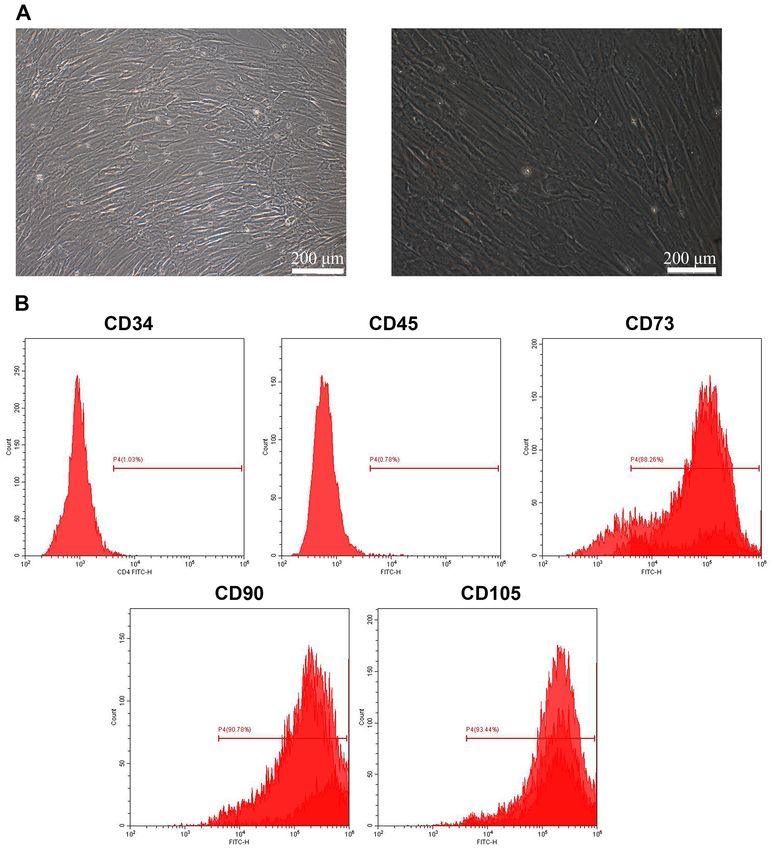
80‑90%. BMSCs were identified using light microscopy (200x alcohol (5 min, 4˚C). After two washes with double‑distilled
magnification) and flow cytometry (CD34, CD45, CD73, water for 2 min each, the slices were placed in toluidine blue
CD90 and CD105) as previously described (18,19). staining solution (cat. no. G3668; Beijing Solarbio Science
& Technology Co., Ltd.) for 30 min. The cells were then
Vector construction and transfection. The Bcl‑xL sequence washed with double‑distilled water, sealed with neutral gum
(NM 138578.1) was obtained from the National Center for and observed under a light microscope (MD1000; Leica
Biotechnology Information database. Since cDNA cannot enter Microsystems, Inc.) to detect the integrated optical density
the cell directly without a corresponding vector (20), the gene values in each group.
fragment was introduced into BMSCs to make it stable and
abundantly expressed. The cDNA of the human Bcl‑xL gene Flow cytometry. The cells (1x10⁶ per group) were resuspended
was inserted into the pcDNA3.1‑Bcl‑xL vector (Addgene, Inc.) in 100 µl of flow buffer (cat. no. PAB180076; Bioswamp WuhanEXPERIMENTAL AND THERAPEUTIC MEDICINE 22: 736, 2021 3
Beinle Biotechnology Co., Ltd.) in an Eppendorf tube and 2 µl Table I. Primer sequences.
of CD45‑FITC (cat. no. 11‑9459‑42, eBioscience; Thermo
Fisher Scientific, Inc.), CD34‑FITC (cat. no. CD34‑581‑01; Primer Sequence (5'→3')
Invitrogen; Thermo Fisher Scientific, Inc.), CD73‑FITC
(cat. no. 11‑0739‑42r; eBioscience; Thermo Fisher Scientific, Bcl‑xL‑F GCCACTTACCTGAATGACC
Inc.), CD90‑FITC (cat. no. 11‑0903‑82; eBioscience; Thermo Bcl‑xL‑R TGAGCCCAGCAGAACC
Fisher Scientific, Inc.) or CD105‑FITC (cat. no. MA1‑19594; TGF‑β‑F ATTCCTGGCGATACCTCA
Invitrogen; Thermo Fisher Scientific, Inc.) was added. The cells TGF‑β‑R GGCGAAAGCCCTCAAT
were incubated in the dark for 45 min at 4˚C, following which, BMP2‑F TGACGAGGTCCTGAGCG
400 µl of flow cytometry dyeing buffer (cat. no. PAB180076; BMP2‑R CCTGAGTGCCTGCGATA
Wuhan Beinlai Biotechnology Co., Ltd.) was added to each GAPDH‑F CCACTCCTCCACCTTTG
tube. The cells were subjected to flow cytometry (CytoFLEX S; GAPDH‑R CACCACCCTGTTGCTGT
Beckman Coulter, Inc.) and the results were analyzed using the
CYEXPERT software (CXP Analysis 2.0; Beckman Coulter, BMP, bone morphogenic protein; F, forward; R, reverse.
Inc.).
Cells were cultured for 24 h at 37˚C, harvested, treated
with 1 ml of pre‑cooled PBS and centrifuged at 1,000 x g for
5 min at 4˚C. Subsequently, 10 µl of Annexin V‑FITC and 10 µl Statistical analysis. Data are expressed as the mean ± SD (n=3).
of PI were added. The cell samples were then analyzed using To analyze the differences between groups, data comparisons
flow cytometry as aforementioned. A one‑step fluorescence were performed using one‑way ANOVAs and subsequent
compensation strategy was used to eliminate interference with Tukey's post‑hoc tests. P4 XIAO et al: Bcl‑xL MUTANT PROMOTES BMSC CARTILAGE DIFFERENTIATION Figure 1. Culture and identification of bone marrow mesenchymal stem cells. (A) Light microscopy of BMSCs (scale bar=100 µm for the left image and 200 µm for the right image). (B) Percentage of CD34‑, CD45‑, CD73‑, CD90‑ and CD105‑positive BMSCs were detected by flow cytometry. BMSC, bone marrow mesenchymal stem cell. Figure 2. Identification of transfection efficiency. (A) Expression of Bcl‑xL after vector construction. (B) Expression of Bax and Bak after pSuper‑Bax and pSuper‑Bak vector construction. *P0.05) (Fig. 3B). These observations and ∆OV+Bax‑/Bak‑staining increased (P
EXPERIMENTAL AND THERAPEUTIC MEDICINE 22: 736, 2021                                                          5

Figure 3. Effect of Bcl‑xL mutant on BMSC apoptosis and cartilage differentiation. (A) Detection of apoptosis by flow cytometry. (B) Cartilage tissue
morphology was observed using saffron and toluidine blue staining (scale bar=50 µm) and the IOD values of each group were statistically analyzed.
*
 P
6                           XIAO et al: Bcl‑xL MUTANT PROMOTES BMSC CARTILAGE DIFFERENTIATION

However, the targeted cartilage differentiation of BMSCs is          Funding
dependent on the local microenvironment and the synergistic
effects of various inducing factors (29). The effective induc‑       This work was supported by financial grants from the
tion of BMSC differentiation into chondrocytes and initiation        Wuhan Applied Foundational Frontier Project (grant
of cartilage tissue formation has become a challenging process       no. 2020020601012309); Science and Technology Department
that needs to be addressed.                                          of Hubei Province (grant no. 2020CFB784); and Hubei
    Bcl‑xL is an important anti‑apoptotic molecule in the            Province health and family planning scientific research project
Bcl‑2 family that inhibits the pro‑apoptotic molecule,               (grant no. WJ2019Q006).
Bax, preventing the apoptosis caused by the release of
cytochrome C (30). Bcl‑xL also blocks apoptosis by inhibiting        Availability of data and materials
the binding of Apaf‑1 and caspase‑9 downstream of Bax
activation (31), as well as by inhibiting apoptosis induced by       The datasets used and/or analyzed during the current study are
the Fas‑FasL pathway (32). Therefore, Bcl‑xL exerts important        available from the corresponding author on reasonable request.
anti‑apoptotic effects by blocking various apoptotic pathways
stimulated by pro‑apoptotic factors, such as hypoxia and             Authors' contributions
inflammation, thereby demonstrating its role in promoting
chondrogenic differentiation (33). Wang et al (34) reported          KX, LY and WX contributed to the conception of the study. KX
an increase in cartilage formation during fracture repair in         and XG designed and performed the experiments, analyzed
mice with Bax gene deletion, suggesting that Bcl‑xL continues        the data and wrote the manuscript. RH and MX analyzed the
to promote cartilage production and repair during chondro‑           data and provided technical support. KX and LY confirm the
genesis, even in the absence of the anti‑apoptotic effect of         authenticity of all the raw data. All authors read and approved
Bcl‑xL (presumably through alternative pathways). Mutants            the final manuscript.
are defined as individuals with mutations showing phenotypes
that differ from the wild type. A previous study have shown          Ethics approval and consent to participate
that the Bcl‑xL mutant, where the GRI amino acid sequence
at positions 138‑140 is replaced by ELN, has no anti‑apoptotic       All operations were approved by the Ethics Review
effect, while its other biological functions remain similar to       Committee of Pu'ai Hospital affiliated with Tongji Medical
those of wild‑type Bcl‑xL (12).                                      College of Huazhong University of Science and Technology
    The present study examined the anti‑apoptotic effect of          [approval no. (2017)IEC(S118)]. Informed consent was
the Bcl‑xL mutant on cartilage differentiation of BMSCs. it          obtained from all patients.
was demonstrated that the Bcl‑xL mutant promoted cartilage
differentiation of BMSCs without affecting BMSC apoptosis,           Patient consent for publication
whereby this effect was not related to the expression of Bax
and Bak. The potential mechanism of action of the Bcl‑xL             Not applicable.
mutant on promoting cartilage differentiation of BMSCs was
further examined. The results showed that the expression of          Competing interests
TGF‑ β and BMP increased significantly after Bcl‑xL and
Bcl‑xL mutant intervention, revealing that the Bcl‑xL mutant         The authors declare that they have no competing interests.
may promote cartilage differentiation of BMSCs by upregu‑
lating TGF‑β/BMP expression levels.                                  References
    A limitation of the present study was that the differentiation
experiments were performed after a long term culture. BMSCs           1. Sandell LJ and Aigner T: Articular cartilage and changes in arthritis:
can differentiate to chondrocytes within two weeks. As such,             Cell biology of osteoarthritis. Arthritis Res 3: 107‑113, 2001.
                                                                      2. Buckwalter JA and Mankin: Articular cartilage: Tissue design
analyzing the effects of overexpression of Bcl‑xL longitudi‑             and chondrocyte‑matrix interactions. Instr Course Lect 47:
nally, examining the expression profiles of this protein in the          477‑486, 1998.
various groups to show the kinetics of expression, is necessary.      3. Natoli RM and Athanasiou KA: Traumatic loading of articular
                                                                         cartilage: Mechanical and biological responses and post‑injury
However, the main aim of the present study was to observe the            treatment. Biorheology 46: 451‑485, 2009.
effect of the Bcl‑xL mutant on cartilage differentiation and the      4. Wang DH, Lin YF, Chen L, Mo YQ, Huang P and Ma RX: Guided
expression of TGF‑β and BMP. As such, further studies should             bone regeneration using a bone tissue engineering complex
                                                                         consisting of a poly‑dl‑lactide membrane and bone mesenchymal
examine the effects of Bcl‑xL in the various BMCS treatment              stem cells. Oncotarget 9: 16380‑16388, 2017.
groups in subsequent experiments.                                     5. Dang M, Saunders L, Niu XF, Fan YB and Ma PX: Biomimetic
    In conclusion, the present study demonstrated that Bcl‑xL            delivery of signals for bone tissue engineering. Bone Res 6: 25, 2018.
                                                                      6. Ding WW, Huang JH and Xu WL: Research progress on the
mutants promoted cartilage differentiation of BMSCs and                  differentiation of bone marrow mesenchymal stem cells into
upregulated TGF‑ β/BMP expression levels, whereby this                   chondrocytes. Med Rev 12: 2148‑2151, 2015.
enhancement of chondrogenic differentiation was not related           7. Yang K, Xie L, Zhu WM, Huang JH, Duan L and Wang DP:
                                                                         Progress in the mechanism of microRNA regulating chondro‑
to the expression of Bax and Bak.                                        genic differentiation of mesenchymal stem cells. Orthopedics 7:
                                                                         219‑221, 2016.
Acknowledgements                                                      8. Cheng EH, Wei MC, Weiler S, Flavell RA, Mak TW, Lindsten T
                                                                         and Korsmeyer SJ: Bcl‑2, Bcl‑X(L) sequester BH3 domain‑only
                                                                         molecules preventing Bax‑ and Bak‑mediated mitochondrial
Not applicable.                                                          apoptosis. Mol Cell 8: 705‑711, 2001.
EXPERIMENTAL AND THERAPEUTIC MEDICINE 22: 736, 2021 7
9. Nakagami H, Kiomy Osako M, Shimizu H, Hanayama R and 23. Tsuchiya A, Kojima Y, Ikarashi S, Seino S, Watanabe Y,
Morishita R: Potential contribution of action of renin angiotensin Kawata Y and Terai S: Clinical trials using mesenchymal stem
system to bone metabolism. Curr Hypertens Rev 3: 129‑132, cells in liver diseases and inflammatory bowel diseases. Inflamm
2007. Regen 37: 16, 2017.
10. Chen CB, Zhou H, Wei F, Jiang L, Liu XG, Liu ZJ and Ma Q: 24. Antoniou KM, Karagiannis K, Tsitoura E, Bibaki E, Lasithiotaki I,
Increased levels of hypoxia‑inducible factor‑1α are associated Proklou A, Spandidos DA and Tzanakis N: Clinical applications
with Bcl‑xL expression, tumor apoptosis, and clinical outcome in of mesenchymal stem cells in chronic lung diseases. Biomed
chondrosarcoma. J Orthop Res 29: 143‑151, 2011. Rep 8: 314‑318, 2018.
11. Ming LH, Wang P, Bank A, Yu J and Zhang L: PUMA dissoci‑ 25. Gafni Y, Turgeman G, Liebergal M, Pelled G, Gazit Z and
ates Bax and Bcl‑xl to induce apoptosis in colon cancer cells. Gazit D: Stem cells as vehicles for orthopedic gene therapy. Gene
J Biol Chem 281: 16034‑16042, 2006. Ther 11: 417‑426, 2006.
12. Cong YM, Liu P, Jin D, Guang WJ and Ma YH: Relationship 26. Djouad F, Mrugala D, Noël D and Jorgensen C: Engineered
between Bcl‑2 family and apoptosis. Chin J Med Res 2008. mesenchymal stem cells for cartilage repair. Regen Med 1:
13. Lee HL, Yu B, Deng P, Wang CY and Hong C: Transforming 529‑537, 2006.
growth factor‑β‑induced KDM4B promotes chondrogenic differ‑ 27. Jin CZ: Application of autologous bone marrow mesenchymal
entiation of human mesenchymal stem cells. Stem Cells 34: stem cell matrix scaffold in cartilage tissue engineering.
711‑719, 2016. CRTER 33: 7864‑7866, 2015.
14. Jin EJ, Park JH, Lee SY, Chun JS, Bang OS and Kang SS: Wnt‑5a 28. Hu B: Bone marrow mesenchymal stem cells and chondrocytes
is involved in TGF‑beta3‑stimulated chondrogenic differentia‑ aggregation and co‑culture to repair osteochondral defects in
tion of chick wing bud mesenchymal cells. Int J Biochem Cell rabbit knee joint. SCU 2016.
Biol 38: 183‑195, 2006. 29. Novak T, Seelbinder B, Twitchell CM, Voytik‑Harbin Prof SL
15. Rueger DC and Chubinskaya S: Bone morphogenetic proteins in and Prof CPN: Dissociated and reconstituted cartilage micropar‑
articular cartilage repair. Prog Inflamm Res 109‑132, 2004. ticles in densified collagen induce local hMSC differentiation.
16. Zhao L, Li G and Zhou GQ: SOX9 directly binds CREB as a novel Adv Funct Mater 26: 5427‑5436, 2016.
synergism with the PKA pathway in BMP‑2‑induced osteochon‑ 30. Eskes R, Desagher S, Antonsson B and Martinou JC: Bid induces
drogenic differentiation. J Bone Miner Res 24: 826‑836, 2009. the oligomerization and insertion of Bax into the outer mitochon‑
17. Retting KN, Song B, Yoon BS and Lyons KM: BMP canonical drial membrane. Mol Cell Biol 20: 929‑935, 2000.
Smad signaling through Smad1 and Smad5 is required for endo‑ 31. Rosśe T, Olivier R, Monney L, Rager M, Conus S, Fellay I,
chondral bone formation. Development 136: 1093‑1104, 2009. Jansen B and Borner C: Bcl‑2 prolongs cell survival after
18. Mahmood SC, Michael B, Angela M, John P and David H: Bax‑induced release of cytochrome C. Nature 391: 496‑499,
Cryopreservation of whole adipose tissue for future use in 1998.
regenerative medicine. J Surg Res 187: 24‑35, 2014. 32. Biswas RS, Cha HJ, Hardwick JM and Srivastava RK: Inhibition
19. Sierra‑Sánchez Á, Ordóñez‑Luque A, Espinosa‑Ibáñez O, of drug‑induced Fas ligand transcription and apoptosis by
Ruiz‑García A and Arias‑Santiago S: Epithelial in vitro differen‑ Bcl‑XL. Mol Cell Biochem 225: 7‑20, 2001.
tiation of mesenchymal stem cells. Curr Stem Cell Res Ther 13: 33. Zhou W, Wu SM, Chen HJ and Pu SX: Expression of Bcl‑x
409‑422, 2018. mRNA after hypoxic‑ischemic brain injury in neonatal rats.
20. Feng Z, Chen Q, Ren MQ, Tian ZG and Gong YP: CD40L J Clin Neurol 13: 7‑9, 2000.
inhibits cell growth of THP‑1 cells by suppressing the PI3K/Akt 34. Wang CY, Chen LL, Kuo PY, Chang JL, Wang YJ and Hung SC:
pathway. Onco Targets Ther 12: 3011‑3017, 2019. Apoptosis in chondrogenesis of human mesenchymal stem
21. Bayer J, Grunwald D, Lambert C, Mayol JF and Maynadié M: cells: Effect of serum and medium supplements. Apoptosis 15:
Thematic workshop on fluorescence compensation settings in 439‑449, 2010.
multicolor flow cytometry. Cytometry B Clin Cytom 72: 8‑13,
2007. This work is licensed under a Creative Commons
22. Livak KJ and Schmittgen TD: Analysis of relative gene expres‑ Attribution-NonCommercial-NoDerivatives 4.0
sion data using real‑time quantitative PCR and the 2(‑Delta Delta International (CC BY-NC-ND 4.0) License.
C(T)) method. Methods 25: 402‑408, 2001You can also read