Muscle genomics and aerobic training - Original Article - Journal of Human ...
←
→
Page content transcription
If your browser does not render page correctly, please read the page content below
Original Article

Muscle genomics and aerobic training
YECID MINA-PAZ1,2 , DIANA CAROLINA ZAMBRANO1,2, ANDRÉS JENUER MATTA1,2, ALEJANDRA
RODRÍGUEZ1, FELIPE GARCIA-VALLEJO1
1Department of Physiological Sciences, School of Basic Sciences, Faculty of Health, University of Valle, Cali,
Colombia
2Faculty of Education and Sports Sciences, University Institution National Sports School, Cali, Colombia

 ABSTRACT

The performance in physical activity is determined not only by physiological processes such as age, body
composition, gender and degree of training, but also by the genomics and even epigenetic events occurring
during the training programs. In this context, using bioinformatics resources, we aimed to analyse the
expression of genes associated with muscle function in vastus lateral samples. We used data from DNA
microarray experiments reported in NCBI's GEO DataSet database under the series number GSE117070.
Differential expression was calculated using the Z-ratio equation. We also used the software Cytoscape 3.6
to build a protein-protein interaction network with over-expressed genes. We found that seven genes out of
the 397 genes analysed in the 41 individuals subjected to aerobic exercise with an increase in training
intensity through the percentage of VO2max, were over-expressed based on the statistical approach. The
Protein-Protein Interaction (PPI) network showed 477 nodes, two connected components, 17 multi-edge
node pairs and an average number of neighbours of 2.092. The node with the highest number of interactions
was TPM1 with 150. GO categories of biological processes most relevant of the network included
indispensable processes for muscle function and contraction such as polymerization of actin filaments and
ATP synthesis from electron transport chain.
Keywords: Aerobic training; Muscle; DNA microarray; Differential expression; Network.

 Cite this article as:
 Mina-Paz, Y., Zambrano, D.C., Matta, A.J., Rodríguez, A., & García-Vallejo, F. (2021). Muscle genomics and
 aerobic training. Journal of Human Sport and Exercise, in press. doi:https://doi.org/10.14198/jhse.2022.173.11

1
 Corresponding author. Molecular Biology and Pathogenesis Research Group, Department of Physiological Sciences, School of
Basic Sciences, Faculty of Health, Universidad del Valle, Cali, Colombia. https://orcid.org/0000-0002-0623-1526
 E-mail: Yecid.mina@correounivalle.edu.co
 Submitted for publication July 28, 2020
 Accepted for publication September 11, 2020
 Published in press January 13, 2021
 JOURNAL OF HUMAN SPORT & EXERCISE ISSN 1988-5202
 © Faculty of Education. University of Alicante
 doi:10.14198/jhse.2022.173.11
 VOLUME -- | ISSUE - | 2021 | 1
Mina-Paz, et al. / Muscle genomics & aerobic training JOURNAL OF HUMAN SPORT & EXERCISE

INTRODUCTION

The type of exercise and the training load during the exercise to which the athlete is subjected modifies
muscle contraction and the VO2max This measurement is defined as the maximum amount of O2 that the body
is able to absorb, transport and is consumed per unit of time (Wasserman, et al.,). VO 2max is regulated by
metabolic changes and by a wide variety of protein isoforms at the fibre level (Di Prampero, et al., 2015)
(Samozino, et al., 2016), caused by functional regulation of the genome. This is determined by variations in
DNA sequences, methylation of the promoter and coding regions of genes, topological changes in the
structure of chromatin, homeotic regulation of genes and by small RNA molecules, also known as microRNAs
in skeletal muscle (Liu, et al., 2010) (Pilegaard, et al., 2000).

The physiological processes that determine the performance in physical activity or sport are the following:
age, body composition, gender, degree of training. However, there are global genomic processes that
participate in the complexity of the manifestations of performance for people who perform a monitoring in the
sports load, these manifestations are mediated by alterations in the number of copies of functional and non-
traditional genomic elements. Associated with this, epigenetic mechanisms could perform transcriptional
modifications of the skeletal muscle through the induction of gene expression changes that do not necessarily
alter the nucleotide sequence, but in a small percentage can generate some polymorphism.

Currently, experiments with DNA microarrays allow examining transcriptional changes in the skeletal muscle
with a previous gene characterization based on age, gender, strength and muscular resistance (Rowlands,
et al., 2011) (Timmons, et al., 2010). It is important to note that the signalling histones, the binding of the
transcription factor and the chemical modifications of the DNA can actively influence or passively reflect the
musculoskeletal expression programs, which can affect the activity of the isoform that performs the
contraction through molecular signalling events (Rockman & Kruglyak, 2006) (Visscher, et al., 2017).
Associated with this, speed exercises allow a percentage development of fast-twitch muscle fibres and
resistance or aerobic exercises allow the development of a high percentage of slow-twitch muscle fibres
(Saltin, et al., 1977) (Trappe, et al., 2015).

Understanding these changes will improve the development of sports planning that will not only take into
account the phenotypic change that, in the case of aerobic exercise, would primarily be oxygen consumption,
but would also make it possible to determine the markers of a type of fibre or other metabolic modifications
in the tissue, reflected by the muscular transcriptome. In this context, we aimed to analyse the gene
expression of genes associated with muscle function in vastus lateral samples, by using data from DNA
microarray experiments recorded in NCBI's GEO DataSet database (https://www.ncbi.nlm.nih.gov/gds/),
GSE117070. Differential expression was calculated using the Z ratio. We found overexpressed and sub
expressed genes associated with muscle function after a 20-week training incrementally.

MATERIALS AND METHODS

To perform the bioinformatic analysis of the present study, we selected 397 genes associated with muscle
function, previously reported in the Gene Entrez of the NCBI database. (https://www.ncbi.nlm.nih.gov/gene),
which are described in Supplementary Table 1. We used the transformed expression values of log2 intensity
of a DNA microarray experiment whose registration code and free access in the GEO database was
GSE117070 (https://www.ncbi.nlm.nih.gov/geo/query/acc.cgi?acc=GSE117070) (Bouchard, 1995). The
selected microarray experiment included gene expression data from more than 43,797 probes from 82
samples of the quadriceps muscle (vastus lateralis) from 90 Caucasians and 40 African American, with a

 2 | 2021 | ISSUE - | VOLUME -- © 2021 University of Alicante
Mina-Paz, et al. / Muscle genomics & aerobic training JOURNAL OF HUMAN SPORT & EXERCISE

range of age between 17 and 65 years old, the selected microarray experiment included gene expression
data from more than 43,797 probes from 82 samples of the quadriceps muscle (vastus lateralis) from 90
Caucasians and 40 African American, with a range of age between 17 and 65 years old, for the category of
"sons" between 17 and 40 years old and for the category of "parents" between 40 and 65 years old. They
could not have done any physical activity in the last 3 months before starting the study, nor have had any
chronic non-contagious disease. The entire population was evaluated before and after the 20-week aerobic
exercise program. There was no group control as in the usual studies, because they were the same cases
at the beginning of the 20-week program and their own controls after the 20 weeks. Regarding the gene
expression profiles, they were determined from the muscle biopsies, to subsequently perform Affymetrix
microarray analysis, at the beginning and at the end of the 20-week aerobic exercise program.

This was developed as follows: the first two weeks at 55% of VO2max, each week 3 sessions of 30 minutes
each, the next two weeks at 55% of VO2max, each week 3 sessions of 35 minutes each. The following two
weeks at 65% of VO2max, each week 3 sessions of 35 minutes each, the next two weeks at 65% of VO 2max,
each week 3 sessions of 40 minutes each. The next two weeks at 70% of VO 2max, each week 3 sessions of
40 minutes each, the next two weeks at 70% of VO2max, each week 3 sessions of 45 minutes each. The next
two weeks at 75% of VO2max, each week 3 sessions of 45 minutes each and at the end, 6 weeks at 75% of
VO2max, each week 3 sessions of 50 minutes each. All this was done on a cycle ergometer with continuous
monitoring of the heart rate (Bouchard, 1995). However, for the present study, we decided to study the genes
associated with muscle function before and after the application of the training program.

Differential gene expression quantification
Raw intensity log2 data for each experiment was used for the calculation of Z-score (Cheadle, et al., 2003).
Z-scores of the protein coding genes analysed, were calculated according to the Equation 1:

 ( − log … )
 − =
 log …

 Equation 1. Z-score formula.

All values of the Z score were normalized on a linear scale -3.0 ≤ 0 ≥ +3.0 (two-tailed p-value
Mina-Paz, et al. / Muscle genomics & aerobic training JOURNAL OF HUMAN SPORT & EXERCISE

 [( − 1 ) − − ( − 1 ) − ]
 − =

 √ 1 + 1

 Equation 3. Z-test formula.

Where G1 represents the average Z score for gene being tested under experimental conditions, for this study,
post-training versus pre-training. The repeated hybridization intensity measurements (expressed as Z scores)
are mean difference corrected by the SE for the difference between means where is the SD, for either
condition 1 or condition 2, and n equals the number of repeated measurements for either condition 1 or
condition 2.

Interaction network
We used the software Cytoscape 3.6 to build a network of expression and interaction entering a matrix
created with the information of physical protein interactions placed in the BioGRID database. Ontological
categories were extracted from the network in the Cytoscape 3.2 program using the plugin BiNGO, which
establishes a remote connection to the database Gene Ontology, reporting the most important biological
processes of the network, with their own p-value (Bonferroni correction). For the present study, we chose 10
processes with the most significant p-values.

Statistical analysis
Analyses were performed to compare the media values of the Z score between post-trained and pre-trained.
The Wilcoxon signed-rank /Two Tailed was used to calculate the differences between the media of two
samples. In all cases we use an alpha .05 to test the importance of H0. On the other hand, to calculate the
statistical differences in the log2 values of Post-trained and Pre-trained, we applied the t test for two paired
samples / two-tailed test with an alpha of .05.

Principal component analysis (PCA) was employed as a computational procedure for the classification of
multiclass gene expression in muscle biopsy samples from individuals in post-trained and pre-trained
conditions. All analyses were performed in the SPSS program, version 25.

RESULTS

Table 1. Over-expressed genes associated with muscle functions.
 Gene ID Symbol Z-score Post Z-score Pre Z-ratio p-value*
 7168 TPM1 0.10790368 -0.10352638 284.064.636 < .00001
 1282 COL4A1 224.952.369 205.107.125 266.628.685 < .00001
 4617 MYF5 -0.0230669 -0.21585979 259.024.856 < .00001
 4478 MSN -0.2643089 -0.44033643 236.499.942 < .00001
 9639 ARHGEF1 -0.14934508 -0.312698 219.471.096 < .00001
 7052 TGM2 -0.48019236 -0.64051953 215.405.883 < .00001
 91746 YTHDC1 -0.14333736 -0.29550284 204.440.332 < .00001
 (*). Is statistically significant p-value.

 4 | 2021 | ISSUE - | VOLUME -- © 2021 University of Alicante
Mina-Paz, et al. / Muscle genomics & aerobic training JOURNAL OF HUMAN SPORT & EXERCISE

Supplementary Table 1. Over-expressed genes associated with muscle function.
 Gene ID Symbol Name Locus Function
 7168 TPM1 Tropomyosin 1 15q22.2 This gene is a member of the tropomyosin family of highly
 conserved, widely distributed actin-binding proteins
 involved in the contractile system of striated and smooth
 muscles and the cytoskeleton of non-muscle cells.
 Tropomyosin is composed of two alpha-helical chains
 arranged as a coiled-coil. It is polymerized end to end
 along the two grooves of actin filaments and provides
 stability to the filaments.
 1282 COL4A1 Collagen type IV 13q34 Collagen alpha-1(IV) chain; Type IV collagen is the major
 alpha 1 chain structural component of glomerular basement
 membranes (GBM), forming a 'chicken-wire' meshwork
 together with laminins, proteoglycans and
 entactin/nidogen.
 4617 MYF5 Myogenic factor 5 12q21.31 Acts as a transcriptional activator that promotes
 transcription of muscle-specific target genes and plays a
 role in muscle differentiation. Together with MYOG and
 MYOD1, co-occupies muscle-specific gene promoter
 core region during myogenesis. Induces fibroblasts to
 differentiate into myoblasts. Probable sequence specific
 DNA-binding protein; Basic helix-loop-helix proteins.
 4478 MSN Moesin Xq12 Probably involved in connections of major cytoskeletal
 structures to the plasma membrane. May inhibit herpes
 simplex virus 1 infection at an early stage. Plays a role in
 regulating the proliferation, migration, and adhesion of
 human lymphoid cells and participates in immunologic
 synapse formation; FERM domain containing.
 9639 ARHGEF10 Rho guanine 8p23.3 May play a role in developmental myelination of
 nucleotide peripheral nerves; Rho guanine nucleotide exchange
 exchange factor 10 factors.
 7052 TGM2 Transglutaminase 20q11.23 Catalyses the cross-linking of proteins and the
 2 conjugation of polyamines to proteins;
 Transglutaminases.
 91746 YTHDC1 YTH domain 4q13.2 Regulator of alternative splicing that specifically
 containing 1 recognizes and binds N6-methyladenosine (m6A)-
 containing RNAs. M6A is a modification present at
 internal sites of mRNAs and some non- coding RNAs and
 plays a role in the efficiency of mRNA splicing, processing
 and stability. Acts as a key regulator of exon-inclusion or
 exon-skipping during alternative splicing via interaction
 with mRNA splicing factors SRSF3 and SRSF10.

Out of 397 genes that were analysed, seven of them were significantly overexpressed in the vastus lateralis
muscle samples analysed. They corresponded to: Tropomyosin 1 (TPM1), Collagen type IV alpha 1 chain
(COL4A1), Myogenic factor 5 (MYF5), Moesin (MSN), Rho Guanine Nucleotide Exchange Factor 10
(ARHGEF10), Transglutaminase 2 (TGM2) and YTH Domain Containing 1 (YTHDC1) were overexpressed
(Table 1). That is, 1.9% of the analysed genes explained the behaviour of VO2 in relation to skeletal muscle
activity, for that population.

 VOLUME -- | ISSUE - | 2021 | 5
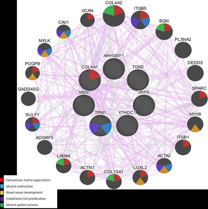
Mina-Paz, et al. / Muscle genomics & aerobic training JOURNAL OF HUMAN SPORT & EXERCISE

The Protein-Protein Interaction (PPI) network made with the seven overexpressed genes associated with
muscle function found in the previous analysis showed a total of 477 nodes, two connected components, 17
multi-edge node pairs and an average number of neighbours of 2.092 (Figure 1). The node with the highest
number of interactions was TPM1 with 150, followed closely by MSN with 142. GO categories of biological
processes most relevant of the network (Table 2) include indispensable processes for muscle function and
contraction such as polymerization of actin filaments (p-value 2.57E-14) and ATP synthesis from electron
transport chain (p-value 1.58E-13). According to the co-expression network made in GENEMANÍA, the
relevant functions associated were muscle contraction and extracellular matrix organization.

Figure 1. Protein - Protein Interaction (PPI) Network with the 7 genes associated with muscle function found
over-expressed (Z-ratio above 1.96) shown in red. Interacting proteins are shown in yellow.

Table 2. Top ten GO categories of biological processes outstanding from the network.
 G.O_ID Description p-value
 16043 Actin filament polymerization 2.57E-14
 9987 ATP synthesis coupled electron transport 1.58E-13
 48518 Positive regulation of L-glutamate imports across plasma membrane 1.33E-12
 10941 Negative regulation of apoptotic process 3.71E-12
 22607 Actin filament bundle assembly 4.49E-12
 43067 Negative regulation of cardiac muscle cell apoptotic process 8.01E-12
 42981 Negative regulation of neuron apoptotic process 1.63E-11
 48522 Activation of MAPKK activity 1.93E-11
 44085 Contractile actin filament bundle assembly 3.00E-10
 42221 Cellular response to calcium ion 7.76E-10
 Note: p-values with Bonferroni correction are shown.

 6 | 2021 | ISSUE - | VOLUME -- © 2021 University of Alicante
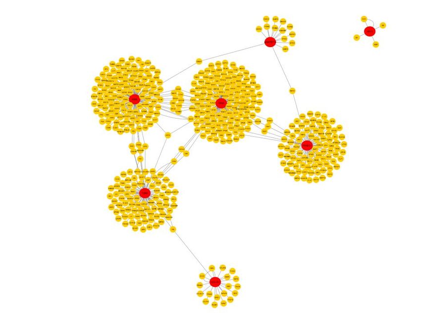
Mina-Paz, et al. / Muscle genomics & aerobic training JOURNAL OF HUMAN SPORT & EXERCISE

Figure 2. Co-expression interaction network with the 7 genes associated with muscle function found over-
expressed.

 Figure 3. Pre-training samples PCA with the seven overexpressed genes shown in green.

 VOLUME -- | ISSUE - | 2021 | 7
Mina-Paz, et al. / Muscle genomics & aerobic training JOURNAL OF HUMAN SPORT & EXERCISE

 Figure 4. Post-training samples PCA with the seven overexpressed genes shown in red.

A 76% of the accumulated variance was reached within two components in the Pre-training samples PCA
(Figure 3), while a 78% of the accumulated variance was reached within two components in the Post-training
samples PCA (Figure 4).

DISCUSSION AND CONCLUSIONS

In the present study, we aimed to analyse the gene expression of genes associated with muscle function in
vastus lateral samples, by performing bioinformatic analysis and identified expressed genes on publicly
available after the exercise of an aerobic exercise for 20 weeks in human skeletal muscle.

The overexpressed genes were TPM1, COL4A1, MYF5, MSN, ARHGEF10, TGM2 and YTHDC1. These
genes are known to be important in the structure / remodelling of the matrix / actin, as well as in mechano-
transduction in skeletal muscle and any dysregulation may affect not only their function but also the one from
the associated genes, as the PPI network shows. This is confirmed by the work of Turner et al. (2019), in
which he performs a comparative transcriptome and analysis of methylome in the anabolism of human
skeletal muscle, hypertrophy and epigenetic memory. They found 13 genes associated with muscular
function in aerobic exercise, and two of them, MSN and COL4A1, are in fact, present in our study. It is
confirmed that some of these genes have a structural relationship and a remodeller that increase after aerobic
exercise and resistance in the expression of genes, protein abundance or activity level, such as; COL4A1
are involved in the regulation of muscle cells or satellites (Kanazawa, et al., 2019) (Xu, et al., 2017).

Adaptations of skeletal muscles dependent on frequency, duration and intensity (Egan & Zierath, 2013), the
characterization of muscle proteins associated with different types of exercise are constantly replanted.
Myofibrillar protein isoforms are the most frequent, mainly in the distribution of myosin heavy chain isoforms,
thin filament isoforms, calcium proteins of the sarcoplasmic reticulum, mitochondrial proteins and some
chaperones involved in the response to the correct one. In our study, the MYF5 gene was over-expressed,
this gene acts as a transcriptional activator that promotes the transcription of muscle-specific genes and

 8 | 2021 | ISSUE - | VOLUME -- © 2021 University of Alicante
Mina-Paz, et al. / Muscle genomics & aerobic training JOURNAL OF HUMAN SPORT & EXERCISE

plays a role in muscle differentiation. However, protein, activity, epigenetic factors, training activities during
the performance of aerobic or glycolytic exercise, production in the muscle fibre, dysregulation in the
distribution of the heavy chain of myosin, regulation of the low of the filament proteins in both the vastus
lateralis and soleus muscles (Gonzalez‐Freire, et al., 2017).

Moreover, mutations in type VI collagen (COL4A1) have shown abundant changes in proteins related to the
cytoskeleton and sarcomere functions (Sela, et al., 2011), patients with skeletal muscle diseases such as
hereditary myopathy of the body of inclusion, myofibrillary myopathies, desminopathies and some muscular
disorders. These changes, not only to the skeletal muscle, but also to the connective tissues that produce
contractions and weak muscle mutations in the genes that encode the sarcomeric and extraarcomeric
proteins (De Palma, et al., 2014), for our study. COL4A1, at 20 weeks of training that contributes to proline
3-hydroxylation of collagen in the tendons, for a development of physical activity.

The most relevant GO categories found for the PPI network were mostly associated with muscular function,
but we highlight the positive regulation of L-glutamate import across plasma membrane. L-glutamine is an
amino acid that the body needs to stay in optimum health, and it has been demonstrated that during exercise,
plasma glutamine levels increases and decreases, these variations are dependent on the type, duration, and
intensity of exercise (Street, et al., 2011). The body naturally produces this amino acid, but it is also often
consumed as a supplement in sports and bodybuilding as a supplement to help with muscle repair and
growth, given the reports that show that L-glutamine might also help with post-exercise recovery (Street, et
al., 2011) (Legault, et al., 2015). Legault et al. (2015), found that the supplementation with this amino acid
resulted in faster recovery of peak torque and diminished muscle soreness following eccentric exercise of 16
healthy volunteers. The authors even concluded that the effect of L-glutamine on muscle force recovery may
be greater in men than women.

Therefore, the present study demonstrates that skeletal muscle genes are regulated epigenetically by training
loads for participants, in which DNA methylation is found, predominantly through hypomethylation and
positive regulation of associated genes with the structure and remodelling of the matrix and the cytoskeleton.
Similarly, the mechanics-transduction for skeletal muscle.

AUTHOR CONTRIBUTIONS

Mina, Matta and Zambrano wrote and verified the abstract and the introduction. Mina, Garcia and Rodriguez
wrote and verified the methods, the results, the discussion and conclusions. All authors reviewed the final
document and assisted in its construction.

SUPPORTING AGENCIES

The project was funded by the vice-rectory of research at the Universidad del Valle through the scholarship
119-2019.

DISCLOSURE STATEMENT

No potential conflict of interest was reported by the authors.

 VOLUME -- | ISSUE - | 2021 | 9
Mina-Paz, et al. / Muscle genomics & aerobic training JOURNAL OF HUMAN SPORT & EXERCISE

REFERENCES

 Bouchard, C. L. (1995). The heritage family study. Aims, design, and measurement protocol. Medicine
 and science in sports and exercise, 27(5), 721-729. https://doi.org/10.1249/00005768-199505000-
 00015
 Cheadle, C. V. (2003). Análisis de datos de microarrays utilizando la transformación de puntuación Z. .
 J Mol Diag: JMD. 5 (2): 73-81.
 Chicharro, J. L., & Vaquero, A. F. (2006). Fisiología del ejercicio. Madrid: Ed. Médica Panamericana.
 Colberg, S. R. (2016). Physical activity/exercise and diabetes: a position statement of the American
 Diabetes Association. Diabetes care, 39(11), 2065-2079. https://doi.org/10.2337/dc16-1728
 De Palma, S., Capitanio, D., Vasso, M., Braghetta, P., Scotton, C., Bonaldo, P., & Gelfi, C. (2014). Muscle
 proteomics reveals novel insights into the pathophysiological mechanisms of collagen VI
 myopathies. Journal of proteome research, 5022-5030. https://doi.org/10.1021/pr500675e
 Di Prampero, P. E., Botter, A., & Osgnach, C. (2015). The energy cost of sprint running and the role of
 metabolic power in setting top performances. European journal of applied physiology, 115(3), 451-
 469. https://doi.org/10.1007/s00421-014-3086-4
 Egan, B., & Zierath, J. R. (2013). Exercise metabolism and the molecular regulation of skeletal muscle
 adaptation. . Cell metabolism, 17(2), 162-184. https://doi.org/10.1016/j.cmet.2012.12.012
 Gonzalez‐Freire, M., Semba, R. D., Ubaida‐Mohien, C., Fabbri, E., Scalzo, P., Højlund, K., & Ferrucci,
 L. (2017). The Human Skeletal Muscle Proteome Project: a reappraisal of the current literature.
 Journal of cachexia, sarcopenia and muscle, 8(1), 5-18. https://doi.org/10.1002/jcsm.12121
 Kanazawa, Y., Ikegami, K., Sujino, M., Koinuma, S., Nagano, M. O., & Shigeyoshi, Y. (2019). Effects of
 aging on basement membrane of the soleus muscle during recovery following disuse atrophy in rats.
 Experimental gerontology, 98, 153-161. https://doi.org/10.1016/j.exger.2017.08.014
 Liu, D., Sartor, M. A., Nader, G. A., Gutmann, L., Treutelaar, M. K., Pistilli, E. E., & Gordon, P. M. (2010).
 Skeletal muscle gene expression in response to resistance exercise: sex specific regulation. BMC
 genomics, 11(1), 659. https://doi.org/10.1186/1471-2164-11-659
 Lortie, G. S. (1984). Responses of maximal aerobic power and capacity to aerobic training. International
 journal of sports medicine, 5(05), 232-236. https://doi.org/10.1055/s-2008-1025911
 Pilegaard, H., Ordway, G., Saltin, B., & Neufer, P. (2000). Transcriptional regulation of gene expression
 in human skeletal muscle during recovery from exercise. Am J Physiol Endocrinol Metab, 279, E806-
 E814. https://doi.org/10.1152/ajpendo.2000.279.4.e806
 Rockman, M. V., & Kruglyak, L. (2006). Genetics of global gene expression. . Nature Reviews Genetics,
 7(11), 862-872. https://doi.org/10.1038/nrg1964
 Rowlands, D. S., Thomson, J. S., Timmons, B. W., Raymond, F., Fuerholz, A., Mansourian, R., &
 Kussmann, M. (2011). Transcriptome and translational signaling following endurance exercise in
 trained skeletal muscle: impact of dietary protein. Physiological Genomics, 43(17), 1004-1020.
 https://doi.org/10.1152/physiolgenomics.00073.2011
 Saltin, B., Henriksson, J., Nygaard, E., Andersen, P., & Jansson, E. (1977). Fiber types and metabolic
 potentials of skeletal muscles in sedentary man and endurance runners. Annals of the New York
 Academy of Sciences,, 1(301), 3-29. https://doi.org/10.1111/j.1749-6632.1977.tb38182.x
 Samozino, P., Rabita, G., Dorel, S., Slawinski, J., Peyrot, N., Saez de Villarreal, E., & Morin, J. B. (2016).
 A simple method for measuring power, force, velocity properties, and mechanical effectiveness in
 sprint running. Scandinavian journal of medicine & science in sports, 26(6), 648-658.
 https://doi.org/10.1111/sms.12490

 10 | 2021 | ISSUE - | VOLUME -- © 2021 University of Alicante
Mina-Paz, et al. / Muscle genomics & aerobic training JOURNAL OF HUMAN SPORT & EXERCISE

 Sela, I., Krentsis, I. M., Shlomai, Z., Sadeh, M., Dabby, R., Argov, Z., & Mitrani-Rosenbaum, S. (2011).
 The proteomic profile of hereditary inclusion body myopathy. PLoS One, 6(1), e16334.
 https://doi.org/10.1371/journal.pone.0016334
 Timmons, J. A., Knudsen, S., Rankinen, T., Koch, L. G., Sarzynski, M., Jensen, T., & Åkerström, T.
 (2010). Using molecular classification to predict gains in maximal aerobic capacity following
 endurance exercise training in humans. Journal of applied physiology, 108(6), 1487-1496.
 https://doi.org/10.1152/japplphysiol.01295.2009
 Trappe, S., Luden, N., Minchev, K., Raue, U., Jemiolo, B., & Trappe, T. A. (2015). Skeletal muscle
 signature of a champion sprint runner. Journal of Applied Physiology, 12 (118), 1460-1466.
 https://doi.org/10.1152/japplphysiol.00037.2015
 Turner, D. C., Seaborne, R. A., & Sharples, A. P. (2019). Comparative Transcriptome and Methylome
 Analysis in Human Skeletal Muscle Anabolism, Hypertrophy and Epigenetic Memory. Scientific
 reports, 9(1), 4251. https://doi.org/10.1101/465708
 Visscher, P. M., Wray, N. R., Zhang, Q., Sklar, P., McCarthy, M. I., Brown, M. A., & Yang, J. (2017). 10
 years of GWAS discovery: biology, function, and translation. The American Journal of Human
 Genetics, 101(1), 5-22. https://doi.org/10.1016/j.ajhg.2017.06.005
 Wasserman, K., Hansen, J. E., Sue, D. Y., Whipp, B. J., & Froelicher, V. F. (1987). Principles of exercise
 testing and interpretation. Journal of Cardiopulmonary Rehabilitation and Prevention(7(4), 189).
 https://doi.org/10.1097/00008483-198704000-00014
 Xu, Q., Wu, N., Cui, L., Wu, Z., & Qiu, G. (2017 ). Filamin B: the next hotspot in skeletal research? Journal
 of Genetics and Genomics, 44(7), 335-342. https://doi.org/10.1016/j.jgg.2017.04.007

 This work is licensed under a Attribution-NonCommercial-NoDerivatives 4.0 International (CC BY-NC-ND 4.0).
 VOLUME -- | ISSUE - | 2021 | 11
You can also read