Altered expression of parvalbumin immunoreactivity in rat main olfactory bulb following pilocarpine-induced status epilepticus
←
→
Page content transcription
If your browser does not render page correctly, please read the page content below
BMB Rep. 2020; 53(4): 234-239
BMB www.bmbreports.org
Reports
Altered expression of parvalbumin immunoreactivity in rat
main olfactory bulb following pilocarpine-induced status
epilepticus
Yeon Hee Yu, Dae-Kyoon Park, Dae Young Yoo* & Duk-Soo Kim*
Department of Anatomy, College of Medicine, Soonchunhyang University, Cheonan 31151, Korea
Epilepsy is a chronic neurological disease characterized by with epilepsy. [BMB Reports 2020; 53(4): 234-239]
spontaneous recurrent seizures and caused by various factors
and mechanisms. Malfunction of the olfactory bulb is frequently
observed in patients with epilepsy. However, the morpho- INTRODUCTION
logical changes in the olfactory bulb during epilepsy-induced
neuropathology have not been elucidated. Therefore, in the Epilepsy is a chronic neurological disorder characterized by
present study, we investigated the expression of parvalbumin spontaneous recurrent seizures. Status epilepticus (SE) is a
(PV), one of the calcium-binding proteins, and morphological seizure that persists for 20 to 30 minutes or repeats without
changes in the rat main olfactory bulb (MOB) following pilo- recovery (1). About 1-2% of people suffer from various epi-
carpine-induced status epilepticus (SE). Pilocarpine-induced SE leptic diseases and treatment is not effective for 20-40% of the
resulted in neuronal degeneration in the external plexiform patients with epilepsy (2). In addition, previous investigators
layer (EPL) and glomerular layer (GL) of the MOB. PV immuno- have reported excitotoxic injury, necrosis, and programmed
reactivity was observed in the neuronal somas and processes apoptotic cell death in the brain induced by epilepsy (3). In
in the EPL and GL of the control group. However, six hours the mouse pilocarpine-induced SE model massive neurode-
after pilocarpine administration, PV expression was remarkably generative lesions were detected in the brain subregions includ-
decreased in the neuronal processes compared to the somas ing the hippocampus and cerebral cortex (4). Behavioral and
and the average number of PV-positive interneurons was sig- cognitive disorders have been observed in epileptic animal
nificantly decreased. Three months after pilocarpine treatment, models, as well as in patients, as a result of seizure-induced
the number of PV-positive interneurons was also significantly neuronal damage (5, 6). Moreover, cognitive impairment by
decreased compared to the 6 hour group in both layers. In significant olfactory dysfunction was frequently detected in pa-
addition, the number of NeuN-positive neurons was also sig- tients with temporal lobe epilepsy (7).
nificantly decreased in the EPL and GL following pilocarpine The olfactory circuitry has been studied for over a hundred
treatment. In double immunofluorescence staining for PV and years and its well-defined structure and connections have been
MAP2, the immunoreactivity for MAP2 around the PV-positive recently clarified (8). The olfactory bulb has a highly layered
neurons was significantly decreased three months after pilocarpine structure containing dendrodendritic synapses of various neurons
treatment. Therefore, the present findings suggest that decreases and is regarded as a favorable region for morphological, electro-
in PV-positive GABAergic interneurons and dendritic density physiological and biochemical analyses (9). In a previous
in the MOB induced impaired calcium buffering and recip- study, parvalbumin (PV) immunoreactivity was detected in the
rocal synaptic transmission. Thus, these alterations may be con- -aminobutyric acid (GABA)ergic interneurons in the external
sidered key factors aggravating olfactory function in patients plexiform layer (EPL) and glomerular layer (GL) of the main
olfactory bulb (MOB) and PV expression was decreased with
aging (9). Calcium-binding proteins, including PV, modulate
*Corresponding authors. Duk-Soo Kim, Tel: +82-41-570-2471; Fax: 2+
+82-41-574-1770; E-mail: dskim@sch.ac.kr; Dae Young Yoo, Tel: Ca concentrations that are crucial for diverse cellular events,
+82-41-570-2472; Fax: +82-41-574-1770; E-mail: dyyoo@sch.ac.kr such as synaptic transmission, cell division, neurite outgrowth
and neurodegeneration (10-12). In the nervous system, PV is
https://doi.org/10.5483/BMBRep.2020.53.4.002 known to be expressed on fast-spiking GABAergic interneurons
(13) and PV-positive interneurons are vital for normal olfactory
Received 3 January 2020, Revised 24 January 2020, function by regulating principal cells in the MOB (14). However,
Accepted 12 March 2020
although many studies have focused on the connections of the
Keywords: Epilepsy, Neurodegeneration, Olfaction, Parvalbumin, olfactory circuitry and its functional odor tuning, little is known
Rat main olfactory bulb about the expression of calcium-binding proteins and morpho-
ISSN: 1976-670X (electronic edition)
Copyright ⓒ 2020 by the The Korean Society for Biochemistry and Molecular Biology
This is an open-access article distributed under the terms of the Creative Commons Attribution Non-Commercial License (http://creativecommons.org/li-
censes/by-nc/4.0) which permits unrestricted non-commercial use, distribution, and reproduction in any medium, provided the original work is properly cited.
PV immunoreactivity in the MOB following SE
Yeon Hee Yu, et al.
logical changes in PV-positive interneurons in pathological states and the average number of PV-positive interneurons was 16.75
such as SE. Therefore, in the present study, we investigated the per 250 × 250 m2 in the EPL (Fig. 2A1 and 2D1). However,
expression of PV in the subregions of the main olfactory bulb PV immunoreactivity in the 6 hour group after pilocarpine was
to identify a correlation between morphological changes in remarkably decreased in the neuronal processes rather than in
PV-positive interneurons and the functional loss of olfaction the somas, and the average number of PV-positive interneurons
following SE. was reduced in the EPL compared to the control group (Fig.
2B1, D1, and E1). In addition, three months after pilocarpine
RESULTS treatment, the number of PV-positive interneurons was severely
decreased in the EPL compared to six hours after pilocarpine
Neuronal degeneration in the MOB after pilocarpine treatment (Fig. 2C1, D1, and E1). Similar to the results
administration observed in the EPL area, the expression of PV-positive
To evaluate the neuronal degeneration in the pilocarpine-in- interneurons in the GL gradually declined with time following
duced SE model, we performed Fluoro-jade b (FJB) staining in the induction of SE. The distribution of PV-immunoreactive
the control and pilocarpine-induced SE rat groups. The struc- interneurons six hours following SE was remarkably decreased
ture of the main olfactory bulb is described in Fig. 1A (George in the GL region of the MOB compared to the control and
Paxinos and Charles Watson) and we focused on changes in decreased expression was also observed in the dendritic pro-
FJB staining in the EPL and GL. In the control group, a few cesses (Fig. 2A2, B2, D2, and E2). Three months after
FJB-positive cells were detected in the EPL and GL of the MOB pilocarpine treatment, PV immunoreactivity was barely detec-
(Fig. 1B1 and C1). However, six hours after pilocarpine table at the recurrent seizure time period following SE (Fig.
treatment, the number of FJB-positive cells was significantly 2C2, D2, and E2). Moreover, immunoblot analysis of PV
increased in both the EPL and GL zones of the MOB compared expression showed results similar to the immunohistochemical
to the control group (Fig. 1B2, C2, D and E). In addition, the data (Fig. 2F1 and F2).
elevated numbers of FJB-positive cells were still present at
three months, as the period of recurrent seizures, after the Effects of long-term epilepsy on NeuN and MAP2
induction of SE by pilocarpine treatment (data not shown). immunoreactivity
To identify the correlation between the neuronal loss in
Altered PV immunoreactivity in the MOB following SE PV-positive and NeuN-positive cells, double immunofluorescence
Immunohistochemistry for PV was performed to identify the staining for these fast-spiking GABAergic interneurons and
expression and morphological changes in PV-positive interneurons neuronal markers, respectively, was performed. In the control
in the EPL and GL of the MOB. In the control group, PV group, the PV immunoreactivity in the NeuN-positive neurons
immunoreactivity was observed in neuronal somas and processes was widespread and distinctly detected in the EPL of the MOB
(Fig. 3A1 - D1). However, six hours after SE induction by
pilocarpine treatment, the double-labeled expression of PV
and NeuN was reduced in the EPL compared to the control
levels (Fig. 3A2 - D2). The quantitative analyses of the immuno-
fluorescence staining results for these two markers were similar
(Fig. 3E1). The expression of PV and NeuN was lower after
three months of recurrent seizures than six hours after pilo-
carpine treatment (Fig. 3A3 - D3), which was supported by the
quantitative analysis of the immunofluorescence staining (Fig.
3E1). Similar to the results in the EPL following SE, in the GL,
double-labeled PV- and NeuN- positive interneurons were
seen in the same patterns within the EPL six hours after SE, but
not in the control groups (Fig. 3A4 - D4 and A5 - D5). The quan-
titative analysis was consistent with the immunofluorescence
data (Fig. 3E2). The PV and NeuN immunoreactive interneurons
Fig. 1. Structure of the rat main olfactory bulb (MOB) adapted from had disappeared three months following pilocarpine (Fig. 3A6
the Rat Brain in Stereotaxic Coordinates (George Paxinos and Charles
Watson) (A). Neuronal degeneration in the EPL (B) and GL (C) of
- D6) and the percentage of PV- and NeuN- positive inter-
the MOB in the control and 6 h after SE. The FJB positive cells neurons was also downregulated (Fig. 3E2).
expression is significantly increased in the EPL and GL regions To identify the structural changes in the dendritic processes
compared to the control (D and E). All data are presented as mean of the PV-positive interneurons, we performed double immuno-
± SEM. ***P < 0.005 vs. control. glome-rular layer; GL, external
plexiform layer; EPL, mitral cell layer; Mi, internal plexiform
fluorescence staining with MAP2. In the control group, MAP2
layer; IPl, granule cell layer of accessory lofactory bulb; GrA, H; was densely expressed in the dendrites of the PV-positive
hours, M; months. Scale bar = 17 m. interneuronal population in the entire EPL and GL regions of
http://bmbreports.org BMB Reports 235
PV immunoreactivity in the MOB following SE
Yeon Hee Yu, et al.
Fig. 2. Immunohistochemistry for PV in the EPL and GL of the
MOB in the control (A), 6 h (B), and 3 M (C) groups following
SE. In the control group, PV immunoreactivity is detected in the
somas and processes (A1-A2). After SE, PV immunoreactivity is
markedly decreased both in the EPL and GL (B1-B2, C1-C2, D,
and E). All data are presented as mean ± SEM. *P < 0.05, **P <
0.01 vs. control. Glomerular layer; GL, external plexiform layer; EPL,
H; hours, M; months. Scale bar = 17 m. Western blot analysis Fig. 3. Double immunofluorescence staining for PV (A, green), NeuN
of PV antibody in MOB following SE (F1). Lane 1, control; Lane (B, red), with DAPI (C), and merged images (D) in the EPL and
2, 6 hr after SE; Lane 3, 3 month after SE. Optical density (O.D.) GL after SE. Some of the PV-positive interneurons are co-localized
analyses of PV protein levels in the MOB (F2). All data are pre- with NeuN (arrows). Similar to the PV immunoreactivity pattern,
sented as mean ± SEM. *P < 0.05, ***P < 0.005, vs. control. H; the NeuN expression in the EPL and GL is significantly decreased
hours, M; months. following SE. All data are presented as mean ± SEM. *P < 0.05,
**P < 0.01 vs. control. Glomerular layer; GL, external plexiform
layer; EPL, H; hours, M; months. Scale bar = 17 m.
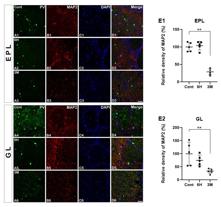
the MOB (Fig. 4A1 - D1 and A4 - D4). Six hours after pilo-
carpine treatment, MAP2 immunoreactivity in both the EPL
and GL was not changed in the PV-positive interneurons (7) and the olfactory bulb volumes were decreased in the tem-
compared to the control group (Fig. 4A2 - D2 and A5 - D5). poral lobe of epilepsy patients (17). Therefore, because stimula-
Quantitative analysis of the double immunofluorescence staining tions of the olfactory bulb can also induce seizures in a certain
data also revealed similar results at the same time periods (Fig. mouse model (18), these phenomena may support a close
4E1 and 4E2). Three months after pilocarpine administration, correlation between olfactory bulb function and epileptogenesis.
at the recurrent seizure time period, however, the double- The MOB is generally known as a useful region for invest-
labeled expression of PV and MAP2 immunoreactivity was igating neuronal interactions due to its distinct structures and
markedly decreased in the EPL and GL (Fig. 4A3 - D3 and A6 - diversity of neuroactive substances (19). Excitatory mitral and
D6). The quantitative analysis of the immunoreactivity of these tufted cells have reciprocal dendrodendritic connections with
two makers showed the same results as the immunostaining inhibitory granule cells (20). Mitral cells receive odor input from
data at this time point (Fig. 4E1 and 4E2). olfactory sensory neurons and glutamate released from the
excited mitral cells induces the GABAergic dendrodendritic
DISCUSSION reversion of granule cells back into mitral cells (21). In the EPL
of the rat MOB, PV is expressed in fast-spiking GABAergic
In the present study, we observed the neuronal degeneration interneurons (9, 13) and mitral cells form a reciprocal
and morphological changes following pilocarpine-induced SE connection with these PV-positive cells. Feedback inhibition
in the rat MOB. Not only PV-positive interneurons but also modulates the output of the mitral cells (14). These structural
NeuN-positive neurons were decreased time-dependently after and functional features enable the fine-tuning of odor process-
pilocarpine treatment. Several previous studies have reported ing. In addition, it has been reported that PV has a crucial role
that SE resulted in oxidative stress and neuroinflammation in in modulating intracellular calcium levels that are important
the brain subregions, including the hippocampus and olfactory for synaptic plasticity (22). In olfactory reciprocal synapses, the
bulb, causing seizure-induced neuronal damage (15, 16). In release of fast neurotransmitters is mediated by calcium influx
addition, functional and morphological abnormalities of the olfac- through the N- and P/Q-type calcium channels and the magni-
tory bulb were observed in the temporal lobe of epilepsy patients tude of the dendrodendritic transmission depends on the level
236 BMB Reports http://bmbreports.org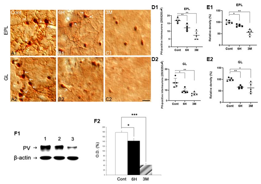
PV immunoreactivity in the MOB following SE
Yeon Hee Yu, et al.
of PV. Morphological changes in the dendritic processes are
closely related to synaptic plasticity (26, 27) and we observed
decreased dendritic density in the MOB of the SE rats.
Decreases in the dendritic density of PV-positive interneurons
important for odor tuning support a functional loss of olfaction.
In the present study, we confirmed morphologically that
PV-positive interneurons were detected in the EPL and GL of
the rat MOB and that pilocarpine-induced SE resulted in
neurodegeneration and a decrease in PV- and NeuN-positive
neurons. In the MOB, the reciprocal interaction of PV-positive
interneurons with mitral cells is crucial for odor tuning (14)
and the roles of PV for modulating calcium levels are essential
for synaptic transmission and various neuronal function. Thus,
the SE-induced changes in PV expression in the MOB strongly
support the loss of olfaction seen in epilepsy patients.
MATERIALS AND METHODS
Experimental animals
All experiments utilized the progeny of Sprague-Dawley rats
Fig. 4. Double immunofluorescence staining for PV (A, green), MAP2 obtained from the Experimental Animal Center, Soonchunhyang
(B, red), with DAPI (C), and merged images (D) in the EPL and University (Cheonan, South Korea). All animals were provided
GL after SE. In the control group, MAP2 expression shows dense with a commercial diet and water ad libitum under controlled
dendritic trees (B1 and B4). Three months following SE, however,
MAP2 expression is remarkably decreased compared to the early temperature, humidity and lighting conditions (light/dark cycle
o
time period (B3 and B6). Relative density for MAP2 in the EPL 12:12, and 22 ± 2 C, 55 ± 5%). All animal protocols were
and GL (E). All data are presented as mean ± SEM. **P < 0.01 approved by the Administrative Panel on Laboratory Animal
vs. control. Glomerular layer; GL, external plexiform layer; EPL, H; Care of Soonchunhyang University (No. SCH16-0051). All
hours, M; months. Scale bar = 17 m.
possible efforts were made to avoid animal suffering and
minimize the number used in the experiments.
of intracellular calcium influx (21). In the SE model of this
study, as the level of PV expression decreased, we predicted Seizure induction
that decreased calcium buffering capability and impaired The SE rat model was induced by treatment with pilocarpine
inhibitory postsynaptic potential occurred, which consequently as previously described (28). The rats were pretreated with lithium-
resulted in olfaction dysfunction. PV is also involved in the chloride before pilocarpine to ensure a high survival rate. Twenty
phosphorylation of protein kinase which has a crucial role in hours after lithium-chloride (127 mg/kg, i.p.; Sigma-Aldrich, St.
2+ 2+
cell signal transduction via modulating Ca and Mg ions, Louis, MO, USA) treatment, scopolamine was injected (2 mg/
indicating that PV is involved in a variety of cellular processes kg, i.p.; Sigma-Aldrich) 30 min prior to pilocarpine (30 mg/kg,
(23, 24). This suggests that in the MOB of SE rats, decreases in i.p.; Sigma-Aldrich) treatment, which induces SE. Two hours
PV-positive interneurons alter not only GABAergic inhibitory following pilocarpine treatment, diazepam was injected for
functions but also various calcium-dependent cell signaling. depression. The seizures were classified into 5 grades according
In a previous study of rat olfactory bulbs, Hwang et al. (9) to behavioral characteristics (28), and the SE animals used in
reported that PV-positive interneurons had branched and the study, consistently exhibited 4 or 5-grade seizures until
well-developed processes with varicosities and spines like they were injected with diazepam. For the study, the animals
calbindin-positive interneurons. The well-developed processes were divided into three groups; the control group, and groups
of PV-positive interneurons have much denser connections at six hours and three months after SE.
with mitral cells than those between granule cells and mitral
cells (14). However, we confirmed that PV immunoreactivity Fluoro-jade b (FJB) staining
and dendritic density was markedly decreased following SE. To identify degeneration, FJB staining was performed (29). Each
Oxidative stress and neuroinflammation induced by SE can section of the MOB was mounted on gelatin-coated slides and
result in neuronal loss in the olfactory bulb, which decreased air-dried overnight. The slides were immersed in 1% NaOH in
dendritic density in this study. In contrast, some studies have 80% alcohol for 5 min, 2 min in 70% alcohol, and 2 min in
suggested that calcium signals are associated with the growth distilled water. Then, the slides were transferred to a 0.06%
and branching of dendrites (25). Therefore, we can consider a potassium permanganate solution for 10 minutes. The slides
correlation between decreased dendritic density and the roles were washed in distilled water for 2 minutes. The staining
http://bmbreports.org BMB Reports 237PV immunoreactivity in the MOB following SE
Yeon Hee Yu, et al.
solution was prepared from a 0.01% stock solution of fluoro- membranes (Pall Crop, East Hills, NY, USA) were incubated
jade b (Millipore, Burlington, MA, USA) in 0.1% acetic acid. with rabbit anti-PV IgG (1:10,000, Swant Switzerland) and the
The slides were incubated in a final concentration of 0.0004% data were normalized compared to the -actin levels as descri-
for 20 min, then dried for 1 hour. After drying, the slides were bed in a previous study (30).
rinsed 1 min in xylene. The slides were mounted in mounting
medium 4′ ,6-diamidine-2′-phenylindole dihydrochloride (DAPI) Quantification of data and statistical analysis
(Vector Laboratories, Burlingame, CA, USA). All images were The cell counts were performed by two different investigators
captured using a model Fluoview FV10i and FV10i software who were blinded to the classification of the tissues. Each
(Olympus, Japan). image was normalized by adjusting the black and white range
of the image using Adobe PhotoShop v. 8.0. Thereafter, 10
2
Immunohistochemistry and double immunofluorescence areas per rat (250 × 250 m for each area) were selected and
The anesthetized animals (urethane 1.5 kg/kg, i.p.; Sigma- the intensity measurements were represented as the mean number
Aldrich) were perfused transcardially with phosphate-buffered of a 256-shade gray-scale (using NIH Image 1.59 software).
saline (PBS), followed by 4% paraformaldehyde in 0.1M The optical density values were corrected by subtracting the
phosphate-buffer (PB) (n = 5 in each group). The brains were average background noise values obtained from five image
removed and post-fixed in the same fixative for 4 h, and rinsed inputs. All data obtained from the quantitative measurements
o
in PB containing 30% sucrose at 4 C for 2 days. Thereafter, the were analyzed using an one-way analysis of variance (ANOVA)
tissues were frozen and sectioned with a cryostat at 30 μm to determine the statistical significance. Bonferroni's test was
thickness and consecutive sections were collected in six-well used for post-hoc comparisons. P-values of < 0.01, 0.05, and
plates containing PBS. The sections were incubated with 0.005 was considered statistically significant (28).
primary rabbit anti-PV antibody(1:10000, Swant, Switzerland)
in PBS containing 0.3% Triton X-100 overnight at room ACKNOWLEDGEMENTS
temperature. The sections were washed 3 times for 10 min with
PBS, incubated sequentially in biotinylated goat anti-rabbit IgG This work was supported by the Soonchunhyang University
(1:200, Vector Laboratories) and ABC complex (Vector Labora- and a Basic Science Research Program (NRF-2017R1D1A1B05
tories). The sections were visualized with 3,3′-diaminobenzidine 036195) through the National Research Foundation of Korea
(DAB) in 0.1 M Tris buffer and mounted on gelatin-coated funded by the Ministry of Science.
slides. The immunoreactions were observed using DMRB
microscope (Leica, Germany) and the images were captured CONFLICTS OF INTEREST
using a model DP72 digital camera and DP2-BSW microscope
digital camera software (Olympus, Japan). To establish the The authors have no conflicting interests.
specificity of the immunostaining, a negative control test was
carried out with pre-immune serum instead of the primary REFERENCES
antibody. The negative control resulted in the absence of immuno-
reactivity in any structures. To identify the morphological changes 1. Al-Mufti F and Claassen J (2014) Neurocritical care: status
induced by SE in the same MOB tissue, double immunofluo- epilepticus review. Crit Care Clin 30, 751-764
rescence staining for PV/NeuN and PV/MAP2 was performed. 2. French JA (2007) Refractory epilepsy: clinical overview.
The MOB tissues were incubated overnight at room temperature Epilepsia 48, 3-7
in a mixture of rabbit anti-PV IgG (1:5,000, Swant)/mouse 3. Henshall D (2007) Apoptosis signaling pathways in seizure-
induced neuronal death and epilepsy. Biochem Soc Trans
anti-NeuN IgG (1:50, Millipore) and rabbit anti-PV IgG (1:
35, 421-423
5,000, Swant)/mouse anti-MAP2 IgG (1:300, Millipore). After 4. Wang L, Liu YH, Huang YG and Chen LW (2008) Time-
washing three times for 10 min with PBS, the sections were course of neuronal death in the mouse pilocarpine model
incubated in a mixture of Cy2- and Cy3-conjugated secondary of chronic epilepsy using Fluoro-Jade C staining. Brain Res
antisera (1:200, Jackson Immunoresearch, Baltimore, PA, USA) J 1241, 157-167
for 1 h at room temperature. The sections were mounted with 5. Helmstaedter C and Kockelmann E (2006) Cognitive out-
DAPI (Vector). All images were captured using a model comes in patients with chronic temporal lobe epilepsy.
Fluoview FV10i and FV10i software (Olympus, Japan). Epilepsia 47, 96-98
6. Mehla J, Reeta K, Gupta P and Gupta YK (2010) Protective
effect of curcumin against seizures and cognitive impair-
Western blot analyses
ment in a pentylenetetrazole-kindled epileptic rat model.
To quantify the expression of PV following pilocarpine-induced Life Sci 87, 596-603
status epilepticus (SE), the rats (n = 5 per group) were euthan- 7. Desai M, Agadi J, Karthik N, Praveenkumar S and Netto A
ized with urethane (1.5 kg/kg, i.p.; Sigma-Aldrich). The olfactory (2015) Olfactory abnormalities in temporal lobe epilepsy.
bulbs were quickly removed and homogenized in a buffer, as J Clin Neurosci 22, 1614-1618
previously described (30). The protein-transferred nitrocellulose 8. Imai T (2014) Construction of functional neuronal cir-
238 BMB Reports http://bmbreports.orgPV immunoreactivity in the MOB following SE
Yeon Hee Yu, et al.
cuitry in the olfactory bulb. Semin Cell Dev Biol 35, in the mouse olfactory bulb. J Comp Neurol 523, 262-280
180-188 20. Egger V and Urban NN (2006) Dynamic connectivity in
9. Hwang IK, Kim DS, Lee HY et al (2003) Age-related Changes mitral cell–granule cell microcircuit. Semin Cell Dev Biol
of Parvalbumin Immunoreactive Neurons in the Rat Main 17, 424-432
Olfactory Bulb. Mol Cells 16, 302-306 21. Isaacson JS and Strowbridge BW (1998) Olfactory reci-
10. Caillard O, Moreno H, Schwaller B, Llano I, Celio MR procal synapses: dendritic signaling in the CNS. Neuron
and Marty A (2000) Role of the calcium-binding protein 20, 749-761
parvalbumin in short-term synaptic plasticity. Proc Natl 22. Schwaller B, Meyer M and Schiffmann S (2002) ‘New’
Acad Sci U S A 97, 13372-13377 functions for ‘old’proteins: the role of the calcium-binding
11. Choi WS, Chun SY, Markelonis GJ, Oh TH and Oh YJ proteins calbindin D-28k, calretinin and parvalbumin, in
(2001) Overexpression of calbindin-D28K induces neurite cerebellar physiology. Studies with knockout mice.
outgrowth in dopaminergic neuronal cells via activation Cerebellum 1, 241-258
of p38 MAPK. Biochem Biophys Res Commun 287, 656- 23. Schwaller B (2009) The continuing disappearance of
661 “pure” Ca 2+ buffers. Cell Mol Life Sci 66, 275-300
12. Mattson MP (2007) Calcium and neurodegeneration. Aging 24. Yu L, Xu L, Xu M, Wan B, Yu L and Huang Q (2011) Role
Cell 6, 337-350 of Mg2+ ions in protein kinase phosphorylation: insights
13. Kim J, Kwak S, Kim D et al (2006) Reduced calcium from molecular dynamics simulations of ATP-kinase com-
binding protein immunoreactivity induced by electrocon- plexes. Mol Simul 37, 1143-1150
vulsive shock indicates neuronal hyperactivity, not neuronal 25. Konur S and Ghosh A (2005) Calcium signaling and the
death or deactivation. Neuroscience 137, 317-326 control of dendritic development. Neuron 46, 401-405
14. Kato HK, Gillet SN, Peters AJ, Isaacson JS and Komiyama 26. Yuste R and Bonhoeffer T (2001) Morphological changes
T (2013) Parvalbumin-expressing interneurons linearly in dendritic spines associated with long-term synaptic
control olfactory bulb output. Neuron 80, 1218-1231 plasticity. Annu Rev Neurosci 24, 1071-1089
15. Kovac S, Domijan A, Walker M and Abramov A (2014) 27. Sjostrom PJ, Rancz EA, Roth A and Hausser M (2008)
Seizure activity results in calcium-and mitochondria-inde- Dendritic excitability and synaptic plasticity. Physiol Rev
pendent ROS production via NADPH and xanthine 88, 769-840
oxidase activation. Cell Death Dis 5, e1442 28. Kim DS, Kim JE, Kwak SE et al (2008) Spatiotemporal
16. González-Reyes S, Santillán-Cigales JJ, Jiménez-Osorio AS, characteristics of astroglial death in the rat hippocampo-
Pedraza-Chaverri J and Guevara-Guzmán R (2016) Glycyrrhizin entorhinal complex following pilocarpine-induced status
ameliorates oxidative stress and inflammation in hippo- epilepticus. J Comp Neurol 511, 581-598
campus and olfactory bulb in lithium/pilocarpine-induced 29. Schmued LC and Hopkinsa CJ (2000) Fluoro-Jade B: a
status epilepticus in rats. Epilepsy Res 126, 126-133 high affinity fluorescent marker for the localization of
17. Hummel T, Henkel S, Negoias S et al (2013) Olfactory neuronal degeneration. Brain Res J 874, 123-130
bulb volume in patients with temporal lobe epilepsy. J 30. Jung HY, Kim DW, Nam SM et al (2017) Pyridoxine
Neurol 260, 1004-1008 improves hippocampal cognitive function via increases of
18. Nguyen MQ and Ryba NJ (2012) A smell that causes serotonin turnover and tyrosine hydroxylase, and its
seizure. PLoS One 7, e41899 association with CB1 cannabinoid receptor-interacting protein
19. Suzuki Y, Kiyokage E, Sohn J, Hioki H and Toida K (2015) and the CB1 cannabinoid receptor pathway. Biochim
Structural basis for serotonergic regulation of neural circuits Biophys Acta Gen Subj 1861, 3142-3153
http://bmbreports.org BMB Reports 239You can also read