Mitochondrial DNA editing in mice with DddA-TALE fusion deaminases - Nature
←
→
Page content transcription
If your browser does not render page correctly, please read the page content below
ARTICLE
                  https://doi.org/10.1038/s41467-021-21464-1                 OPEN

                  Mitochondrial DNA editing in mice with
                  DddA-TALE fusion deaminases
                  Hyunji Lee1,3, Seonghyun Lee1,3, Gayoung Baek1, Annie Kim1,2, Beum-Chang Kang1, Huiyun Seo1 &
                  Jin-Soo Kim 1,2 ✉
1234567890():,;

                  DddA-derived cytosine base editors (DdCBEs), composed of the split interbacterial toxin
                  DddAtox, transcription activator-like effector (TALE), and uracil glycosylase inhibitor (UGI),
                  enable targeted C-to-T base conversions in mitochondrial DNA (mtDNA). Here, we
                  demonstrate highly efficient mtDNA editing in mouse embryos using custom-designed
                  DdCBEs. We target the mitochondrial gene, MT-ND5 (ND5), which encodes a subunit of
                  NADH dehydrogenase that catalyzes NADH dehydration and electron transfer to ubiquinone,
                  to obtain several mtDNA mutations, including m.G12918A associated with human mito-
                  chondrial diseases and m.C12336T that incorporates a premature stop codon, creating
                  mitochondrial disease models in mice and demonstrating a potential for the treatment of
                  mitochondrial disorders.

                  1 Center for Genome Engineering, Institute for Basic Science, Daejeon, Republic of Korea. 2 Department of Chemistry, Seoul National University,

                  Seoul, Republic of Korea. 3These authors contributed equally: Hyunji Lee, Seonghyun Lee. ✉email: jskim01@snu.ac.kr

                  NATURE COMMUNICATIONS | (2021)12:1190 | https://doi.org/10.1038/s41467-021-21464-1 | www.nature.com/naturecommunications                          1
ARTICLE NATURE COMMUNICATIONS | https://doi.org/10.1038/s41467-021-21464-1
M
itochondrial DNA plays a critical role in cellular oxidoreductase chain 5 protein to demonstrate in vivo mtDNA
respiration via the mitochondrial oxidative phosphor- editing using our Golden-Gate assembly system. The ND5 pro-
ylation (OXPHOS) system. Because the OXPHOS system tein is a core subunit of NADH dehydrogenase (ubiquinone),
is essential for survival, mutations in mtDNA cause severe mal- which catalyzes the transfer of electrons from NADH to the
functions in multiple organs and muscles, especially in high-energy respiratory chain. In humans, mutations in the ND5 gene are
demand tissues1. Typically, in humans with a mitochondrial dis- known to be related to mitochondrial encephalomyopathy, lactic
ease, wild-type (WT) and mutant mtDNA with single-base muta- acidosis, and stroke-like episodes (MELAS), as well as some
tions coexist in a cell, resulting in a heteroplasmic state of the symptoms of Leigh’s syndrome and Leber’s hereditary optic
mtDNA population2. The balance between mutant and WT neuropathy (LHON)10. We sought to generate mouse models
mtDNA determines the development of mitochondrial diseases with genetic variations in the mitochondrial gene to mimic the
with clinical phenotypes3. Programmable nucleases have been used dysfunctions in humans.
to cleave a mutant mtDNA, but not the WT mtDNA to reduce the First, we assembled several DdCBE plasmids that were
mutant mtDNA population in vitro and in vivo4,5. But these designed to induce two possible silent mutations, m.C12539T
nucleases cannot induce or revert a specific mutation in mtDNA, and m.G12542A, in the ND5 gene. We transfected these plasmids
possibly because DNA double-strand breaks are not efficiently into the NIH3T3 mouse cell line, and measured editing
repaired in mitochondria by nonhomologous end joining or frequencies at day 3 post transfection. As expected, cytosine
homologous recombination, unlike those in the nucleus. bases in the editing window were successfully converted into
Mok et al. recently developed a base editing approach using the thymine with editing efficiencies of up to 19% (Fig. 2a). In line
bacterial cytidine deaminase toxin, DddAtox, to demonstrate with the previous report showing that DddAtox exclusively
efficient C-to-T base conversions in vitro6. In this approach, split deaminates cytosine in a “TC” motif6, only the two cytosine
DddAtox nontoxic halves fused to transcription activator-like bases in a TC context were edited. Indels and other types of point
effector (TALE) proteins, which can be custom-designed to mutations were not detectably induced in the editing window.
recognize predetermined target DNA sequences7, form a func-
tional cytosine deaminase within the editing window to induce
C-to-T base editing at the target site in mtDNA. Mitochondrial DNA editing in vivo. We chose the most active
In this study, we investigate whether DdCBEs can achieve DdCBE pair (left-G1397-N and right-G1397-C) for an in vivo
mtDNA base editing in vivo to create animal models with study. At day 4 post microinjection of in vitro transcripts encoding
mitochondrial mutations and to show germline transmission of this DdCBE pair into one-cell stage C57BL6/J embryos, we were
the resulting mitochondrial mutations in mice. able to obtain nine edit-positive embryos out of a total of 32
embryos (28%; Table 1). The TALE–DddAtox deaminase efficiently
induced C ∙ G to T ∙ A transitions, with frequencies that ranged from
Results
2.2 to 25% at the m.C12539 position and from 0.63 to 5.8% at the
Assembly of DdCBE plasmids. To facilitate the assembly of
m.G12542 position (Fig. 2b). Next, we implanted DdCBE-injected
custom-designed TALE arrays in DdCBEs, we constructed
embryos into surrogate mothers, and obtained offspring carrying m.
expression plasmids encoding split DddAtox halves and used our
C12539T and m.G12542A mutations (Supplementary Fig. 2a).
Golden-Gate cloning system, which employs a total of 424 (=6 ×
Three out of four newborn (F0) mice harbored C ∙ G to T ∙ A
64 tripartite + 2 × 16 bipartite + 2 × 4 monopartite) modular
conversions with frequencies that ranged from 1 to 27% (Fig. 2c).
TALE array plasmids8,9 (Fig. 1a). We mixed six TALE array
Two pups showed similar mutation frequencies in the toe and the
plasmids and an expression vector in a single Eppendorf tube to
tail, which were retained for at least 14 days after birth. Further-
construct a ready-to-use DdCBE plasmid that encodes 15.5–18.5
more, these mtDNA mutations were detected in various tissues
repeat variable diresidue arrays (Supplementary Fig. 1). The
obtained from a fully grown adult F0 mouse at day 50 post birth
resulting DdCBEs recognized DNA sequences of 17–20 base pairs
(Fig. 2d). This result suggests that mtDNA heteroplasmy induced
(bps) in length, including a conserved T at the 5′ terminus
by DdCBEs in one-cell stage zygotes can be maintained throughout
(Supplementary Table 1). As a result, a functional DdCBE pair
development and differentiation.
recognized 32- to 40-bp DNA sequences (Fig. 1b).
To investigate whether the DdCBE-induced mutations can be
transmitted to the next generation, we crossed the female F0
Mitochondrial DNA editing in vitro. We chose the Mus mus- mouse with a WT C57BL6/J male and obtained F1 offspring. The
culus mitochondrial ND5 gene encoding NADH-ubiquinone m.C12539T and m.G12542A mutations were observed in two
Fig. 1 Schematic illustration for assembling DdCBE and its mitochondrial DNA editing. a Scheme of one-pot Golden-Gate assembly for efficient DdCBE
construction. A total of 424 arrays (64 tripartite arrays × 6 + 16 bipartite arrays × 2 + 4 monopartite arrays × 2) and expression vector were mixed to
generate left and right modules for the final plasmid constructs. b Illustration of DdCBE interacting with mouse mitochondrial DNA target ND5. TALE-
binding regions are shown in gray and base editing windows are depicted in black. Different repeat variable diresidue modules are shown in orange, blue,
green, and yellow, which represent “NI” for adenine, “NG” for thymine, “NN” for guanine, and “HD” for cytosine recognition, respectively.
2 NATURE COMMUNICATIONS | (2021)12:1190 | https://doi.org/10.1038/s41467-021-21464-1 | www.nature.com/naturecommunicationsNATURE COMMUNICATIONS | https://doi.org/10.1038/s41467-021-21464-1 ARTICLE
Fig. 2 Mouse mitochondrial ND5 point mutation generated by DdCBE-derived base editing. a DdCBE deaminase-mediated cytosine-to-thymine base
editing target and efficiency in NIH3T3 cells. In the target sequence, translation codons are underlined and possible editing loci are shown in red. Transfected
combinations of DdCBE are annotated as left or right, -G1333 or -G1397, and -N or -C. P values of left-G1333-N + right-G1333-C, left-G1333-C + right-G1333-N,
left-G1397-N + right-G1397-C, and left-G1397-C + right-G1397-N for C10 mutation are 0.0012, 0.0003, 0.0014, and 0.0009, and for C13 mutation are 0.0116,
0.0076, 0.0030, and 0.0003, respectively (*p < 0.05 and **p < 0.01 using Student’s two-tailed t test). b Corresponding base editing efficiency in mouse
blastocysts. The sequencing data were obtained from blastocysts that developed after zygotes were microinjected with mRNA encoding the left-G1397-N and
right-G1397-C DdCBE. c Alignments of mutant sequences from newborn pups. Targeted deep sequencing was performed using genomic DNA isolated from the
tail of the newborns immediately after birth, and that from the toe 7 and 14 days after birth. Edited bases are shown in red. Editing frequencies in the mutant
mitochondrial genome are shown. d Editing efficiencies in various tissues of an adult F0 mouse (sipup-1). The sequencing data were obtained from each tissue
50 days after birth. In all graphs, the dark and light gray bars represent the frequency of m.C12539T (C10) and m.G12542A (C13) mutations, respectively. Error
bars are the standard error of the mean (s.e.m.) for n = 3 biologically independent samples. Source data are provided in the Source data file.
Table 1 Summary of the numbers of blastocysts used and mutants obtained.
Type of Number of Number of Number of Number of Number of edited/ Number of edited/
mutagenesis examined blastocysts (%) transferred offspring (%) total blastocysts (%) total offspring (%)
embryos embryos
N/A (buffer 20 11 (55) NA NA 0/11 (0) NA
injection)
ND5 silent 56 32 (57) 30 4 (13) 9 (28) 3 (75)
ND5 G12918A 79 44 (55) 50 11 (22) 11 (25) 4 (36)
ND5 STOP 68 37 (54) 120 27 (23) 19 (51) 9 (33)
newborns with frequencies that ranged from 6 to 26% (Fig. 3a). to 23% (Fig. 4c). Next, we implanted DdCBE-microinjected
Furthermore, these two mtDNA edits were detected at compar- embryos into surrogate mothers to obtain the G12918A mutant
able frequencies in 11 different tissues (Fig. 3b). offspring (Supplementary Fig. 2b). Four out of 11 newborn mice
harbored the mutant allele with frequencies of 3.9–31.6% (Fig. 4d).
Although no apparent phenotypes were observed right after birth,
DdCBE-mediated MT-ND5 G12918A mutation. Next, we possibly because the pups were still very young and also because
sought to produce the m.G12918A point mutation, which can the WT mitochondrial DNA coexists with the mutant DNA in a
cause mitochondrial disorders in humans. Note that mutations at heteroplasmic state, these data suggest that DdCBEs can be used
this position give rise to multiple mitochondrial diseases, such as to create animal models with mitochondrial disorders.
Leigh disease, MELAS syndrome, and LHON syndrome10,11. The
cytosine base at this position has adjacent thymine, amenable for
DdCBE-mediated base editing (Fig. 4a). We assembled four MT-ND5 nonsense mutation. Last but not the least, we investi-
DdCBE pairs and confirmed successful base editing at this posi- gated whether a loss-of-function mutation in ND5 would be tol-
tion in NIH3T3 cells with up to 6.4% efficiency (Fig. 4b). We then erated in mice by creating a nonsense mutation in the gene. We
microinjected the best-performing DdCBE combination into chose m.C12336 as a target cytosine for incorporation of a pre-
mouse zygotes and measured editing efficiencies in the resulting mature stop codon at the 199th position of the ND5 protein
blastocysts. Eleven out of 44 (25%) embryos harbored the m. (Q199*; Fig. 5a). We first determined the editing activity of four
G12918A mutation with editing efficiencies that ranged from 0.25 custom-designed DdCBE combinations in transfected NIH3T3
NATURE COMMUNICATIONS | (2021)12:1190 | https://doi.org/10.1038/s41467-021-21464-1 | www.nature.com/naturecommunications 3ARTICLE NATURE COMMUNICATIONS | https://doi.org/10.1038/s41467-021-21464-1
Fig. 3 Germline transmission of mutant mtDNA. a To observe the germline transmission of mtDNA mutations, the female F0 (sipup-3) mouse was crossed
with a wild-type C57BL6/J male to obtain F1 pups (101, 102), after which targeted deep sequencing was performed. Edited bases are shown in red. Editing
frequencies in the mutant mitochondrial genome are shown. b Base editing efficiencies in various tissues from an F1 newborn pup (101), obtained using targeted
deep sequencing of genomic DNA. Dark and light gray bars represent the frequency of m.C12539T (C10) and m.G12542A (C13) mutations, respectively. Error
bars are the standard error of the mean (s.e.m.) for n = 3 biologically independent samples. Source data are provided in the Source data file.
Fig. 4 Mouse mitochondrial ND5 G12918A mutation induced by DdCBE. a The DdCBE target for generating the m.G12918A point mutation, which would
create a D393N change in the ND5 protein. The target codon is underlined and the possible editing locus is shown in red. b The efficiency of cytosine-to-thymine
base editing with DdCBE in NIH3T3 cells. The annotations indicate the combination of DdCBE pairs that were co-transfected. Error bars are s.e.m. for n = 3
biologically independent samples (n.s. not significant, *p < 0.05, and **p < 0.01 using Student’s two-tailed t test). P values of left-G1333-N + right-G1333-C, left-
G1333-C + right-G1333-N, left-G1397-N + right-G1397-C, and left-G1397-C + right-G1397-N for C6 mutation are 0.0052, 0.0099, 0.0027, and 0.0040,
respectively. P values for n.s. is 0.4971. c m.G12918A point mutation base editing efficiency in mouse blastocysts. The sequencing data were obtained from
cultured blastocysts that developed after one-cell stage embryos were microinjected with mRNA encoding the left-G1397-C and right-G1397-N DdCBE. d Mice
(F0) carrying an ND5 point mutation. F0 pups, which harbor an ND5 point mutation, that developed after microinjection of the DdCBE mRNAs. Corresponding
alignment of mutant sequences from newborn pups. Edited bases are shown in red, and the column on the right indicates the editing frequencies in the mutant
mitochondrial genome. Source data are provided in the Source data file.
cells and found that the most effective DdCBE pair induced the substitutions in the nuclear genome without double-stranded
nonsense mutation at an editing efficiency of 5.7% (Fig. 5b). This DNA cleavage12,13. Previously, we were able to use both cytosine
DdCBE also induced a C-to-T conversion, albeit less efficiently, at and adenine base editors to create animal models with point
m.G12341A in the editing window, which created a silent muta- mutations in the nuclear genome and to correct them in vivo14,15.
tion (Q200Q). In 19 out of 37 (=51%) mouse embryos, we also Unlike the nuclear genome, however, mitochondrial DNA has
observed the two m.C12336T and m.G12341A mutations with never been successfully edited in vitro or in vivo using the Cas9
editing frequencies of up to 32% and 23%, respectively (Fig. 5c). nickase–deaminase fusion proteins, possibly because it is difficult
Encouraged by these results, we implanted mouse embryos into to deliver both the protein component and guide RNA to mito-
surrogate mothers, and obtained offspring with m.C12336T and chondria at the same time. Mok et al. demonstrated that a base
m.G12341A mutations (Supplementary Fig. 2c). A total of 9 out of editing system, free of guide RNA, composed of a TALE array
27 F0 mice (23%) harbored C ∙ G to T ∙ A conversions with fused to DddAtox enables mtDNA base editing in cell lines. In this
frequencies that ranged from 0.22 to 57% (Fig. 5d, e), showing that study, we used DdCBEs to create various point mutations,
the ND5 nonsense mutation did not lead to embryonic lethality. including silent, nonsense, and missense mutations, in the
mitochondrial ND5 gene in mice for the first time (Table 1). To
Discussion this end, we developed a Golden-Gate cloning system that con-
Base editing, catalyzed by a Cas9 nickase fused to a deaminase sists of a total of 432 plasmids (8 expression plasmids plus 424
protein, is a powerful method for inducing point mutations or TALE array plasmids) to assemble DdCBE plasmids rapidly.
4 NATURE COMMUNICATIONS | (2021)12:1190 | https://doi.org/10.1038/s41467-021-21464-1 | www.nature.com/naturecommunicationsNATURE COMMUNICATIONS | https://doi.org/10.1038/s41467-021-21464-1 ARTICLE
Fig. 5 Mouse mitochondrial ND5 nonsense mutation generated via cytidine-deaminase-mediated base editing. a The DdCBE target for generating the m.
C12336T nonsense mutation and m.G12341A silent mutation. The m.C12336T (C9) mutation creates a Q199stop mutation in the ND5 protein, whereas m.
G12341A (C14) causes a silent Q200Q mutation. Translation triplets are underlined and possible editing loci are shown in red. b The efficiency of the
cytosine-to-thymine base editing that creates a nonsense mutation in NIH3T3 cells. The annotations indicate the combination of DdCBE pairs that were co-
transfected into cells. Dark and light gray bars represent the frequency of m.C12336T (C9) and m.G12341A (C14) mutations, respectively. Error bars
represent s.e.m. for n = 3 biologically independent samples (n.s. not significant, *p < 0.05, and **p < 0.01 using Student’s two-tailed t test). P values of left-
G1333-N + right-G1333-C, left-G1333-C + right-G1333-N, left-G1397-N + right-G1397-C, and left-G1397-C + right-G1397-N for C9 mutation are 0.0065,
0.1143, 0.0266, and 0.0037, and for C14 mutation are 0.0077, 0.0144, 0.0406, and 0.0214, respectively. c Base editing efficiency in mouse blastocysts. The
sequencing data were obtained from blastocysts that developed after zygotes were microinjected with mRNA encoding left-G1333-N and right-G1333-C
DdCBE. Dark and light gray bars represent the frequency of the C9 and C14 mutations, respectively. d Alignment of mutant sequences from newborn pups.
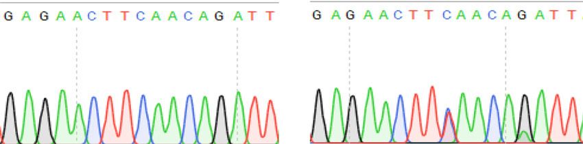
Edited bases are shown in red, and the column on the right indicates the editing frequencies in the mutant mitochondrial genome. e Sanger sequencing
chromatograms from non-edited and edited mice. The red arrows indicates the substituted nucleotides. Source data are provided in the Source data file.
MtDNA mutations induced by the resulting DdCBEs were assembly kit (NEB); assembled plasmids were chemically transformed into Escherichia
detected in various tissues in an adult mouse, showing that coli DH5ɑ (Enzynomics), and their identity confirmed by Sanger sequencing.
Thereby, we obtained eight expression plasmids, which include BsaI restriction
mtDNA heteroplasmy induced by DdCBEs was maintained enzyme sites between regions encoding the N-terminal domain and C-terminal half
throughout the development and differentiation. We also showed domain (NG) of TALE for Golden-gate assembly. For DdCBE plasmid assembly, each
successful germline transmission of mtDNA edits induced by expression plasmid was mixed with six module vectors (each encoding a TALE array),
DdCBEs, suggesting that it is possible to create animal models BsaI (10 U), T4 DNA ligase (200 U), and reaction buffer in a single tube (Supple-
mentary Fig. 1). Next, restriction–ligation reactions were performed in a thermo-
with mitochondrial disorders. We propose that our Golden-Gate cycler, with 20 cycles of 37 °C and 50 °C for 5 min each, followed by final incubations
plasmids are valuable resources for studying the functions of at 50 °C for 15 min and 80 °C for 5 min. Ligated plasmids were chemically trans-
mitochondrial genes in vitro and in vivo, and to correct patho- formed into E. coli DH5ɑ, and subjected to Sanger sequencing to confirm the identity
genic mutations for the treatment of mitochondrial genetic dis- of the constructs. Correct plasmids were midi-prepped (Qiagen) for cell transfection.
orders in the future.
Mammalian cell culture and transfection. NIH3T3 (CRL-1658, American Type
Culture Collection) cells were cultured and maintained at 37 °C with 5% CO2. Cells
Methods were grown in DMEM supplemented with 10% (v/v) bovine calf serum (Gibco)
Plasmid construction. We adapted our transcription activator-like effector nucleases without any antibiotics. For lipofection, cells were seeded in 12-well cell culture
(TALEN) assembly system to construct expression vectors for the split DddA halves, plates (SPL, Seoul, Korea) at a density of 1.5 × 10⁴ cells per well, 18–24 h before
as well as final TALE–DddAtox constructs8. Beginning with the expression vector transfection. Lipofection using Lipofectamine 3000 (Invitrogen) was performed
from the TALEN system, we replaced the fragments encoding the nuclear localization with 500 ng of each TALE half monomer plasmid to make up 1000 ng of total
sequence and FokI obligatory heterodimeric halves with fragments encoding mito- plasmid DNA. Cells were harvested at day 3 post transfection.
chondrial translocation sequences (MTS), DddA deaminase dimeric halves, and uracil
glycosylase inhibitor (UGI). The MTS-, DddA-, and UGI-encoding sequences were
synthesized by IDT. To construct expression plasmids, DNA fragments for Gibson mRNA preparation. The mRNA templates were prepared by PCR using Q5 High-
assembly were amplified using Q5 DNA Polymerase (NEB), and subjected to PCR Fidelity DNA Polymerase (NEB) with the following primers (F: 5′-CATCAA
and gel purification. Purified gene fragments were assembled with a HiFi DNA TGGGCGTGGATAG-3′, R: 5′-GACACCTACTCAGACAATGC-3′). DdCBE
NATURE COMMUNICATIONS | (2021)12:1190 | https://doi.org/10.1038/s41467-021-21464-1 | www.nature.com/naturecommunications 5ARTICLE NATURE COMMUNICATIONS | https://doi.org/10.1038/s41467-021-21464-1
mRNAs were synthesized using an in vitro RNA transcription kit (mMESSAGE 4. Hashimoto, M. et al. MitoTALEN: a general approach to reduce mutant
mMACHINE T7 Ultra kit, Ambion) and purified with a MEGAclear kit (Ambion). mtDNA loads and restore oxidative phosphorylation function in
mitochondrial diseases. Mol. Ther. 23, 1592–1599 (2015).
Animals. Experiments involving mice were approved by the Institutional Animal 5. Bacman, S. R., Williams, S. L., Pinto, M., Peralta, S. & Moraes, C. T. Specific
Care and Use Committee of Institute for Basic Science. Super ovulated C57BL/6 elimination of mutant mitochondrial genomes in patient-derived cells by
J females were mated to C57BL/6 J males, and females from the ICR strain were mitoTALENs. Nat. Med. 19, 1111–1113 (2013).
used as foster mothers. Mice were maintained in a specific pathogen-free facility 6. Mok, B. Y. et al. A bacterial cytidine deaminase toxin enables CRISPR-free
under a 12 h dark–light cycle, and constant temperature (20–26 °C) and humidity mitochondrial base editing. Nature 583, 631–637 (2020).
maintenance (40–60%). 7. Boch, J. et al. Breaking the code of DNA binding specificity of TAL-type III
effectors. Science 326, 1509–1512 (2009).
8. Kim, Y. et al. A library of TAL effector nucleases spanning the human
Microinjection of mouse zygotes. Steps prior to microinjection, including
genome. Nat. Biotechnol. 31, 251–258 (2013).
superovulation and embryo collection, as well as microinjection itself, were per-
9. Sung, Y. H. et al. Knockout mice created by TALEN-mediated gene targeting.
formed as described previously16. For microinjection, a mixture containing left
Nat. Biotechnol. 31, 23–24 (2013).
DdCBE mRNA (300 ng/μl) and right DdCBE mRNA (300 ng/μl) was diluted in
10. Chol, M. et al. The mitochondrial DNA G13513A MELAS mutation in the
DEPC-treated injection buffer (0.25 mM EDTA, 10 mM Tris, pH 7.4), and injected
into the cytoplasm of zygotes using a Nikon ECLIPSE Ti micromanipulator and a NADH dehydrogenase 5 gene is a frequent cause of Leigh-like syndrome with
FemtoJet 4i microinjector (Eppendorf). After injection, embryos were cultured in isolated complex I deficiency. J. Med. Genet. 40, 188–191 (2003).
micro drops of KSOM + AA (Millipore) at 37 °C for 4 days in a humidified 11. Shanske, S. et al. The G13513A mutation in the ND5 gene of mitochondrial
atmosphere containing 5% CO2. Two-cell-stage embryos were implanted into the DNA as a common cause of MELAS or Leigh syndrome: evidence from 12
oviducts of 0.5-d.p.c. pseudo-pregnant foster mothers. cases. Arch. Neurol. 65, 368–372 (2008).
12. Komor, A. C., Kim, Y. B., Packer, M. S., Zuris, J. A. & Liu, D. R.
Programmable editing of a target base in genomic DNA without double-
Genotyping. Blastocyst stage embryos and tissues were incubated in lysis buffer stranded DNA cleavage. Nature 533, 420–424 (2016).
(25 mM NaOH, 0.2 mM EDTA, pH 10) at 95 °C for 20 min, after which the pH was 13. Gaudelli, N. M. et al. Programmable base editing of A•T to G•C in genomic
adjusted to 7.4 using HEPES (free acids, without pH adjustment) at a final con- DNA without DNA cleavage. Nature 551, 464–471 (2017).
centration of 50 mM. Genomic DNA was extracted from pups for PCR genotyping 14. Ryu, S. M. et al. Adenine base editing in mouse embryos and an adult mouse
using DNeasy Blood & Tissue Kits (Qiagen), and subjected to Sanger and targeted
model of Duchenne muscular dystrophy. Nat. Biotechnol. 36, 536–539 (2018).
deep sequencing.
15. Kim, K. et al. Highly efficient RNA-guided base editing in mouse embryos.
Nat. Biotechnol. 35, 435–437 (2017).
Mitochondrial DNA isolation for high-throughput sequencing. To isolate 16. Hur, J. K. et al. Targeted mutagenesis in mice by electroporation of Cpf1
mitochondria from NIH3T3 cells in 12-well cell culture plates, the culture medium ribonucleoproteins. Nat. Biotechnol. 34, 807–808 (2016).
was aspirated, and 200 µl of Mitochondrial isolation buffer A (ScienCell) was added 17. Park, J., Lim, K., Kim, J. S. & Bae, S. Cas-analyzer: an online tool for assessing
to each well. Cells were scraped with cell lifter, collected into microtubes, and genome editing results using NGS data. Bioinformatics 33, 286–288 (2017).
homogenized with a disposable pestle designed for cell grinding. After 15 strokes,
the homogenate was centrifuged at 1000 × g for 5 min at 4 °C. The supernatant was
transferred to a clean microtube and centrifuged at 10,000 × g for 20 min at 4 °C. Acknowledgements
The pellet was resuspended in 20 µl of lysis solution (25 mM NaOH, 0.2 mM EDTA, This work was supported by the Institute for Basic Science (IBS-R021-D1 to J.-S.K).
pH 10), and incubated at 95 °C for 20 min. To lower the pH, we added 2 µl of 1 M
HEPES (free acids, without pH adjustment) to the lysed mitochondrial solution. A
total of 1 µl of lysate was used as a template for high-throughput sequencing. Author contributions
H.L., S.L., and J.-S.K. designed the research. H.L., S.L., G.B., A.K., B.-C.K., and H.S.
performed the experiments. J.-S.K. supervised the research. All authors discussed the
Targeted deep sequencing. To create a high-throughput sequencing library, results and commented on the manuscript.
nested first PCR and second PCR were performed, and final index sequences were
incorporated, using Q5 DNA Polymerase. The library was subjected to paired-end
read sequencing using MiniSeq (Illumina). In all cases, the paired-end sequencing Competing interests
results were joined into a single fastqjoin file and analyzed via CRISPR RGEN J.-S.K. is a cofounder of and holds stock in ToolGen. The other authors declare no
Tools (http://www.rgenome.net/)17. competing interests.
Data analysis and display. Microsoft Excel (2019) and Powerpoint (2019) was Additional information
used for drawing figures, graphs, and tables. Genome alignment, primer design, Supplementary information The online version contains supplementary material
and cloning design were performed with Geneious (version 2021.0.1) and Snapgene available at https://doi.org/10.1038/s41467-021-21464-1.
5.2.3, using NC_005089 genome as a reference.
Correspondence and requests for materials should be addressed to J.-S.K.
Reporting summary. Further information on research design is available in the Nature
Research Reporting Summary linked to this article. Peer review information Nature Communications thanks the anonymous reviewer(s) for
their contribution to the peer review of this work. Peer reviewer reports are available.
Data availability Reprints and permission information is available at http://www.nature.com/reprints
The data that support the findings of this study are available from the corresponding
author upon request. The high-throughput sequencing data from this study have been
Publisher’s note Springer Nature remains neutral with regard to jurisdictional claims in
deposited in the NCBI Sequence Read Archive (SRA) database under the accession codes
published maps and institutional affiliations.
PRJNA694733 and PRJNA695094. Source data are provided with this paper.
Received: 3 November 2020; Accepted: 28 January 2021; Open Access This article is licensed under a Creative Commons
Attribution 4.0 International License, which permits use, sharing,
adaptation, distribution and reproduction in any medium or format, as long as you give
appropriate credit to the original author(s) and the source, provide a link to the Creative
Commons license, and indicate if changes were made. The images or other third party
material in this article are included in the article’s Creative Commons license, unless
References indicated otherwise in a credit line to the material. If material is not included in the
1. Craven, L., Alston, C. L., Taylor, R. W. & Turnbull, D. M. Recent advances in article’s Creative Commons license and your intended use is not permitted by statutory
mitochondrial disease. Annu. Rev. Genomics Hum. Genet. 18, 257–275 regulation or exceeds the permitted use, you will need to obtain permission directly from
(2017). the copyright holder. To view a copy of this license, visit http://creativecommons.org/
2. Schon, E. A., DiMauro, S. & Hirano, M. Human mitochondrial DNA: roles of licenses/by/4.0/.
inherited and somatic mutations. Nat. Rev. Genet. 13, 878–890 (2012).
3. Gorman, G. S. et al. Mitochondrial diseases. Nat. Rev. Dis. Prim. 2, 16080
(2016). © The Author(s) 2021
6 NATURE COMMUNICATIONS | (2021)12:1190 | https://doi.org/10.1038/s41467-021-21464-1 | www.nature.com/naturecommunicationsYou can also read