A novel mutation in the NR2E3 gene associated with Goldmann-Favre syndrome and vasoproliferative tumor of the retina
←
→
Page content transcription
If your browser does not render page correctly, please read the page content below
Molecular Vision 2014; 20:724-731                                     © 2014 Molecular Vision
        Received 12 January 2014 | Accepted 27 May 2014 | Published 29 May 2014

A novel mutation in the NR2E3 gene associated with Goldmann-
Favre syndrome and vasoproliferative tumor of the retina
    George J. Manayath,1 Prasanthi Namburi,2 Sundaresan Periasamy,2 Jeevan A. Kale,1 Venkatapathy
    Narendran,1 Anuradha Ganesh3

    1
     Department of Retina and Ocular Oncology, Aravind Eye Hospital and Postgraduate Institute of Ophthalmology, Coimbatore,
    Tamilnadu, India; 2Department of Genetics, Aravind Medical Research Foundation, Aravind Eye Hospital, Madurai, Tamilnadu,
    India; 3Department of Ophthalmology, Sultan Qaboos University Hospital, Muscat, Oman

         Purpose: Various autosomal recessive retinal dystrophies are reported to be associated with mutations in nuclear re-
         ceptor subfamily 2, group E, member 3 (NR2E3, also called PNR) gene. The present study proposed to understand the
         clinical and genetic characteristics of the family of a patient with an ocular phenotype consistent with Goldmann-Favre
         syndrome (GFS) and vasoproliferative tumors of the retina (VPTRs).
         Methods: Twelve family members of the proband from three generations underwent complete ophthalmic examina-
         tion, including best-corrected visual acuity with Snellen optotypes, tonometry, biomicroscopic examination, indirect
         ophthalmoscopy after pupillary dilatation, computerized perimetry, optical coherence tomography, fundus photography,
         intravenous fluorescein angiography, and electroretinography (ERG). All the study subjects underwent genetic analysis
         of the entire coding region of the NR2E3 gene with the bidirectional DNA sequencing approach. Hundred healthy
         individuals were screened for the variant.
         Results: The phenotype of the proband had features of GFS with VPTRs. The tumors showed complete resolution with
         cryotherapy and transpupillary thermotherapy (TTT). Sequencing of the entire coding region of the NR2E3 gene in
         the proband revealed a novel homozygous c.1117 A>G variant that led to the amino acid change from aspartic acid to
         glycine at position 406 (p.D406G). This change was present in the homozygous state in affected family members and in
         the heterozygous state in unaffected family members, and was undetectable in the control subjects. The identified novel
         p.D406G homozygous mutation was at an evolutionarily highly conserved region and may possibly affect the protein
         function (Sorting Intolerant From Tolerant [SIFT] score = 0.00).
         Conclusions: Patients with GFS may present with retinal VPTRs that respond to therapy with cryotherapy and TTT.
         Molecular genetic studies helped to identify a novel p.D406G mutation in the affected members, which will aid in
         confirming the diagnosis, for genetic counseling of family members and potentially provide some form of therapy for
         the affected patients.

     Goldmann-Favre syndrome (GFS) is a vitreoretinal                      on ERG of absent rod activity and large S cone–mediated
dystrophy that manifests with early onset of night blind-                  responses under photopic and scotopic conditions, known as
ness, atypical pigmentary dystrophy of the retina, degen-                  the enhanced S-cone response [6].
erative changes in the vitreous humor, peripheral and, less                     Vasoproliferative tumors of the retina (VPTRs) are
often, central retinoschisis, lens opacities, and an enhanced              benign tumors of retinal vascular origin. These masses may
S-cone response on electroretinography (ERG) [1]. Various                  be idiopathic or secondary to predisposing conditions such
autosomal recessive retinal dystrophies including enhanced                 as intermediate uveitis, retinitis pigmentosa, ocular toxoca-
S-cone syndrome (ESCS), GFS, and clumped pigmentary                        riasis, Coats disease, chronic retinal detachment, and other
retinal degeneration have been described as associated with                traumatic or inflammatory diseases [7-10].
mutations in nuclear receptor subfamily 2, group E, member
                                                                                We report a patient with clinical features of GFS with
3 (NR2E3 also called PNR [NCBI Reference Sequence:
                                                                           secondary VPTRs successfully treated with a combination
NM_014249.3]) gene [2–5]. The NR2E3 gene codes for
                                                                           of transpupillary thermotherapy (TTT) and cryotherapy.
a nuclear receptor that is specific to photoreceptors [2]. A
                                                                           The molecular genetic evaluation of the family revealed a
common feature of these syndromes is a unique abnormality
                                                                           novel mutation in the NR2E3 gene. To our knowledge, this
Correspondence to: George J Manayath, Department of Retina
                                                                           is the first report of VPTRs in association with GFS, and
and Ocular Oncology, Aravind Eye Hospital and Postgraduate                 the VPTRs successfully resolved with TTT and cryotherapy.
Institute of Ophthalmology, Coimbatore, Tamilnadu, India; Phone:
+918903219137; FAX: +914222594344; email: gjmanayath@yahoo.
com

                                                                     724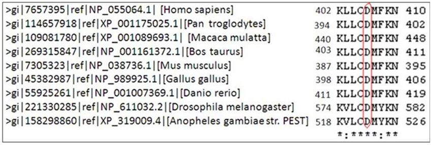
Molecular Vision 2014; 20:724-731                                              © 2014 Molecular Vision

                                                                                                         Figure 1. Pedigree of the family
                                                                                                         in this study. The asterisk symbol
                                                                                                         represents the sequenced individ-
                                                                                                         uals in this study. The genotyping
                                                                                                         of alleles were shown below the
                                                                                                         individuals.

                           METHODS                                         Sample collection and DNA preparation: Approximately
The study was performed after receiving approval from the                  5 ml intravenous blood was collected in a anticoagulant
institutional ethics review board and in accordance with                   (EDTA) coated tubes from all 12 study subjects and 100
the Declaration of Helsinki. The nature of the study was                   Indian controls. Genomic DNA was prepared from peripheral
conveyed, and informed consent was obtained from all study                 blood leukocytes with salting-out method [11], by dehydration
subjects. We certify that all applicable institutional and                 and precipitation of cellular proteins in a saturated sodium
government regulations concerning the ethical use of human                 chloride solution. The isolated DNA will be dissolved in TE
volunteers were followed during this research. The proband                 buffer (1 M Tris-pH 8.0; 0.5 M EDTA-pH 8.0) and stored at
along with 11 family members from three generations were                   -20 °C until use.
included in the study (Figure 1).                                          Polymerase chain reaction and DNA sequencing: Eight sets
Ophthalmological and electrophysiological studies: All study               of primers were used to amplify the entire coding region
subjects underwent a complete ophthalmic examination that                  of NR2E3 gene [12]. The PCR products were gel eluted
included evaluation of best-corrected visual acuity (BCVA)                 and column purified using an EZ-10 spin-column DNA gel
with Snellen optotypes, Goldman applanation tonometry,                     extraction kit (Bio Basic, East Markham, Canada). A total of
biomicroscopic examination, indirect ophthalmoscopy                        20 μl master mix was prepared using 50–100 ng of genomic
after pupillary dilatation, computerized perimetry, optical                DNA, 1×PCR buffer, 200 μM of dNTPs (Medox Biotech India
coherence tomography (OCT; Zeiss Cirrus HD OCT –4000,                      Pvt. Ltd, Chennai, India), 50% dimethyl sulphoxide (DMSO;
Carl Zeiss meditec, Inc. Dublin, CA), fundus photography                   Merck, Mumbai, India), 0.25 picomoles of each primer, and
(TOPCON TRC 50 DX), and intravenous fluorescein angi-                      1 unit of Taq DNA polymerase (Sigma, Saint Louis, MO), to
ography (IVFA; TOPCON TRC 50 DX, Tokyo, Japan). ERG                        perform PCR. The conditions followed were initial denatur-
(LKC Technologies, Gaithersburg, MD) was performed                         ation at 95 °C for 5 min, followed by 32 cycles (95 °C for 30 s,
according to standard testing protocols recommended by the                 55 °C for 30 s, and 72 °C for 1 min) and final extension at 72
International Society for Clinical Electrophysiology of Vision             °C for 10 min. Bidirectional sequencing was performed using
(ISCEV).                                                                   the ABI 3130 genetic analyzer (Applied Biosystems, Foster
                                                                           City, CA) with dye- termination chemistry.
Molecular genetic studies: A detailed family history and
pedigree was constructed. One hundred unrelated ethnic- and                Bioinformatics assessment: The evolutionary conservation
age-matched control subjects were recruited for this study.                of the identified NR2E3 mutation was checked using the
                                                                           clustalW multiple sequence alignment tool. Sorting Intolerant
                                                                           From Tolerant (SIFT) was used to assess the likely phenotypic

                                                                       725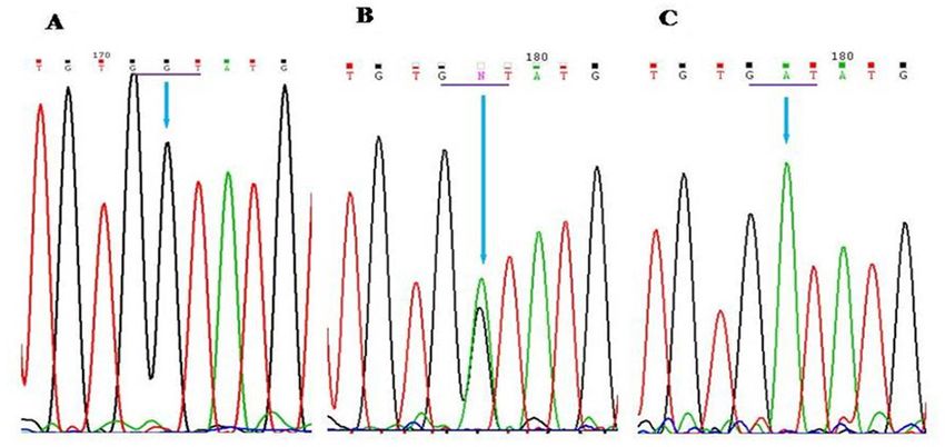
Molecular Vision 2014; 20:724-731 © 2014 Molecular Vision
effect of the identified missense mutation. PolyPhen analysis arterial attenuation (Figure 2A). Three raised dome-shaped,
was performed to calculate the probability of the identified yellowish-pink, vascular masses 4 DD in extent were noted
mutation being deleterious toward disease pathogenesis. in the peripheral retina in the superior, superotemporal, and
inferotemporal quadrants. These masses were associated with
RESULTS subretinal exudation (Figure 2B,C). No dilated or tortuous
feeder vessels were noted. IVFA showed rapid filling of the
Patient 1: The proband on presentation had a BCVA of
dye in the early phase, with the lesions becoming increas-
perception of light in the right eye (RE) and 6/12 in the left
ingly hyperfluorescent and leaking diffusely in the late phase.
eye (LE). Anterior segment examination of the RE showed
Telangiectatic and dilated vessels were observed within the
severe corneal edema, neovascularization of the iris, poste-
tumor masses (Figure3). Full-field ERG showed a severe
rior subcapsular cataract, and intraocular pressure (IOP) of
decrease in the rod and cone responses in both eyes (BE).
50 mmHg suggestive of neovascular glaucoma, and the LE
Based on the clinical, angiographic, and electrophysiological
showed a clear cornea and mild lens opacities due to posterior
findings, a diagnosis of secondary VPTRs associated with
subcapsular cataract. The RE fundus details were not clearly
retinal dystrophy was made. All the tumors were treated with
visible through the hazy media due to corneal edema and
transpupillary thermotherapy (TTT) in two sittings. Periph-
lenticular opacity. However, peripheral retinal exudation and
eral tumors were treated with cryotherapy with the triple
mass lesions similar to those in the LE could be seen hazily.
freeze thaw technique. Complete regression of the vascular
The LE fundus examination showed vitreous degenerative
masses was noted over 6 months (Figure 2D), and the final
changes, a healthy optic disc, and nummular pigment clumps
BCVA was 6/9 in the LE. Notably, there was an absence of
at the level of the RPE in the midperiphery and along the
macular or peripheral retinal schisis in this patient.
vascular arcades, associated with diffuse RPE atrophy.
The opaque, white dendritic appearance of the peripheral Patient 2: The 43-year-old healthy elder brother of our proband
vessels was present throughout the midperiphery with mild had complained of defective night vision since childhood.
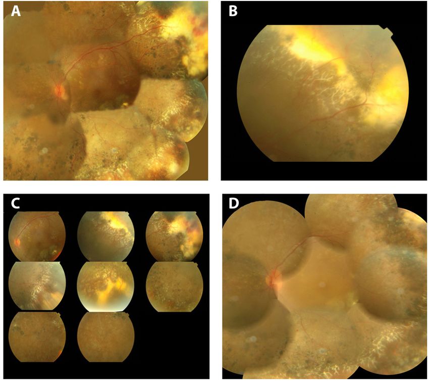
Figure 2. Pre and Post treatment
LE color fundus photograph of
the proband A: Pre-treatment
montage fundus photograph of left
eye. LE Fundus shows a healthy
appearing optic nerve head with
diffuse RPE degeneration with
nummular pigment clumps and
white dentritic peripheral vessels
in mid peripheral region . Vascu-
larized tumor mass with exudation
and absent feeder vessels is seen in
the supro-temporal periphery. B:
Vascularized peripheral tumor with
profuse exudation. C: 9 Up fundus
photograph of LE showing vasop-
roliferative tumors with exudation
in superior, suprotemporal, infero-
temporal and inferior quadrants. D:
Post-treatment fundus photograph.
LE fundus showing complete
regression of the tumor masses with
treatment. (6 month post treatment).
726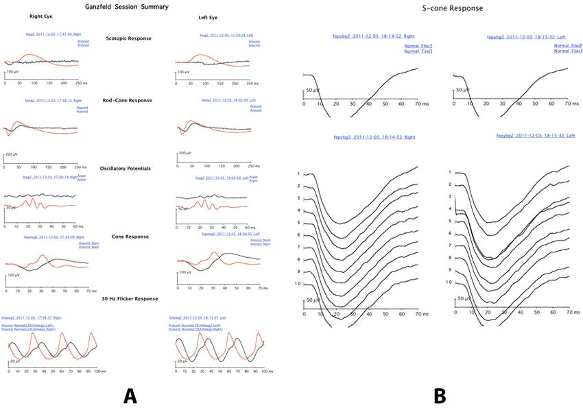
Molecular Vision 2014; 20:724-731                                             © 2014 Molecular Vision

                                                                                                        Figure 3. Fundus f luorescein
                                                                                                        angiography of vasoproliferative
                                                                                                        tumor. FFA shows rapid filling of
                                                                                                        the dye in the early phase, with
                                                                                                        the lesion becoming increasingly
                                                                                                        hyperfluorescent, and profuse and
                                                                                                        diffuse leakage of dye in the late
                                                                                                        phase of angiogram.

His BCVA was 6/6 BE. Anterior segment examination was                      the p.D406G variant. The identified novel p.D406G homo-
normal. Dilated fundus examination showed a healthy optic                  zygous mutation was evolutionarily highly conserved in
disc and normal caliber of the retinal vessels. There was a                different species (Figure 6). The SIFT score was 0.00, and
mild diffuse RPE change with few pigment deposits in the                   the PolyPhen score was 0.998. These features suggest the
midperipheral and peripheral retina. ERG showed decreased                  pathologic nature of the identified genetic variation.
rod-specific responses, the response to a standard flash was
delayed with low amplitude waveform under photopic and                                            DISCUSSION
scotopic conditions, and the 30 Hz flicker response was                    Patients with GFS typically present with early onset night
delayed and of decreased amplitude. The responses to long                  blindness, atypical pigmentary retinal dystrophy, degenera-
duration stimulus using a blue flash with orange background                tive changes of the vitreous, peripheral or macular retinos-
(S-cone ERG response) showed an abnormally large delayed                   chisis, lens opacities, and characteristic ERG abnormalities
waveform typical of the enhanced S-cone response (Figure                   [1]. The presenting features and findings of the ophthalmic
4A,B). Ocular history and examination of the other ten                     examination in the proband in this study led to the clinical
members of the family were unremarkable.                                   diagnosis of GFS. Although ERG responses were severely
     The findings of the clinical, angiographic, and electro-              reduced in the proband due to advanced disease, the proband’s
physiological examinations suggested a possible diagnosis of               brother displayed a hypersensitive response to blue light. The
GFS. Thus, full sequencing of the NR2E3 gene was under-                    maximal response to short wavelengths, virtually no change
taken in the proband. This revealed a novel, homozygous                    in the response waveform to light and dark adaptation, some
c.1117 A>G variant in exon 8 of the gene, a substitution muta-             long- and middle-wavelength–sensitive cone dysfunction, and
tion. This change leads to an amino acid change (aspartic acid             high subjective S-cone spectral sensitivity has been demon-
to glycine) at position 406 of the gene. The p.D406G change                strated in patients with GFS, and is termed the enhanced
was detected in the homozygous state in the proband’s brother              S-cone response [1,2].
(affected) and in the heterozygous state in eight unaffected                   The NR2E3 gene responsible for causing ESCS is located
family members (Figure 5; Table 1).                                        on chromosome 15q23 [12]. The gene coding region contains
    Screening of the NR2E3 gene in 100 unrelated Indian                    eight exons and spans a 7 kb region [13]. The gene contains
control samples of the same ethnic background did not show                 the DNA-binding domain (DBD) at the N-terminus end and

                                                                       727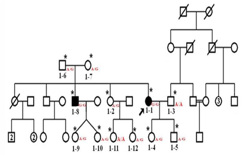
Molecular Vision 2014; 20:724-731                                          © 2014 Molecular Vision

Figure 4. Electroretinography and S-Cone ERG findings in case 2. A: ERG findings in case 2. ERG shows severely reduced rod specific
responses, response to standard flash was delayed with low amplitude waveform under photopic and scotopic conditions, the 30 Hz flicker
was delayed and of lower amplitude. B: S-Cone ERG with blue flash and orange background showing abnormally large, delayed, simplified
waveform as enhanced S cone ERG responses.

                                                                                                    Figure 5. Chromatogram repre-
                                                                                                    senting the p.Asp406Gly mutation
                                                                                                    in the NR2E3 gene. A: p.Asp406Gly
                                                                                                    mutation in the homozygous state.
                                                                                                    B: p.Asp406Gly mutation in the
                                                                                                    heterozygous state. C: Wild type.
                                                                                                    The underline marks the mutated
                                                                                                    codon. The arrow indicates the
                                                                                                    position of the mutation.

                                                                       728
Molecular Vision 2014; 20:724-731 © 2014 Molecular Vision
Figure 6. Evolutionary conservation
of p.Asp406Gly (D406G) mutation
in different species. Amino acid
residues highly conserved in all
species are indicated by asterisk,
and lower identity are shown using
colon. Red color box indicate
NR2E3 mutations analyzed in this
study.
ligand-binding domain (LBD) at the C-terminus end. Most show characteristic features of GFS, as in our patient, or may
of the human NR2E3 variants and mutations are identified in have the ERG pattern typically seen in ESCS or clumped
these two domains [14]. In the present study, we identified a pigmentary retinal degeneration.
novel mutation in the LBD of the C-terminus end. To our knowledge, the p.D406G homozygous mutation is
Thus far, nearly 50 NR2E3 gene variations and mutations novel. It was not seen in any of the control samples screened.
have been reported in patients with various retinal degenera- The p.D406G homozygous mutation was also observed in the
tive disorders. Gerber et al. identified a p.R311Q mutation proband’s brother who has clinical features in a less severe
in the NR2E3 gene in the patients who may have ESCS [15]. form and did not show the presence of VPTRs. Both parents
Later, Haider et al. reported the same p.R311Q mutation of the proband were heterozygous for the p.D406G mutation,
with 44.8% frequency in patients with ESCS [12]. This same which indicates that the parents were carriers for the disease
p.R311Q mutation in the NR2E3 gene was also reported with and transferred the risk allele (G) to the proband. Out of the
homozygosity in a patient with GFS [16]. In our study, we 12 family members who participated in this study, only two
identified a novel p.D406G (c.1117A>G) mutation with homo- carry the normal allele (A). The remaining family members
zygous state in the patient and in the affected sibling. carry the risk allele with heterozygosity (A/G) that indicated
The variability of clinical features and the severity of they are carriers for the disease. A similar condition was
retinal degeneration produced by NR2E3 mutations may often reported by Haider et al., in the ESCS case. The p.M407K
complicate the diagnosis. Patients with NR2E3 mutations may mutation was observed with the homozygous condition in the
Table 1. Genotype of the family members in the study.
Family member’s rela-
Individual ID Age /Sex Disease state Genotype Genotype Status
tion with the Proband
1–1 40/F Proband Affected GG Affected homozygote
1–2 41/F Sister Normal AG Carrier heterozygote
1–3 48/M Husband Normal AA Normal homozygous
1–4 12/F Daughter Normal AG Carrier heterozygote
1–5 14/M Son Normal AG Carrier heterozygote
1–6 75/M Father Normal AG Carrier heterozygote
1–7 70/F Mother Normal AG Carrier heterozygote
Similar clinical features of
1–8 42/M Brother GG Affected homozygote
proband
1–9 9/F Brother’s daughter Normal AG Carrier heterozygote
1–10 9/F Brother’s daughter Normal AG Carrier heterozygote
1–11 16/F Sister’s daughter Normal AA Normal homozygous
1–12 13/F Sister’s daughter Normal AG Carrier heterozygote
The p.D406G change is detected in the homozygous state in the proband and proband’s brother and in the heterozygous state in eight
unaffected family members.
729Molecular Vision 2014; 20:724-731 © 2014 Molecular Vision
patient, whereas two unaffected siblings carry the risk allele is nummular pigmentary deposition at the level of the RPE,
with heterozygous condition in their study [12]. usually located in the midperiphery and often associated
Atsuhiro Kanda et al. showed that the NR2E3 sequence is with RPE atrophy. This characteristic pigmentary clumping
highly conserved during evolution and the residue p.M407K was noted in our proband, along with degenerative vitreous
was conserved in all NR2E3 orthologs [14]. The identified changes, lenticular opacity, and the abnormal ERG pattern.
p.D406G mutation in the proband was also highly conserved In our patient, opaque white dentritic retinal vessel changes
during evolution (Figure 6).The SIFT score (0.00) showed were present throughout the midperiphery extending anteri-
that this mutation affected the protein function. The PolyPhen orly between the equator and ora serrata. Since the fundus
analysis also predicted that this mutation probably damaging features vary among patients, Fishman et al. concluded that a
the protein function with a score of 0.998. The amino acid diagnosis of GFS should be considered in patients presenting
aspartic acid/aspartate that is acidic polar and has been with an early history of poor night vision, bilateral atypical
changed by the substitution of a nonpolar amino acid glycine pigmentary changes in the retina, and degenerative changes
at the 406 conserved position might have altered the NR2E3 in the vitreous humor. Additional diagnostic findings include
protein structure and led to the disease occurrence in this retinoschisis, opaque dentritic retinal vessels, diffuse leakage
case. The SIFT and PolyPhen scores support the deleterious from retinal capillaries, and cystoid macular edema [1].
nature of the p.D406G mutation. Further functional studies Molecular genetic testing is essential for establishing the
are required to confirm the pathogenesis of the p.D406G correct diagnosis in patients with NR2E3 mutations because
mutation. of the variable phenotype associated with these degenerations.
Further research may shed light on the association between
VPTRs may be primary or secondary. In the largest the genetic mutation seen in our family with the observed
review of 295 patients with 334 VPTRs by Shields et al. in phenotype.
2012, 80% were idiopathic, and 20% were secondary. The
most common preexisting ocular disease included retinitis In conclusion, we have described a heretofore unreported
pigmentosa (22%), pars planitis (21%), Coats disease (16%), association of retinal VPTRs in GFS. The tumors regressed
previous retinal detachment surgery (12%), idiopathic periph- with standard treatment modalities. Detection of a novel
eral retinal vasculitis (6%), and familial exudative vitreoreti- p.D406G mutation in the NR2E3 gene helped to confirm
nopathy (4%). Other retinal lesions that might predispose to the diagnosis. Genetic testing also helped in detecting the
the development of VPTRs include toxoplasmic retinitis, presymptomatic carrier and thus proved to be of immense
toxocariasis, and traumatic choroidopathy [8]. A literature value in informed genetic counseling of the family members
review showed no previous documentation of VPTRs in and would potentially provide some form of therapy for the
patients with GFS. affected patients.
Primary tumors tend to be solitary, small, and located
ACKNOWLEDGMENTS
near the inferotemporal portion of the fundus. Secondary
VPTRs are more often multifocal, bilateral, and believed We sincerely thank Dr. Rodney John Morris, for helping with
to be a reactive vascular response to various ocular insults. the electrophysiological analysis of the patients involved in
The vascularized retinal nodules may threaten vision due to this study.
retinal exudation, macular edema, intraretinal or vitreous
hemorrhage, and formation of epiretinal membranes [8].
REFERENCES
These benign but vision-threatening tumors have been
1. Fishman GA, Jampol L, Goldberg M. Diagnostic features of
shown to respond to various treatment modalities such as
the Favre - Goldmann syndrome. Br J Ophthalmol 1976;
cryotherapy, TTT, brachytherapy, and tumor resection [9]. 60:345-[PMID: 1085161].
The proband lost vision in her RE due to neovascular glau-
2. Pachydaki SI, Klaver CC, Barbazetto IA, Roy MS, Allikmets
coma (NVG) secondary to VPTRs, a described complication R, Yannuzzi LA. Phenotypic Features of Patients With
of VPTRs [8]. The tumors in the LE were treated with TTT NR2E3 Mutations. Arch Ophthalmol 2009; 127:71-5.
(posterior tumors) and cryotherapy with the triple freeze thaw [PMID: 19139342].
technique (extreme peripheral tumors). Complete regression 3. Sharon D, Sandberg MA, Caruso RC, Berson EL, Dryja TP.
of the vascular masses in our patient was observed within 6 Shared Mutations in NR2E3 in Enhanced S-cone Syndrome,
months. Goldmann-Favre Syndrome, and Many Cases of Clumped
Pigmentary Retinal Degeneration. Arch Ophthalmol 2003;
Retinoschisis, a feature of GFS, was not seen in our 121:1316-23. [PMID: 12963616].
patient. However, the distinctive ophthalmoscopic feature
730Molecular Vision 2014; 20:724-731 © 2014 Molecular Vision
4. Udar N, Small K, Chalukya M, Garcia RS, Marmor M. Devel- 12. Haider NB, Jacobson SG, Cideciyan AV, Swiderski R, Streb
opmental or degenerative - NR2E3 gene mutations in two LM, Searby C, Beck G, Hockey R, Hanna DB, Gorman S,
patients with enhanced S cone syndrome. Mol Vis 2011; Duhl D, Carmi R, Bennett J, Weleber RG, Fishman GA,
17:519-25. [PMID: 21364904]. Wright AF, Stone EM, Sheffield VC. Mutation of a nuclear
5. Bandah D, Merin S, Ashhab M, Banin E, Sharon D. The receptor gene, NR2E3, causes enhanced S cone syndrome,
Spectrum of Retinal Diseases Caused by NR2E3 Mutations a disorder of retinal cell fate. Nat Genet 2000; 24:127-31.
in Israeli and Palestinian patients. Arch Ophthalmol 2009; [PMID: 10655056].
127:297-302. [PMID: 19273793]. 13. Kobayashi M, Takezawa S, Hara K, Yu RT, Umesono Y, Agata
6. Chavala SH, Sari A, Lewis H, Simpson E, Hagstrom SA, K, Taniwaki M, Yasuda K, Umesono K. Identification of a
Traboulsi EI. An Arg311Gln NR2E3 mutation in a family photoreceptor cell-specific nuclear receptor. Proc Natl Acad
with classic Goldmann – Favre syndrome. Br J Ophthalmol Sci USA 1999; 96:4814-9. [PMID: 10220376].
2005; 89:1065-6. [PMID: 16024868]. 14. Kanda A, Anand S. A comprehensive analysis of sequence
7. Shields CL, Shields JA, Barrett J, Potter P. Vasoprolifera- variants and putative disease –causing mutations in photo-
tive tumors of the ocular fundus. Arch Ophthalmol 1995; receptor - specific nuclear receptor NR2E3. Mol Vis 2009;
113:615-23. [PMID: 7748132]. 15:2174-84. [PMID: 19898638].
8. Shields CL, Kaliki S, Al-Dahmash S, Rojanaporn D, Shukla 15. Gerber S, Rozet JM, Takezawa SI, dos Santos LC, Lopes
SY, Reilly B, Shields JA. Retinal vasoproliferative tumors: L, Gribouval O, Penet C, Perrault I, Ducroq D, Souied E,
comparative clinical features of primary Vs secondary Jeanpierre M, Romana S, Frézal J, Ferraz F, Yu-Umesono
tumors in 334 cases. JAMA Ophthalmol 2013; 131:328-34. R, Munnich A, Kaplan J. The photoreceptor cell-specific
[PMID: 23494037]. nuclear receptor gene (PNR) accounts for retinitis pigmen-
tosa in the Crypto-Jews from Portugal (Marranos), survivors
9. Heimann H, Bornfeld N, Vij O, Coupland SE, Bechrakis NE, from the Spanish Inquisition. Hum Genet 2000; 107:276-84.
Kellner U, Foerster MH. Vasoproliferative tumours of the [PMID: 11071390].
retina. Br J Ophthalmol 2000; 84:1162-9. [PMID: 11004104].
16. Bernal S, Solans T, Gamundi MJ, Hernan I, de Jorge L,
10. Rennie IG. Retinal vasoproliferative tumours. Eye (Lond) Carballo M, Navarro R, Tizzano E, Ayuso C, Baiget M.
2010; 24:468-71. [PMID: 20075974]. Analysis of the involvement of the NR2E3 gene in autosomal
11. Miller SA, Dykes DD, Polesky HF. A simple salting out recessive retinal dystrophies. Clin Genet 2008; 73:360-6.
procedure for extracting DNA from human nucleated cells. [PMID: 18294254].
Nucleic Acids Res 1988; 16:1215-[PMID: 3344216].
Articles are provided courtesy of Emory University and the Zhongshan Ophthalmic Center, Sun Yat-sen University, P.R. China.
The print version of this article was created on 29 May 2014. This reflects all typographical corrections and errata to the article
through that date. Details of any changes may be found in the online version of the article.
731You can also read