Germline mutation of ARF in a melanoma kindred
←
→
Page content transcription
If your browser does not render page correctly, please read the page content below
# 2002 Oxford University Press                                   Human Molecular Genetics, 2002, Vol. 11, No. 11             1273–1279

Germline mutation of ARF in a melanoma kindred
Chelsee Hewitt1,{, Chu Lee Wu1,{, Gareth Evans1, A. Howell2, Robert G. Elles1, Richard
Jordan3, Philip Sloan4, Andrew P. Read1 and Nalin Thakker1,*
1
 University of Manchester Department of Medical Genetics and Regional Genetic Service, Central Manchester
Healthcare Trust, St. Mary’s Hospital, Manchester, M13 OJH, UK, 2Centre for Cancer Epidemiology, Christie Hospital,
Manchester, M20 4BX, UK, 3Department of Stomatology, University of California San Francisco, San Francisco,
CA 94143-0424, USA and 4University of Manchester Dental Hospital, Manchester, M15 6FH, UK

Received December 3, 2001; Revised and Accepted March 13, 2002

Familial melanoma predisposition is associated with germline mutations at the CDKN2A/ARF locus in up to
40% of families. The exact role of the two proteins encoded by this complex locus in this predisposition is

                                                                                                                                         Downloaded from http://hmg.oxfordjournals.org/ by guest on October 12, 2015
unclear. Most mutations affect either CDKN2A only or products of both genes. Recently a deletion affecting
ARF-specific exon 1b was reported in a family with melanoma and neural tumours. However, the possibility of
this deletion also altering the CDKN2A transcript could not be excluded. More convincingly, a 16 base pair
insertion in exon 1b has been reported in an individual with multiple melanomas suggesting a direct role for
ARF in melanoma predisposition. We report here a splice mutation in exon 1b in a family with melanoma that
results in ARF haploinsufficiency. The mutation was observed in a mother and daughter with melanoma. A
sibling of the mother with breast cancer also had this mutation. Analysis of the melanoma from one individual
revealed a 62 bp deletion in exon 3 of the wildtype allele and loss of the mutant allele; these somatic changes
would affect both CDKN2A and ARF. These somatic events suggest that concomitant inactivation of both
ARF and CDKN2A may be necessary for melanoma development and that mutations in ARF and CDKN2A
possibly confer different levels of susceptibility to melanoma, with the former associated with lesser
predisposition. In this situation, the events follow a ‘three-hit’ model as observed in tumours from FAP
patients with an attenuated phenotype. Overall, the data suggest a direct role for ARF haploinsufficiency in
melanoma predisposition and co-operation between ARF and CDKN2A in tumour formation, consistent with
recent observations in Cdkn2a-specific knockout mice.

                                                                     progression through the G1–S checkpoint. CDKN2A is a
INTRODUCTION                                                         specific inhibitor of CDK4 and 6 (4). Thus inactivation of
The CDKN2A–ARF gene on chromosome 9p21 is implicated in              CDKN2A allows cells to escape cell cycle arrest in G1.
the development of a variety of sporadic malignant tumours (1)         The other product of the CDKN2A–ARF locus, ARF, also
and also of familial melanoma (2). This gene encodes two             acts as a tumour suppressor (5,6). Mice lacking Arf but with
structurally distinct tumour suppressor proteins by virtue of        intact Cdkn2a develop tumours (7), while transfection of ARF
different 50 exons spliced in different reading frames to            into some carcinoma cell lines results in marked growth
common exons 2 and 3. Exons 1a, 2 and 3 encode CDKN2A                inhibition (8,9). ARF mediates G1 and G2 arrest at least partly
(p16INK4a), while exon 1b, spliced to exons 2 and 3 in a             by its interaction with MDM2, a protein that binds to both
different reading frame and transcribed using a different            TP53 and pRb. MDM2 targets TP53 for degradation by
promoter, encodes ARF (also called p19ARF in mice, p14ARF            ubiquitination (10) and also inhibits pRb growth regulatory
in humans) protein (3).                                              function (11). The N-terminal domain of ARF, encoded by
  CDKN2A is a part of the G1–S cell cycle checkpoint                 exon 1b, binds to MDM2 and promotes its degradation (12,13).
mechanism that involves the retinoblastoma-susceptibility            This results in stabilisation and accumulation of TP53 protein
tumour suppressor protein (pRb). Rb protein, in its unphos-          and also of its downstream target CDKN1A, an inhibitor not
phorylated state, inhibits progression of the cell cycle from G1     only of CDK4 and 6 but also of other CDKs. Degradation of
into S phase by sequestering the transcription factor E2F1.          MDM2 also removes the functional inhibition of pRb.
Phosphorylation of pRb by the cyclin dependent kinases CDK4            Thus this complex locus codes for two distinct proteins that
and 6 (CDK4–6/D kinases) releases E2F1 and allows                    are important and overlapping regulators of two key (RB- and

*To whom correspondence should be addressed. Tel: þ 44 161276 6604/6720; Fax: þ 44 161 276 6606; Email: nthakker@man.ac.uk
{
 The authors wish it to be known that, in their opinion, the first two authors should be regarded as joint First Authors.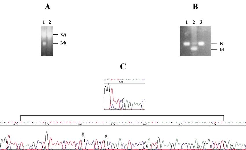
1274     Human Molecular Genetics, 2002, Vol. 11, No. 11

TP53-dependent) cell cycle regulatory pathways. Moreover, it         This base substitution creates a novel BsaAI restriction site
is now apparent that ARF intimately links the two pathways.          (Fig. 2B) and this mutation-specific restriction fragment length
E2F1 and products of oncogenes such as RAS, MYC and v-ABL            polymorphism (RFLP) was used to test control DNA samples
that act in the retinoblastoma pathway have been shown to            from 125 unrelated individuals (250 chromosomes). The change
induce expression of ARF with subsequent stabilisation of            was not observed in any of these controls (data not shown).
TP53 and expression of CDKN1A (p21) (6).                               The 334G > C missense change results in substitution of
   Because a single gene complex codes for two distinct              glycine by arginine at codon 122 (G122R). More importantly,
proteins and mutational events can cause loss of function of         this change of the terminal nucleotide of exon 1b is likely to
either or both proteins, the specific role of the individual         affect the splicing of the ARF mRNA (29). In order to identify
proteins in development of neoplasia can be difficult to assess.     any aberrant transcripts resulting from this, RT–PCR analysis
Inactivation of CDKN2A–ARF in sporadic tumours can occur             was performed on total RNA from individual II-3 using
by mutation (1), methylation of the promoters (14,15) or             forward primers complementary to upstream exon 1b sequence
deletion of the genes (16). In melanoma, point mutations have        and reverse primers complementary to exon 3 sequence. An
been reported in the CDKN2A-specific exon 1a and in                  aberrant-sized transcript was not detected (Fig. 2C). Analyses
sequences encoding both CDKN2A and ARF, but not in the               of the normal sized transcript for the mutation-specific RFLP
ARF-specific exon 1b, while the methylation affects a CpG            revealed loss of the transcript from the mutant allele (Fig. 2C).
island in the CDKN2A-specific promoter. Additionally, it has         This was confirmed by direct sequencing of the normal-sized
been reported that most mutations in exon 2 (common to both          transcript (data not shown). Analysis of the CDKN2A transcript

                                                                                                                                          Downloaded from http://hmg.oxfordjournals.org/ by guest on October 12, 2015
CDKN2A and ARF) appear not to affect ARF function (17).              revealed only the normal transcript (data not shown).
Thus, it is believed that the primary effect of methylation and        To determine the fate of the wild type ARF allele in the
the reported point mutations is on CDKN2A, whereas deletions         tumour, LOH analysis was performed in matched normal and
usually affect both products of the locus. It may be significant     tumour tissues from both individuals with melanoma. We
that deletion appears to be the commonest mode of inactivation       were unable to amplify any sequences (CDKN2A–ARF related
of this locus in many tumours, and homozygous deletion is a          and control) from the tumour from individual II-3, despite
frequent event (18). ARF-specific genetic alterations have been      repeated attempts, probably because of poor or excessive
reported in human T-cell acute lymphoblastic leukaemia (19), a       fixation. In the tumour from her daughter (III-1), LOH was
small number of metastatic melanoma cell lines (20) and also         observed at all informative loci encompassing the CDKN2A–
in chemically-induced murine lymphomas (13). Additionally,           ARF locus except for D9S1747 (Fig. 3), indicating two
reduced or absent expression of ARF without any evident              distinct regions of LOH, one centromeric and the other
genetic alterations has been observed in non-small cell lung         telomeric to D9S1747. The centromeric region involves exon
cancer (21,22).                                                      1b and also possibly the remainder of the CDKN2A–ARF
   Germline CDKN2A inactivation is seen in approximately 25–         locus. Surprisingly, when exon 1b PCR amplification product
50% of kindreds with an autosomal dominant predisposition to         from tumour DNA was tested for the mutation-specific RFLP,
melanoma (2). A very small number of additional melanoma             only non-digested product was observed, indicating loss
families have mutations in the CDK4 gene, which alter the            of the mutant rather than wild-type allele in the tumour
binding of CDK4 to CDKN2A (23). Previous studies failed to           (Fig. 4A). Further investigation showed that the retained
identify germline mutations of ARF-specific exon 1b in               wild-type allele had a deletion of 62 bp that included the
familial melanoma kindreds (24–26). However, recently a              whole of exon 3 (Figs 4B and C). Exons 1b, 1a and 2 were
germline deletion involving exon 1b of the CDKN2A–ARF                successfully amplified and were of the expected size (data not
locus was reported in a melanoma–neural system tumour                shown). Similarly, no mutations were detected in exons 4–9 of
family (27). Whilst providing a strong indication that ARF           TP53 gene.
haploinsufficiency could predipose to melanoma, this study             Analysis of the breast carcinoma from the mother’s sibling
was not able to conclusively exclude the role of CDKN2A or           (II-4) revealed LOH at D9S1747 (Fig. 3) but retention of
confirm the role of ARF in melanoma predisposition. More             heterozygosity at the flanking loci. This deletion definitely does
convincingly, a 16 base pair insertion in exon 1b has been           not include exon 1b but could possibly include the remainder of
reported in an individual with multiple melanomas (28).              the CDKN2A–ARF locus. We did not detect any sequence
   Here we report a germline splice site mutation in exon 1b that    alterations at the CDKN2A–ARF locus. Analyses of methylation
results specifically in the loss of the ARF transcript from this     of CpG islands in the promoters of both ARF and CDKN2A were
allele in a mother and daughter with melanoma. We also               unsuccessful due to the limited amount of tissue available.
demonstrate that somatic inactivation of both the alleles of
CDKN2A and possibly the wild type allele of ARF are likely to
be necessary for tumour development in these patients.
                                                                     DISCUSSION
                                                                     We have demonstrated a germline ARF-specific mutation in a
                                                                     melanoma kindred and analysed two tumours (one melanoma
RESULTS                                                              and one breast carcinoma) from two individuals with the
Sequence analysis of exon 1b revealed a base substitution of the     germline mutation. The melanoma that we analysed had
terminal nucleotide (334G > C) in individuals II-3, II-4 and III-1   suffered two somatic mutations at the CDKN2A/ARF locus in
(Fig. 1) but not individual II-5 (Fig. 2). No changes were           addition to the inherited constitutional mutation that alters the
identified in any of the other exons of the CDKN2A–ARF gene.         terminal nucleotide of the ARF-specific exon 1b. One somatic
Human Molecular Genetics, 2002, Vol. 11, No. 11 1275
Downloaded from http://hmg.oxfordjournals.org/ by guest on October 12, 2015
Figure 1. Pedigree of family with melanoma and breast cancer. Black shading indicates individuals with melanoma and stripes indicate individuals with breast
carcinoma. The tumour, age of onset and genotypes (where DNA available for analyses) is indicated. þ / þ wildtype homozygous; þ / 7 , heterozygous for
the 334G > C mutation.
mutation was a deletion of the whole gene that contained the in familial melanoma clearly do affect both gene products
ARF point mutation and the other was a deletion of exon 3 of (31,32), and somatic mutations that specifically inactivate ARF
the constitutionally wild-type allele. Although the ARF open have been reported in a number of tumour types including
reading frame terminates in exon 2, deletion of exon 3 is melanoma cell lines (see above). A role for both ARF and
expected to prevent production of a stable ARF transcript CDKN2A is strongly suggested by recent evidence that
through loss of the 30 UTR and polyadenylation site. Thus the predisposition to tumours including melanoma in Cdkn2a-null
deletions in the tumour are likely to abolish all expression of mice is greatly enhanced by haploinsufficiency for Arf (33).
both CDKN2A and ARF. Two clinical cases (27,28) with germline mutations involving
There are two possible explanations for these observations. ARF support this view (see above). Functional studies and
First, it is possible that mother and daughter had no inherited spontaneous occurrence of tumours in Arf-specific knockout
predisposition and coincidentally carry a harmless ARF variant. mice also demonstrate that ARF functions as a tumour
Even though we did not find the inherited variant in 250 control suppressor protein.
chromosomes, it could conceivably still be a rare non- If ARF haploinsufficiency is indeed associated with tumour
pathogenic variant. However, there are many precedents to predisposition then we need to explain why the tumour from
suggest that a change in the 30 terminal nucleotide of an exon our family has suffered three hits. A simple explanation is that
will affect splicing and so destabilise the mRNA (29). both CDKN2A and ARF need to be inactivated and a further
Consistent with this, no message from the mutant allele could two hits are necessary to achieve this on a background of a
be detected in peripheral blood lymphocytes from individual II- constitutional ARF mutation. It is also possible that the
3. Expression and function of CDKN2A is unaffected by this constitutional ARF mutation is a weak mutation. Cells with
mutation and normal CDKN2A mRNA could be amplified from this mutation gain a growth advantage when they acquire a
her lymphocytes. Additionally, the family meets the criteria for second hit inactivating both CDKN2A and ARF on the other
a melanoma kindred (30) and neither patient has a history of chromosome, but this is less than the advantage enjoyed by
excess exposure to sunlight. cells that completely inactivate both copies of CDKN2A and
Thus the alternative explanation, that constitutional ARF ARF. Thus there is selective pressure for a third hit. There is a
haploinsufficiency predisposes to tumour formation, seems very good precedent for such a three-hit model in familial
more plausible. Whilst it has been reported that most CDKN2A adenomatous polyposis. In this disease certain germline APC
mutations do not affect ARF (17), many mutations at this locus mutations (APCAP) confer an attenuated phenotype with fewer1276 Human Molecular Genetics, 2002, Vol. 11, No. 11
Downloaded from http://hmg.oxfordjournals.org/ by guest on October 12, 2015
Figure 2. ARF mutation in melanoma-breast cancer family. (A) Forward (F, left panels) and reverse (R, right panels) ARF exon 1b sequences from individual II-3
(top panels) and control (C, bottom panels) showing the 334G > C change. (B) 334G > C mutation-specific RFLP in a control (lane 1) and individuals II-3, II-4,
III-1. This base substitution creates a novel BsaAI restriction site and all three individuals from the family show both the wildtype (Wt) and mutant (Mt) alleles and
the control sample shows only the wildtype allele. (C) ARF transcript analysis in individual II-3. Part of the ARF transcript including exons 1b, 2 and 3 was
amplified using exonic primers from cDNA prepared from peripheral blood lymphocytes from a control sample and individual II-3. Only normal sized transcript
(NT) was seen in both control (C) and II-3 (Lane 1). Testing for the mutation specific BsaAI restriction polymorphism in II-3 revealed the wild type transcript only
(Lane 2). This was confirmed by sequencing (data not shown). Exon 1 product amplified from the matching genomic DNA from II-3 and restriction digested with
BsaAI was used as a positive control for this (Lane 3) and shows both the wildtype (Wt) and mutant (Mt) alleles (the smaller product from the restriction digestion
of the mutant allele is not shown).
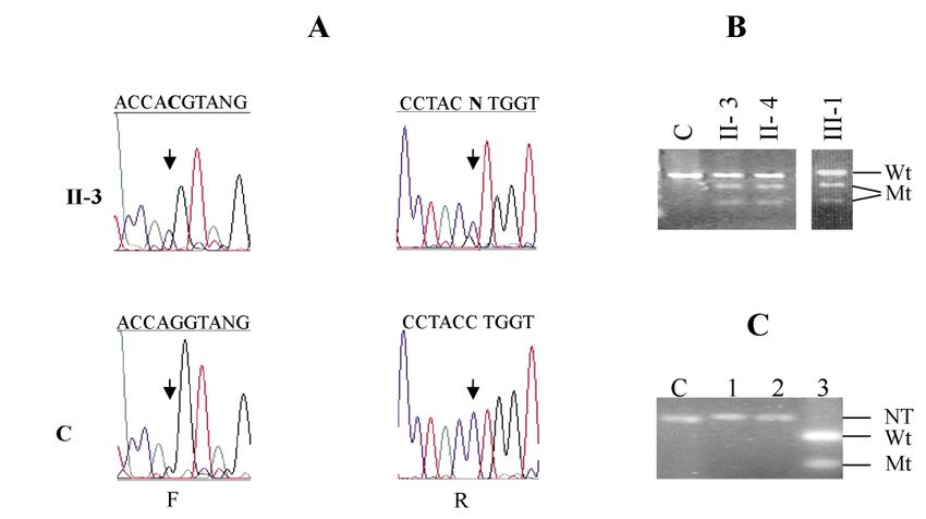
Figure 3. LOH analyses in tumours from individuals II-4 and III-1. (A) Results of LOH analysis in melanoma and breast carcinoma derived from individuals III-1 and
II-4 respectively. The loci are arranged left to right from chromosome 9p telomere to centromere. The shaded areas indicate contiguous areas of LOH. 1Position of
exon 1a, exon 2 and exon 3 of CDKN2A/ARF; 2position of exon 1b of CDKN2A/ARF; LOH was detected here using mutation-specific RFLP; *, LOH, s, ROH, U,
uninformative, ND, not done. (B) Silver-stained denaturing polyacrylamide gels showing products of PCR-amplified chromosome arm 9p STRPs from matched nor-
mal (N) and tumour (T) DNA in individual III-1. The loci tested are indicated above the gels and the arrows indicate the position of the alleles missing in tumours.Human Molecular Genetics, 2002, Vol. 11, No. 11 1277
Downloaded from http://hmg.oxfordjournals.org/ by guest on October 12, 2015
Figure 4. Fate of the wildtype and mutant CDKN2A–ARF alleles in melanoma from individual III-1. (A) Exon 1b was amplified using flanking primers from DNA
from peripheral blood lymphoctyes (Lane 1) and tumour (Lane 2) and restriction digested with BsaAI. The DNA from the tumour shows the presence of only the
wildtype (Wt) allele indicating deletion of the mutant allele (Mt) (the smaller product from the restriction digestion of the mutant allele is not shown). Note: this
figure is the same as that shown in Figure 3 for Exon1b LOH analyses. (B) Exon 3 was amplified using flanking primers from DNA from peripheral blood lym-
phoctyes (Lane 1), tumour (Lane 2) from the patient and from control peripheral blood lymphocytes (Lane 3). The tumour DNA (Lane 2) shows a smaller PCR
product (M) compared to the matching lymphocyte and control DNA (N). (C) Sequence analysis of the tumour (top panel) reveals deletion of 62 bp compared to
normal sequence (bottom panel).
polyps. APCAP mutations lie outside the mutation cluster Arf knock-out mice is somewhat similar to that observed in
region where conventional (APCP) mutations occur (34,35). Tp53 knock-out mice. The three families with exon 1b
Adenomas from such patients often have two somatic mutations show some phenotypic overlap with Li Fraumeni
mutations, an APCP mutation of the wild-type allele and loss syndrome. Both melanoma and neural tumours can occur in
of the germline APCAP allele by chromosomal deletion. It is individuals with germline TP53 mutations. A role for ARF
suggested that APCAP/APCP cells have a weak growth mutations in predisposition to neural tumours is strongly
advantage compared to APCP/APCP cells. suggested by the demonstration of deletions either involving
It is likely that dual inactivation of both RB and TP53 only exon 1b (27) or involving the whole locus (37) in
pathways is necessary in many tumours, including melanomas melanoma families with neural tumours. These tumours appear
(12). Mouse models of cutaneous melanoma clearly show that not to occur in families with conventional germline CDKN2A
both TP53 and RB pathways can act independently to suppress mutations.
melanocyte transformation (36). A large body of evidence Whether predisposition to breast cancer is also part of this
suggests that ARF acts upstream of TP53 and inactivation of overlap is less clear. In melanoma families carrying mutations
TP53 and ARF in tumourigenesis may be mutually exclusive that affect both CDKN2A and ARF there is an increased risk of
(6). Inactivation of either ARF or TP53 could equally serve to other cancers including those of the breast (32). With regard to
disable the ARF–MDM2–TP53 pathway, and the required our individual II-4, our limited observations leave open several
inactivation of both the RB and TP53 pathways could be explanations of the somatic events in her breast carcinoma. The
achieved either through inactivation of CDKN2A and TP53 or loss of heterozygosity on 9p definitely does not involve
alternatively via concomitant inactivation of both CDKN2A and CDKN2A–ARF exon 1b, but may or may not involve the
ARF. The genetic alterations observed in our melanoma are remainder of the CDKN2A–ARF locus. Deletion of the wild-
consistent with the latter. The tumour lacks mutations in type copy of CDKN2A would produce a cell with no ARF and a
conserved exons of TP53, but has lost all CDKN2A and ARF. half dose of CDKN2A. We found no sequence alterations in
Several lines of evidence suggest that ARF insufficiency may CDKN2A (but we do not know whether our experiments
result in phenotypes that partially but not completely overlap amplified one or both copies of exons 1a, 2 and 3) and we
TP53 insufficiency (12). ARF plays no part in the TP53 could not test for promoter methylation. Thus it remains
response to DNA damage, but the tumour spectrum observed in unclear whether the breast tumour had one, two or three hits on1278 Human Molecular Genetics, 2002, Vol. 11, No. 11
CDKN2A–ARF. The lack of germline mutation in individual primer sequences used were as follows: Exon 4aF: 50 -CTG
II-5 is also inconclusive. The breast carcinoma was of relatively CGCGGCGGCTATGACTGCTCTTTTCACCCATC-30 ; Exon
late-onset and there was no history of breast cancer in her 4aR: 5 0 -GGTGTAGGAGCTGCTGGTG-3 0 ; Exon 4bf:
mother. Thus, she may represent a coincidental sporadic breast 50 -AGAATGCCAGAGGCTGCTC-30 ; Exon 4bR: 50 -TCCGC-
cancer case. We were unable to obtain breast cancer samples for GGGCCCGAATGGAAGCCAGCCCCTCAG-30 ; Exon 5aF:
analysis from individual I-2, which might have proved more 50 -TCGGGCCCGCGGAAACTTGTGCCCTGACTTTCAAC-
informative. 30 ; Exon 5aR: 50 -CCATGGCCATCTACAAGCA-30 ; Exon
In conclusion, the families reported here and previously 5bF: 50 -GGGGGTGTGGAATCAACC-30 ; Exon 5bR: 50 -AGC-
(27,28) indicate that germline ARF mutations not involving CCTGTCGTCTCTCCAG-30 ; Exon 6F: 50 -CTGCGCGGCG-
CDKN2A may be associated with predisposition to melanoma GCTATCCCCAGGCCTCTGATTC-30 ; Exon 6R: 50 -TCCGC-
and possibly neural and breast cancer. If ARF links both TP53 GGGCCCGAACTTAACCCCTCCTCCCAGAGAG-30 ; Exon
and RB pathways (see above), then ARF mutations may 7F: 5 0 -TCGGGCCCGCGGAACTGCTTGCCACAGGTC-
predispose to tumours through multiple mechanisms. Studies TCC-30 ; Exon 7R: 50 -GTGTGCAGGGTGGCAAGT-30 ; Exon
of more families, patients and their tumours are necessary to 8F: 50 -TACTGCCTCTTGCTTCTCTTT-30 ; Exon 8R: 50 -AGC-
confirm this and to elucidate further the particular roles of the CGCCGCGCAGACATAACTGCACCCTTGGTCT-30 ; Exon
two proteins encoded by the CDKN2A–ARF gene. 9F: 5 0 -AGCCGCCGCGCAGAGGCATTTTGAGTGTTA-
GACTGG-30 ; Exon 9R: 50 -AGACCAAGGGTGCAGTTATG-30 .
Downloaded from http://hmg.oxfordjournals.org/ by guest on October 12, 2015
MATERIALS AND METHODS RT–PCR
Subjects 1 mg Total RNA was subjected to reverse transcription with
oligo(dT)15 primers using Reverse Transcription System
The pedigree shown in Figure 1 meets the criteria for diagnosis (Promega, Madison, USA) as per manufacturer’s instructions.
of hereditary or familial melanoma (30) with two affected PCR amplification of the ARF cDNA fragment was performed
individuals in the kindred. There was also a history of breast with the same primers that were used for sequencing (see
cancer in three other members of the family (with ages of onset above). The product was size fractionated by gel electrophor-
between 4th and 6th decades) and two members had history of esis through 2% agarose gel and visualised by ethidium
cancers of lung or liver. Samples for analysis however, were bromide staining. CDKN2A was amplified from the cDNA
only available from the mother (II-3, Fig. 1) and daughter with using forward primers complementary to exon 1a(50 -ATG-
melanoma (III-1, Fig. 1), and from the mother’s sister (II-4, GAGCCTTCGGCTGACTGG-30 ) and reverse primers comple-
Fig. 1) and mother’s first cousin (II-5) with breast cancer. mentary to exon 3 (see above).
Sample preparation Loss of heterozygosity (LOH)
Genomic DNA was prepared from peripheral blood lympho- Seven short tandem repeat polymorphisms (STRPs): D8S157,
cytes using conventional procedures. Total RNA was prepared IFNA, D9S1747, D9S942, D9S1748, D9S1752 and D9S171
from a lymphoblastoid cell line from individual II-3 (Fig. 1) encompassing the CDKN2A–ARF gene on chromosome 9p,
using TRIzolä reagent (Gibco, Maryland, USA) as per the were used for LOH analyses of matched normal and tumour
manufacturer’s instructions. DNA from microdissected 10 mm tissues from the subjects. The STRPs were amplified, size
thick sections of formalin-fixed, paraffin-embedded tumour fractionated by gel electrophoresis and visualised by silver
tissue was extracted exactly as previously described (18). staining as described previously (18).
Mutation analysis of CDKN2A–ARF and TP53 ACKNOWLEDGEMENTS
Genomic and/or tumour DNA was screened for mutations in We thank the patients and their families for all their help with
exons la, lb, 2 and 3 of the CDKN2A–ARF gene and exons 4–9 this study.
of the TP53 gene by direct cycle sequencing of both forward
and reverse strands using the Applied Biosystems Prism Ready
Reaction DyeDeoxy Terminator Cycle Sequencing Kit and ABI REFERENCES
373 sequencer (Perkin Elmer, Foster City, USA). All results 1. Pollock, P.M., Pearson, J.V. and Hayward, N.K. (1996) Compilation of
were confirmed by repeat analyses with independent PCR somatic mutations of the CDKN2 gene in human cancers: non-random
products. Flanking intronic primers for the CDKN2A–ARF distribution of base substitutions. Genes Chromosomes Cancer, 15, 77–88.
gene (details referenced in 18) were used for sequencing of the 2. Hussussian, C.J., Struewing, J.P., Goldstein, A.M., Higgins, P.A., Ally, D.S.,
Sheahan, M.D., Clark, W.H. Jr, Tucker, M.A. and Dracopoli, N.C.
genomic/tumour DNA. Sequencing of the cDNA was per- (1994) Germline CDKN2A mutations in familial melanoma. Nat. Genet., 8,
formed with forward primers complementary to exon 1b (50 - 15–21.
AGTGGCGCTGCTCACCTC-3 0 ) and reverse primers 3. Haber, D.A. (1997) Splicing into senescence: the curious case of CDKN2A
complementary to exon 3 sequence (50 -TTCTTTCAATCGGG- and p19ARF. Cell, 91, 555–558.
4. Weinberg, R.A. (1985) The retinoblastoma protein and cell cycle control.
GATGT-30 ). For TP53 analyses of tumour DNA, flanking Cell, 81, 323–330.
intronic primers were used to amplify exons 6–9. Exons 4 and 5. Prives, C. (1998) Signaling to p53: breaking the MDM2-p53 circuit. Cell,
5 were amplified in two overlapping segments (a and b). The 95, 5–8.Human Molecular Genetics, 2002, Vol. 11, No. 11 1279
6. Sherr, C.J. (2000) The Pezcoller lecture: cancer cell cycles revisited. Cancer 23. Zuo, L., Weger, J., Yang, Q., Goldstein, A.M., Tucker, M.A., Walker, G.J.,
Res., 60, 3689–3695. Hayward, N. and Dracopoli, N.C. (1996) Germline mutations in the
7. Kamijo, T., Zindy, F., Roussel, M.F., Quelle, D.E., Downing, J.R., p16(INK4a) binding domain of CDK4 in familial melanoma. Nat. Genet.,
Ashmun, R.A., Grosveld, G., Sherr, C.J. (1997) Tumor suppression at the 12, 97–99.
mouse INK4a locus mediated by the alternative reading frame product 24. FitzGerald, M.G., Harkin, D.P., Silva-Arrieta, S., MacDonald, D.J.,
p19ARF. Cell, 91, 649–659. Lucchina, L.C., Unsal, H., O’Neill, E., Koh, J., Finkelstein, D.M.,
8. Simon, M., Koster, G., Menon, A.G., Schramm, J. (1999) Functional Isselbacher, K.J. et al. (1996) Prevalence of germ-line mutations in p16,
evidence for a role of combined CDKN2A (p16–p14(ARF))/CDKN2B p19ARF, and CDK4 in familial melanoma: analysis of a clinic-based
(p15) gene inactivation in malignant gliomas. Acta Neuropathol., 98, population. Proc. Natl Acad. Sci. USA, 93, 8541–8545.
444–452. 25. Liu, L., Goldstein, A.M., Tucker, M.A., Brill, H., Grui, N.A., Hogg, D. and
9. Yang, C.T., You, L., Yeh, C.C., Chang, J.W., Zhang, F., McCormick, F. and Lassam, N.J. (1997) Affected members of melanoma-prone families with
Jablons, D.M. (2000) Adenovirus-mediated p14 (ARF) gene transfer in linkage to 9p21 but lacking mutations in CDKN2A do not harbor mutations
human mesothelioma cells. J. Natl Cancer Inst., 92, 636–641. in the coding regions of either CDKN2B or p19ARF. Genes Chromosomes
10. Momand, J., Wu, H.H. and Dasgupta, G. (2000) MDM2—master regulator Cancer, 19, 52–54.
of the p53 tumor suppressor protein. Gene, 242, 15–29. 26. Fargnoli, M.C., Chimenti, S., Keller, G., Soyer, H.P., Dal Pozzo, V., Hofler,
11. Xiao, Z.X., Chen, J., Levine, A.J., Modjtahedi, N., Xing, J., Sellers, W.R. H. and Peris, K. (1998) CDKN2a/p16INK4a mutations and lack of
and Livingston, D.M. (1995) Interaction between the retinoblastoma protein p19ARF involvement in familial melanoma kindreds. J. Invest. Dermatol.,
and the oncoprotein MDM2. Nature, 375, 694–698. 111, 1202–1206.
12. Pomerantz, J., Schreiber-Agus, N., Liegeois, N.J., Silverman, A., Alland, L., 27. Randerson-Moor, J.A., Harland, M., Williams, S., Cuthbert-Heavens, D.,
Chin, L., Potes, J., Chen, K., Orlow, I., Lee, H.W. et al. (1998) The Ink4a Sheridan, E., Aveyard, J., Sibley, K., Whitaker, L., Knowles, M., Bishop,
tumor suppressor gene product, p19Arf, interacts with MDM2 and J.N. and Bishop, D.T. (2001) A germline deletion of p14(ARF) but not
neutralizes MDM2’s inhibition of p53. Cell, 92, 713–723. CDKN2A in a melanoma-neural system tumour syndrome family. Hum.
Downloaded from http://hmg.oxfordjournals.org/ by guest on October 12, 2015
13. Zhuang, S.M., Schippert, A., Haugen-Strano, A., Wiseman, R.W. and Mol. Genet., 10, 55–62.
Soderkvist, P. (1998) Inactivations of p16INK4a-alpha, p16INK4a-beta and 28. Rizos, H.L., Puig, S., Badenas, C., Malvehy, J., Darmanian, A.P., Jiménez,
p15INK4b genes in 20 ,30 -dideoxycytidine- and 1,3-butadiene-induced L., Montserrat, M. and Kefford, R.F. (2001) A melanoma associated
murine lymphomas. Oncogene, 16, 803–808. germline mutation in exon 1b inactivates p14ARF. Oncogene, 20,
14. Gonzalez-Zulueta, M., Bender, C.M., Yang, A.S., Nguyen, T., Beart, R.W., 5543–5547.
Van Tornout, J.M. and Jones, P.A. (1995) Methylation of the 50 CpG island 29. Krawczak, M., Reiss, J. and Cooper, D.N. (1992) The mutational spectrum
of the CDKN2A/CDKN2 tumor suppressor gene in normal and transformed of single base-pair substitutions in mRNA splice junctions of human genes:
human tissues correlates with gene silencing. Cancer Res., 55, 4531–4535. causes and consequences. Hum. Genet., 90, 41–54.
15. Merlo, A., Herman, J.G., Mao, L., Lee, D.J., Gabrielson, E., Burger, P.C., 30. Barnhill, R.L., Rousch, G.C. and Titus-Ernstoff, L. (1992) Comparison of
Baylin, S.B., and Sidransky, D. (1995) 50 CpG island methylation is nonfamilial and familial melanoma. Dermatology, 184, 2–7.
associated with transcriptional silencing of the tumor suppressor CDKN2A/ 31. Petronzelli, F., Sollima, D., Coppola, G., Martini-Neri, M.E., Neri, G. and
CDKN2/MTS1 in human cancers. Nat. Med., 1, 686–692. Genuardi, M. (2001) CDKN2A germline splicing mutation affecting both
16. Cairns, P., Polascik, T.J., Eby, Y., Tokino, K., Califano, J., Merlo, A., Mao, p16(ink4) and p14(arf) RNA processing in a melanoma/neurofibroma
L., Herath, J., Jenkins, R., Westra, W. et al. (1995) Frequency of kindred. Genes Chromosomes Cancer, 31, 398–401.
homozygous deletion at CDKN2A/CDKN2 in primary human tumors. Nat. 32. Rizos, H., Darmanian, A.P., Holland, E.A., Mann, G.J. and Kefford, R.F.
Genet., 11, 210–212. (2001) Mutations in the INK4a/ARF melanoma susceptibility locus
17. Arap, W., Knudsen, E., Sewell, D.A., Sidransky, D., Wang, J.Y., Huang, H.J. functionally impair p14ARF. J. Biol. Chem., 276, 41424–41434.
and Cavenee, W.K. (1997) Functional analysis of wild-type and malignant 33. Krimpenfort P., Quon K.C., Mooi, W.J., Loonstra, A. and Berns, A. (2001)
glioma derived CDKN2Abeta alleles: evidence for an RB-independent Loss of p16Ink4a confers susceptibility to metastatic melanoma in mice.
growth suppressive pathway. Oncogene, 15, 2013–2020. Nature, 413, 83–86.
18. Wu, C.L., Roz, L., McKown, S., Sloan, P., Read, A.P., Holland, S., Porter, S., 34. Spirio, L.N., Samowitz, W., Robertson, J., Robertson, M., Burt, R.W.,
Scully, S., Paterson, I., Tavassoli, M. and Thakker, N. (1999) DNA studies Leppert, M. and White, R. (1998) Alleles of APC modulate the
underestimate the major role of CDKN2A inactivation in oral and orophary- frequency and classes of mutations that lead to colon polyps. Nat. Genet.,
ngeal squamous cell carcinomas. Genes Chromosomes Cancer, 25,16–25. 20, 385–388.
19. Gardie, B., Cayuela, J.M., Martini, S. and Sigaux, F. (1998) Genomic 35. Lamlum, H., Ilyas, M., Rowan, A., Clark, S., Johnson, V., Bell, J., Frayling,
alterations of the p19ARF encoding exons in T-cell acute lymphoblastic I., Efstathiou, J., Pack, K., Payne, S. et al. (1999) The type of somatic
leukemia. Blood, 91, 1016–1020. mutation at APC in familial adenomatous polyposis is determined by the
20. Kumar, R., Sauroja, I., Punnonen, K., Jansen, C. and Hemminki, K. (1998) site of the germline mutation: a new facet to Knudson’s ‘two-hit’
Selective deletion of exon 1beta of the p19ARF gene in metastatic hypothesis. Nat. Med., 5, 1071–1075.
melanoma cell lines. Genes Chromosomes Cancer, 23, 273–277. 36. Bardeesy, N., Bastian, B.C., Hezel, A., Pinkel, D., DePinho, R.A. and Chin,
21. Gazzeri, S., Della Valle, V., Chaussade, L., Brambilla, C., Larsen, C.J. and L. (2001) Dual inactivation of RB and p53 pathways in RAS-induced
Brambilla, E. (1998) The human p19ARF protein encoded by the beta melanomas. Mol. Cell Biol., 21, 2144–2153.
transcript of the p16INK4a gene is frequently lost in small cell lung cancer. 37. Bahuau, M., Vidaud, D., Jenkins, R.B., Bieche, I., Kimmel, D.W.,
Cancer Res., 58, 3926–3933. Assouline, B., Smith, J.S., Alderete, B., Cayuela, J.M., Harpey, J.P. et al.
22. Vonlanthen, S., Heighway, J., Tschan, M.P., Borner, M.M., Altermatt, H.J., (1998) Germ-line deletion involving the INK4 locus in familial
Kappeler, A., Tobler, A., Fey, M.F., Thatcher, N., Yarbrough, W.G. and proneness to melanoma and nervous system tumors. Cancer Res., 58,
Betticher, D.C. (1998) Expression of p16INK4a/p16alpha and p19ARF/ 2298–2303.
p16beta is frequently altered in non-small cell lung cancer and correlates
with p53 overexpression. Oncogene, 17, 2779–2785.You can also read