Versican G1 Fragment Establishes a Strongly Stabilized Interaction with Hyaluronan-Rich Expanding Matrix during Oocyte Maturation - MDPI
←
→
Page content transcription
If your browser does not render page correctly, please read the page content below
International Journal of
            Molecular Sciences

Article
Versican G1 Fragment Establishes a Strongly
Stabilized Interaction with Hyaluronan-Rich
Expanding Matrix during Oocyte Maturation
Eva Nagyova 1, *, Antonietta Salustri 2 , Lucie Nemcova 1 , Sona Scsukova 3 , Jaroslav Kalous 1
and Antonella Camaioni 2
 1    Institute of Animal Physiology and Genetics, Czech Academy of Sciences, 27721 Libechov, Czech Republic;
      nemcova@iapg.cas.cz (L.N.); kalous@iapg.cas.cz (J.K.)
 2    Department of Biomedicine and Prevention, University of Rome Tor Vergata, 00133 Rome, Italy;
      salustri@med.uniroma2.it (A.S.); camaioni@med.uniroma2.it (A.C.)
 3    Institute of Experimental Endocrinology, Biomedical Research Center, Slovak Academy of Sciences,
      84505 Bratislava, Slovakia; scsukova@hotmail.com
 *    Correspondence: nagyova@iapg.cas.cz

 Received: 26 February 2020; Accepted: 23 March 2020; Published: 25 March 2020

 Abstract: In the mammalian ovary, the hyaluronan (HA)-rich cumulus extracellular matrix (ECM)
 organized during the gonadotropin-induced process of oocyte maturation is essential for ovulation of
 the oocyte-cumulus complex (OCC) and fertilization. Versican is an HA-binding proteoglycan that
 regulates cell function and ECM assembly. Versican cleavage and function remain to be determined
 in ovarian follicle. We investigated versican expression in porcine ovarian follicles by real-time
 (RT)-PCR and western blotting. The aims of the present work were to determine whether (1) versican
 was produced and cleaved by porcine OCCs during gonadotropin stimulation; (2) these processes
 were autonomous or required the participation of mural granulosa cells (MGCs); and (3) versican
 cleavage was involved in the formation or degradation of expanded cumulus ECM. We demonstrate
 two cleavage products of G1 domain of versican (V1) accumulated in the HA-rich cumulus ECM.
 One of them, a G1-DPEAAE N-terminal fragment (VG1) of ~70 kDa, was generated from V1
 during organization of HA in in vivo and in vitro expanded porcine OCCs. Second, the V1-cleaved
 DPEAAE-positive form of ~65 kDa was the only species detected in MGCs. No versican cleavage
 products were detected in OCCs cultured without follicular fluid. In summary, porcine OCCs are
 autonomous in producing and cleaving V1; the cleaved fragment of ~70 kDa VG1 is specific for
 formation of the expanded cumulus HA-rich ECM.

 Keywords: versican; hyaluronan; oocyte-cumulus complex; extracellular matrix

1. Introduction
      In mammals, growth and development of the ovarian follicles requires a coordinated series of
events controlled by circulating gonadotropins (follicle-stimulating hormone, FSH, and luteinizing
hormone, LH) and locally produced growth factors leading to somatic cell proliferation, oocyte
development, and steroidogenesis [1]. The cumulus matrix newly organized in oocyte-cumulus
complex (OCC) is essential for successful ovulation and fertilization [2,3]. This expanded cumulus
extracellular matrix (ECM) is formed by interaction of high molecular weight hyaluronan (HA) and at
least three proteins: the serum-derived inter-alpha-trypsin inhibitor (IαI), and two factors produced by
cumulus cells, tumor necrosis factor-alpha-induced protein 6 (TNFAIP6) and pentraxin 3 (PTX3) [4–6].
Deletion of these corresponding genes in mice produces female infertility due to disorganization
of the oocyte-cumulus ECM [3]. Another HA-binding component which may affect the stability of

Int. J. Mol. Sci. 2020, 21, 2267; doi:10.3390/ijms21072267                          www.mdpi.com/journal/ijms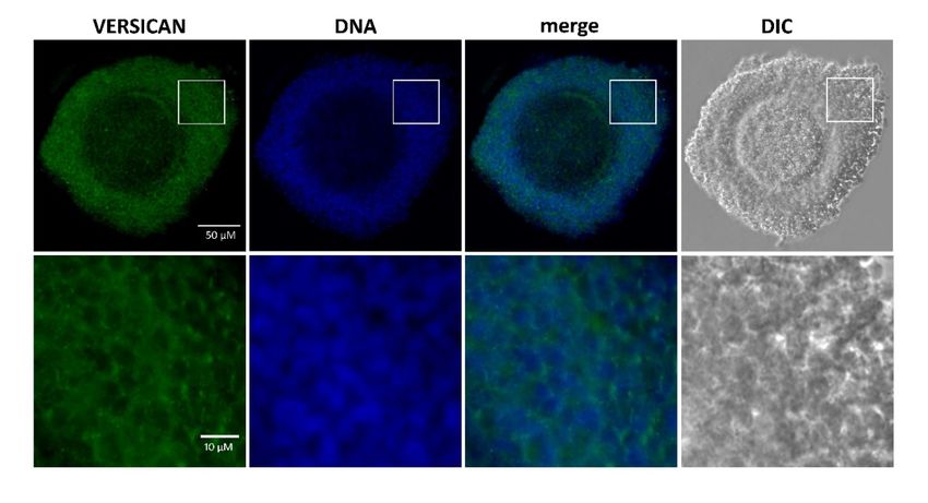
Int. J. Mol. Sci. 2020, 21, 2267                                                                    2 of 14

the oocyte-cumulus ECM is the proteoglycan versican [7–9]. This proteoglycan has a complex structure
constituted by a high-affinity HA binding N-terminal globular domain (G1), a chondroitin sulfate
substituted midsection (αGAG and βGAG), and a cell surface- and matrix-interacting C-terminal
globular domain (G3) [10]. Alternative splicing events of a single gene [11] generate either a full-length
protein possessing both αGAG and βGAG domains (V0~370 kDa), or shorter proteins with the αGAG
alone (V2~180 kDa), or the βGAG alone (V1~263 kDa), or neither GAG domains (V3~74 kDa). These
forms of splicing produce different effects in the cell environment [12–15]. In addition, versican is
a preferred substrate of specific proteases, a disintegrin and metalloproteinase with thrombospondin
like repeats (ADAMTS) [16]. A subset of them, including ADAMTS1, ADAMTS4, and ADAMTS5,
cleave versican V0 and V1 in the βGAG domain generating HA-binding fragments ending with
the neoepitope DPEAAE at the C-terminus. The degradation product of V0 has large size consisting of
a fragment of ~220 kDa carrying several GAG side chains linked to the αGAG domain, while cleavage
of V1 generates a small fragment of ~70 kDa consisting mainly of the HA-binding region (G1) of the core
protein without GAG side chains attached [16]. The versican G1-DPEAAE, also called versikine [17], is
a biologically active molecule in the cell environment affecting apoptosis and angiogenesis during
development and tissue remodeling [16,18]. Its fundamental role in the organization of provisional HA
matrix is supported by the evidence that mice null for versican as well as for HA synthase 2 presented
an identical embryonic lethality due to a defect in the developing of heart matrix [19]. Moreover,
analysis of whole rodent ovaries showed upregulated expression levels of versican V0/V1 and their
degradation enzymes ADAMTS1 and ADAMTS4 after LH ovulatory surge [8,20]. In agreement, human
granulosa cells stimulated in vivo contained abundant ADAMTS1 and Versican mRNA [21], and also
porcine OCCs significantly up-regulated the ADAMTS1 mRNA level [22]. However, versican cleavage
and function in ovarian follicle remain to be determined, since it is not known whether cumulus cells
are independent in generating versican fragments or require the stimulus or direct participation of
granulosa cells. The follicular fluid (FF) contains the heavy chains (HCs) of Iαl that are covalently
linked to HA during formation of HA-rich cumulus ECM [23]. It has been proposed that complexes
of HC (of IαI)-HA play a central role in matrix organization because they serve as docking sites for
the attachment of other proteins to support matrix stabilization [24].
      The aims of the present work were to determine whether versican was produced and its G1
domain cleaved by porcine cumulus cells, whether these processes were autonomous or required
the participation of mural granulosa cells (MGCs), and whether versican cleavage was involved in
the formation or degradation of the porcine oocyte-cumulus ECM.

2. Results

2.1. Versican Fragments Containing the Neo-Epitope DPEAAE in OCCs Expanded in Vivo
     In order to detect the DPEAAE-positive species of the N-terminal versican fragments generated by
ADAMTS cleavage in the ECM of in vivo expanded porcine OCCs, we performed western blot analyses.
The previous published experiments performed with purified molecules showed that the N-terminal
fragment of ADAMTS-cleaved V1 does not carry GAGs, while that from V0 has an elevated molecular
weight for the presence of several chondroitin sulfate chains and does not enter the gel. Therefore, in
order to detect both core protein fragments (~220 and ~70 kDa for V0 and V1, respectively), the OCCs
expanded in vivo were isolated from naturally cycling gilts and treated with ChABC. The total extracts
were resolved by SDS-PAGE under reducing conditions and probed with the DPEAAE neo-epitope
antibody. The results reported in Figure 1 clearly show a positive band of ~65 kDa, approximately
the expected molecular weight of G1 fragment from V1 (VG1), while no positivity was detected in
the gel at higher molecular weight. However, an evident positive signal was present at the top of
the gel, indicating that some versican fragments were prevented from entering. Interestingly, when
the HA-rich ECM of the in vivo matured OCCs was digested with Hyal, the material from the top of
the gel disappeared and a new positive band appeared in the gel at ~70 kDa. This suggests that a highly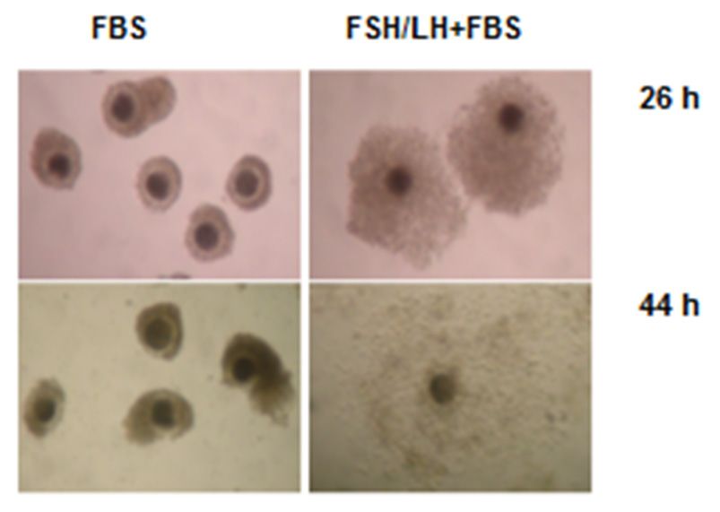
Int. J. Mol. Sci. 2020, 21, x FOR PEER REVIEW
 Int. J. Mol. Sci. 2020, 21, 2267                                                                            3 of 14

unstimulated, transiently increased VCAN V1 expression. The increase was statistically significant
starting  from
 stabilized      8 h of resistant
             linkage,   culture and     followed a temporal
                                     to denaturating          patternconditions,
                                                       and reducing    similar to that observed
                                                                                    occurs betweenin OCCs.
                                                                                                       the ~70 kDa
      Then,  we   analyzed    the pattern   of V1 cleavage  by MGCs    during  in vitro culture.
 DPEAAE-positive versican fragments and HA. This species was mostly present in the matrix        To  thisextract
                                                                                                          purpose,
                                                                                                                 but
MGCs    freshly   isolated  (0   h)  or cultured  for 44 h in medium     supplemented    with  FBS   and  FSH/LH
 also detectable in the cell fraction of in vivo stimulated OCCs, suggesting that it is strongly associated
were   digested
 to either        with either or
           the intercellular     ChABC     or Hyal,
                                    pericellular    and the extracts
                                                 compartment.        were analyzed
                                                                The intensity   of the by
                                                                                       ~65western
                                                                                           kDa band blotwas
                                                                                                         using  the
                                                                                                             similar
anti-DPEAAE       neo-epitope     antibody    (Figure 4B). Interestingly,  both  enzymatic   digestions
 in ChABC and Hyal digested protein extracts indicating that this versican fragment, as the ~70 kDa,      gave  the
same
 does pattern,
       not carrywith   onlyInthe
                   GAGs.           ~65 kDa versican
                                conclusion,            species
                                              V1, but not      present in
                                                          V0, cleavage     the totalwere
                                                                         products    and generated
                                                                                          matrix extracts  (Figure
                                                                                                      by ADAMTS
4B;  lanes  1–3,  5, and  8).  The   ~70 kDa   V1 fragment   found  in cumulus    ECM    was not
 during OCC expansion. A very faint positive band of ~150 kDa also appeared in the matrix extract detected   in the
cultured
 after HyalMGCs,     even but
             digestion,   afteritsHyal   digestion.
                                    identity is unknown.

      Figure 1. Versican fragments containing the neo-epitope DPEAAE are detected in OCCs expanded
     Figure 1. Versican fragments containing the neo-epitope DPEAAE are detected in OCCs expanded in
      in vivo. The complexes expanded in vivo were isolated from naturally cycling gilts (two porcine
     vivo. The complexes expanded in vivo were isolated from naturally cycling gilts (two porcine OCCs
      OCCs for each lane). Total (T), matrix (M) and cell (C) extracts from expanded OCCs digested with
     for each lane). Total (T), matrix (M) and cell (C) extracts from expanded OCCs digested with
      Streptomyces hyaluronidase (H) or chondroitinase ABC (CH) were analyzed by western blot with
     Streptomyces hyaluronidase (H) or chondroitinase ABC (CH) were analyzed by western blot with anti-
      anti-DPEAAE versican antibody. Actin immunodetection was performed to compare sample loading.
     DPEAAE versican antibody. Actin immunodetection was performed to compare sample loading. A
      A representative blot of four independent experiments is shown.
     representative blot of four independent experiments is shown.
2.2. Versican Fragments Produced by Gonadotropin-Stimulated OCCs Cultured in Vitro
      The process of cumulus expansion during oocyte maturation is induced in vitro by gonadotropin
stimulation. To verify whether porcine OCCs upregulate versican V1 during in vitro cumulus
expansion, as it occurs in vivo, we cultured OCCs in FBS-supplemented medium in the absence or
in the presence of FSH/LH. The results reported in Figure 2 showed that the cumulus expansion
induced by FSH/LH (Figure 2A) was accompanied by an increase in VCAN V1 mRNA levels in OCCs
analyzed by real-time (RT)-PCR (Figure 2B). A significant increase in the expression was detected at
4 h, peaked at 8-16 h, and returned toward the basal level at 26 h. Then, we investigated whether
versican V1 was cleaved in vitro, like in vivo, during the process of cumulus expansion. Groups of
porcine OCCs were cultured as above for 44 h (i.e., the time required for in vivo and in vitro full
OCC expansion), as shown in Figure 2A. Complexes not stimulated (unexpanded) or stimulated with
FSH/LH (expanded) were digested with Hyal or ChABC and analyzed by western blot (Figure 2C). No
versican-derived fragments were detected in protein extracts of unstimulated OCCs. In agreement with
the results obtained from in vivo expanded OCCs, the complexes stimulated to expand in vitro showed
the ~70 kDa versican fragment in the cumulus ECM   (A) only after the digestion with Hyal. Moreover, as
observed in vivo, the ~65 kDa band of versican fragment was present in these samples and did not
require Hyal digestion to enter the gel, since it was present also after ChABC digestion. However, in
this in vitro condition, the ~65 and ~70 kDa bands showed similar intensities. We conclude that OCCs
induced to expand in vitro with gonadotropins (FSH/LH) in serum-supplemented medium increase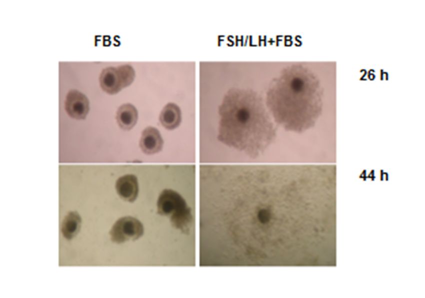
Int. J. Mol. Sci. 2020, 21, 2267                                                                                        4 of 14

             the versican V1 expression and accumulate its N-terminal cleaved forms in the HA-rich cumulus ECM,
             closely resembling the in vivo condition.

                                                A

Int. J. Mol. Sci. 2020, 21, 2267                                                                                   5 of 14

                                      B
                               Figure 2. Effects of gonadotropins on cumulus expansion and versican expression oocyte-cumulus
                               complexes (OCCs) cultured in vitro. (A) Morphology of OCCs stimulated with gonadotropins (100
                               ng/mL follicle-stimulating hormone (FSH) + 100 ng/mL luteinizing hormone (LH)) cultured in vitro
                               for 26 and 44 h in medium supplemented with 5% FBS. Note that the size of OCCs cultured in the
                               presence of gonadotropins was larger than that of OCCs cultured only in basal medium (FBS alone).
                               Full expansion was reached at 44 h when cumulus cells became scattered in an abundant extracellular
                               matrix (ECM). (B) Versican V1 mRNA (VCAN V1) expression investigated by RT-PCR in hormone-
                               stimulated OCCs cultured in vitro. Groups of 30 OCCs were immediately processed (0 h; white bar)
                               or cultured in FBS-supplemented medium without (light grey bars) or with (grey bars) FSH/LH
                               stimulation, as above, for the indicated time points. Three independent experiments were performed.
                               Bars show means ± SEM. In the time course of gonadotropin-stimulated OCCs; different superscripts
                               (a–c) indicate statistically significant differences compared to 0 h sample (p < 0.05). Asterisks indicate
                               statistically significant differences between unstimulated and stimulated groups of OCCs at the same
                               time point (** p < 0.01, *** p < 0.001). (C) Versican 1 fragments containing the neo-epitope DPEAAE
                               accumulated in the cumulus matrix of hormone-stimulated OCCs cultured in vitro. Groups of 15
                               OCCs were cultured for 44 h in FBS-supplemented medium in the absence/presence of FSH/LH.
                               Complexes were digested with Streptomyces hyaluronidase (H) or chondroitinase ABC (CH) and total
                               (T), matrix (M), and cell (C) extract from not expanded (without hormones; lanes 1 and 2) or fully
                               expanded (with hormones, lanes 3-8) were analyzed by western blot with anti-DPEAAE versican

                                      C

                   Figure 2. Effects of gonadotropins on cumulus expansion and versican expression oocyte-cumulus
      Figure 2. Effects of gonadotropins on cumulus expansion and versican expression oocyte-cumulus
      complexes (OCCs) cultured in vitro. (A) Morphology of OCCs stimulated with gonadotropins (100
      ng/mL follicle-stimulating hormone (FSH) + 100 ng/mL luteinizing hormone (LH)) cultured in vitro
      for 26 and 44 h in medium supplemented with 5% FBS. Note that the size of OCCs cultured in the
      presence of gonadotropins was larger than that of OCCs cultured only in basal medium (FBS alone).
      Full expansion was reached at 44 h when cumulus cells became scattered in an abundant extracellular
Int. J. Mol. Sci. 2020, 21, 2267 5 of 14
complexes (OCCs) cultured in vitro. (A) Morphology of OCCs stimulated with gonadotropins
(100 ng/mL follicle-stimulating hormone (FSH) + 100 ng/mL luteinizing hormone (LH)) cultured
in vitro for 26 and 44 h in medium supplemented with 5% FBS. Note that the size of OCCs cultured
in the presence of gonadotropins was larger than that of OCCs cultured only in basal medium (FBS
alone). Full expansion was reached at 44 h when cumulus cells became scattered in an abundant
extracellular matrix (ECM). (B) Versican V1 mRNA (VCAN V1) expression investigated by RT-PCR in
hormone-stimulated OCCs cultured in vitro. Groups of 30 OCCs were immediately processed (0 h;
white bar) or cultured in FBS-supplemented medium without (light grey bars) or with (grey bars)
FSH/LH stimulation, as above, for the indicated time points. Three independent experiments were
performed. Bars show means ± SEM. In the time course of gonadotropin-stimulated OCCs; different
superscripts (a–c) indicate statistically significant differences compared to 0 h sample (p < 0.05). Asterisks
indicate statistically significant differences between unstimulated and stimulated groups of OCCs at
the same time point (** p < 0.01, *** p < 0.001). (C) Versican 1 fragments containing the neo-epitope
DPEAAE accumulated in the cumulus matrix of hormone-stimulated OCCs cultured in vitro. Groups
of 15 OCCs were cultured for 44 h in FBS-supplemented medium in the absence/presence of FSH/LH.
Complexes were digested with Streptomyces hyaluronidase (H) or chondroitinase ABC (CH) and total
(T), matrix (M), and cell (C) extract from not expanded (without hormones; lanes 1 and 2) or fully
expanded (with hormones, lanes 3-8) were analyzed by western blot with anti-DPEAAE versican
antibody. Actin immunodetection was performed to compare sample loading. A representative blot of
three independent experiments is shown.
2.3. Temporal Pattern of Versican Cleavage during in Vitro Expansion
The follicular fluid (FF) constitutes the physiological environment in which cumulus expansion
occurs. Therefore, with the aim to investigate the time course of versican cleavage in the cumulus
ECM, groups of 15 porcine OCCs were stimulated for 26 or 44 h with FSH/LH in the presence of
FF (Figure 3A). Then, they were digested with Hyal, and matrix and cell extracts were analyzed by
western blot. The results clearly indicated that the versican cleaved product of ~70 kDa was already
present in the ECM of OCCs cultured for 26 h, and its amount remained apparently unchanged at 44 h.
Conversely, the ~65 kDa species accumulated in the cumulus ECM over time, being barely visible at
26 h but quite evident at 44 h. It is known that OCCs stimulated with gonadotropins in the absence of
serum or FF did not organize HA-rich cumulus ECM [5]. Importantly, in these culture conditions, no
versican fragments were associated with OCCs (Figure 3A, lanes 1 and 2).
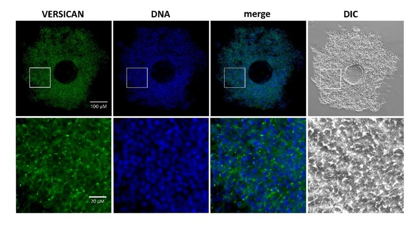
Localization of N-terminal fragments of V1 in the expanded cumulus ECM was analyzed by
immunostaining. The anti-DPEAAE antibody showed a bright punctuated positivity throughout
the matrix of gonadotropin-stimulated porcine OCCs (Figure 3C), while no signal was detected in
the unstimulated OCCs (Figure 3B).OCCs were cultured for 44 h in FBS-supplemented medium in the absence/presence of FSH/LH.
Complexes were digested with Streptomyces hyaluronidase (H) or chondroitinase ABC (CH) and total
(T), matrix (M), and cell (C) extract from not expanded (without hormones; lanes 1 and 2) or fully
expanded (with hormones, lanes 3-8) were analyzed by western blot with anti-DPEAAE versican
antibody. Actin immunodetection was performed to compare sample loading. A representative blot
Int. J. Mol. Sci. 2020, 21, 2267 6 of 14
of three independent experiments is shown.
A
B
Figure 3. Cont.Int. J. Mol. Sci. 2020, 21, 2267 7 of 14
Int. J. Mol. Sci. 2020, 21, x FOR PEER REVIEW
C
Figure 3. (A) Temporal pattern of versican V1 cleavage in hormone-stimulated OCCs cultured in vitro
in the absence
Figureand in Temporal
3. (A) the presence of follicular
pattern fluid
of versican V1(FF). Groups
cleavage of 15 OCCs were stimulated
in hormone-stimulated for 26inorvitro
OCCs cultured
44 h withinFSH/LH in the
the absence andabsence (PVP) orofpresence
in the presence follicularof 5% (FF).
fluid FF. At the indicated
Groups of 15 OCCstimes of culture,
were stimulatedtheyfor 26
or 44 h
were digested with
with FSH/LH in hyaluronidase
Streptomyces the absence (PVP) (H), or
andpresence of 5%
matrix (M) andFF.cell
At (C)
theextracts
indicated times
were of culture,
analyzed
they
by western were
blot withdigested with Streptomyces
anti-DPEAAE-V1 hyaluronidase
antibody. (H), and matrixwas
Actin immunodetection (M)performed
and cell (C)
toextracts
comparewere
analyzedAby
sample loading. western blot with
representative blot anti-DPEAAE-V1
of three independent antibody. Actin immunodetection
experiments was performed
is shown. Immunostaining of to
compare
porcine OCCs sample
cultured forloading. A representative
26 h without (B) and withblot of three inindependent
(C) FSH/LH the presenceexperiments
of FBS. The isOCCsshown.
Immunostaining
were labeled with anti-DPEEAEof porcine OCCs primary
versican cultured antibody,
for 26 h without (B) secondary
and with and with (C) FSH/LHAlexa
antibody in theFluor
presence
of FBS. The OCCs were labeled with anti-DPEEAE versican primary antibody,
488. Nuclei were co-stained with DAPI. The OCCs were mounted on glass slides and examined with and with secondary
antibody
an Olympus AX70Alexa Fluor 488.
microscope Nuclei were
equipped withco-stained
a DP30BW with
CCDDAPI. The OCCs
camera. Note were mounted on
the punctuate glass slides
positivity
and examined with an Olympus AX70 microscope
only in the matrix of expanded OCCs (panel C, higher magnification). equipped with a DP30BW CCD camera. Note the
punctuate positivity only in the matrix of expanded OCCs (panel C, higher magnification).
2.4. Versican Fragments Produced by Mural Granulosa Cells after in Vitro Gonadotropin Stimulation
First, we analyzed by RT-PCR whether porcine MGCs, as cumulus cells, upregulated VCAN V1
mRNA following in vitro gonadotropin stimulation. Mural GCs were isolated from naturally cycling
gilts and incubated in culture medium supplemented with 5% FBS in the presence or absence of
FSH/LH. Results reported in Figure 4A showed that gonadotropin-stimulated MGCs, unlike those
unstimulated, transiently increased VCAN V1 expression. The increase was statistically significant
starting from 8 h of culture and followed a temporal pattern similar to that observed in OCCs.
Then, we analyzed the pattern of V1 cleavage by MGCs during in vitro culture. To this purpose,
MGCs freshly isolated (0 h) or cultured for 44 h in medium supplemented with FBS and FSH/LH
were digested with either ChABC or Hyal, and the extracts were analyzed by western blot using
the anti-DPEAAE neo-epitope antibody (Figure 4B). Interestingly, both enzymatic digestions gave
the same pattern, with only the ~65 kDa versican species present in the total and matrix extracts
(Figure 4B; lanes 1–3, 5, and 8). The ~70 kDa V1 fragment found in cumulus ECM was not detected in
the cultured MGCs, even after Hyal digestion.Int. J. Mol. Sci. 2020, 21, 2267 8 of 14
Int. J. Mol. Sci. 2020, 21, 2267 8 of 15
A
B
Figure 4. (A) Versican 1 mRNA (VCAN V1) expression in hormone-stimulated mural granulosa cells
(MGCs) cultured in vitro. Fifteen small pieces of follicle wall consisting of MGC aggregates were either
Figure 4. (A) Versican 1 mRNA (VCAN V1) expression in hormone-stimulated mural granulosa cells
(MGCs) cultured in vitro. Fifteen small pieces of follicle wall consisting of MGC aggregates were
either immediately processed (0 h, white bar) or cultured in 5% FBS-supplemented medium without
(light grey bars) or with (grey bars) FSH/LH for the indicated time points. The data shown areInt. J. Mol. Sci. 2020, 21, 2267 9 of 14
immediately processed (0 h, white bar) or cultured in 5% FBS-supplemented medium without (light
grey bars) or with (grey bars) FSH/LH for the indicated time points. The data shown are representative
of three independent experiments. Bars show means ± SEM. Different superscripts indicate statistically
significant differences compared to 0 h sample (p < 0.05). Asterisks indicate statistically significant
differences between unstimulated and hormone-stimulated MGCs at the same time point (** p < 0.01,
*** p < 0.001). (B) Versican fragments containing the neo-epitope DPEAAE in mural granulosa cells
(MGCs) after in vitro stimulation with gonadotropins. Porcine MGCs were collected before (0 h) or
after 44 h of culture in 5% FBS-supplemented medium in the presence of FSH/LH. Total (T), matrix
(M) and cell (C) extracts from collected MGCs were digested with Streptomyces hyaluronidase (H)
or chondroitinase ABC (CH) and analyzed by western blot with anti-DPEAAE versican antibody.
Actin immunodetection was performed to compare sample loading. A representative blot of three
independent experiments is shown.
3. Discussion
The ECM organized by cumulus cells around the mammalian oocyte during oocyte maturation is
essential for oocyte release from the follicle, its transfer to the oviduct, and for sperm migration and
fertilization [3,25,26]. In the present study, we demonstrate for the first time that the cumulus ECM
contains two types of DPEAAE-positive G1 fragments derived from versican 1 (VG1): one is specific to
cumulus cells while the other is in common with granulosa cells.
Previously, it has been shown that the ADAMTS family of proteinases cleave proteoglycan
versican, and its fragments have distinct functions in matrix organization [27]. In order to detect
the DPEAAE-positive species of the VG1 fragments generated by ADAMTS cleavage in in vivo
expanded porcine OCCs, we performed western blot analyses. We found that a product of cleavage,
DPEAAE-positive ~70 kDa fragment of VG1, entered into the gel only when OCCs were digested with
Hyal. This suggests that a highly stabilized linkage, resistant to denaturating and reducing conditions,
occurs between the ~70 kDa DPEAAE-positive versican fragments and HA. This observation provided
evidence that disruption of disulfide bonds per se was not sufficient, and enzymatic HA degradation
was necessary to release the fragment from the cumulus ECM. Our results suggest that the link module
in the G1 domain is not the only domain involved in the integration of the V1 fragment into the cumulus
ECM, but that there are present supplementary interactions with HA and other molecules.
To clarify whether the in vitro process mimics the in vivo situation, both in terms of gene expression
and protein cleavage, porcine OCCs were cultured with gonadotropins in the presence of serum or
follicular fluid. Both the latter are necessary as a source of IαI, which provides the HCs that are
covalently linked to the synthesized HA during organization of cumulus ECM. Under these in vitro
culture conditions, porcine OCCs up-regulated VCAN V1 mRNA and generated a ~70 kDa VG1
fragment. Further, we followed in vitro the cleavage of versican by OCCs over time and found that
the ~70 kDa VG1 fragment was clearly evident at 26 and 44 h of culture. The time course of V1 cleavage
correlates with the expression and activation of ADAMTS1, reported by Shimada et al. [22]. Our
observations that the ~70 kDa VG1 fragment associates to HA during formation of the cumulus ECM
strongly support the hypothesis that synthesis of V1 and its processing is related to proper HA matrix
organization. A role of the VG1 fragment in HA-rich cumulus ECM organization is reinforced by
findings that a versican fragment is able to bind to HCs of IαI [18]. The HCs are transferred from
the chondroitin sulphate of IαI to HA through a trans-esterification reaction and become covalently
linked to HA during cumulus expansion in all analyzed species [5,23,28]. The temporal pattern of HCs
(of IαI) incorporation into the cumulus ECM [6] was similar to the ~70 kDa VG1 fragment here. In
agreement with the possible interaction of these two types of components in the cumulus ECM, we
found no versican cleavage products in the porcine OCCs stimulated in vitro in the absence of FF, i.e.,
without a source of HCs of IαI. The immunofluorescence analysis on expanded OCCs with the anti-G1
DPEAAE antibody showed a punctuated positivity in the cumulus, suggesting the presence of VG1
fragments. In agreement, Murasawa et al. [18] identified a macromolecular complex formed by VG1
aggregates associated to HCs (of IαI) translocated onto HA as provisional matrices formed in inflamedInt. J. Mol. Sci. 2020, 21, 2267 10 of 14
skin. As second cleavage product, we found a lower molecular weight VG1 fragment (~65 kDa) in
the OCC matrix extract that entered into the gel independently from Hyal treatment. It was present in
OCCs expanded in vivo and at 44 h in gonadotropin-stimulated OCCs, when the cumulus expansion
was completed and remodeling of cumulus ECM began. This lower molecular weight fragment is
likely the result of further degradative processes taking place at the time of ovulation, when massive
synthesis and activation of several hydrolytic enzymes occur in granulosa and cumulus cells [29],
leading to the detachment of OCC from the wall and follicle rupture. Since the fragment is recognized
by the antibody against the DPEAAE at the C-terminus, it likely derives from the proteolysis of VG1
N-terminus, a G1 subdomain which is known to enhance binding capacity of versican to HA [30].
Then, the N-terminus degradation of VG1 fragment can determine its decreased ability to become
firmly associated to the HA in the cumulus ECM. Finally, MGCs cultured in vitro under the same
conditions for 44 h produced only the ~65 kDa VG1 fragment. Accordingly, MGCs do not produce HA
and release versican into the FF in a form unable to bind to HA [31,32].
In summary, we provide evidence that porcine OCCs are autonomous in producing and cleaving
Versican 1 (V1) during the process of oocyte maturation. We show two distinct cleavage products of
G1 domain of V1 accumulated in the cumulus ECM. One of them, the cleaved fragment of ~70 kDa
VG1, interacted strongly with the HA-rich cumulus ECM. As second cleavage product, VG1 fragment
(~65 kDa) was the only one species detected in MGCs.
Results reported in the present study strongly suggest that this VG1 fragment is involved in
stabilizing the HA-rich cumulus ECM structure by interacting with HA-HCs (of IαI) complexes. Since
infertility is a serious health problem in females, to clarify molecular and cellular mechanisms in
ovarian follicles is an urgent need. The results reported here could help in diagnosis and therapy of
female infertility.
4. Materials and Methods
4.1. Isolation and Culture of Oocyte-Cumulus Complexes and Mural Granulosa Cells
Porcine ovaries were collected at a local abattoir and immediately transported to the laboratory.
Oocyte-cumulus complexes (OCCs) were aspirated from medium-sized antral follicles about 3–5 mm
in diameter. The culture medium was M199 (Gibco, Invitrogen) supplemented with 20 mM NaHCO3 ,
6.25 mM HEPES, 0.91 mM sodium pyruvate, 1.62 mM calcium lactate, and antibiotics (all from Sigma,
Prague, Czech Republic) as well as with recombinant human follicle-stimulating hormone (FSH;
100 ng/mL; Gonal, Merck Serono, Modugno, Italy), recombinant human luteinizing hormone (LH;
100 ng/mL; Luveris, Merck Serono, Modugno, Italy), and 5% fetal bovine serum (FBS; Sigma-Aldrich,
Schnelldorf, Germany) or PVP (polyvinylpyrolidone; 3 mg/mL; Sigma, Prague, Czech Republic).
Groups of 30 OCCs were cultured in 300 µL of medium in 4-well dishes (Nunclon, Roskilde, Denmark)
at 38.5 ◦ C in an atmosphere of 5% CO2 in air for 26 or 44 h.
Mural granulosa cells (MGCs) were aspirated from medium-sized porcine ovarian follicles
(3–5 mm in diameter). Fifteen clumps of MGCs were incubated in the same culture conditions as above
for 44 h.
4.2. RNA Isolation
Total RNA from 30 OCCs or 15 clumps of MGCs either unstimulated (controls) or stimulated
in vitro with FSH/LH (2, 4, 8, 16, 26, and 44 h) were extracted using an RNeasy Mini Kit (Qiagen,
Hilden, Germany) following the manufacturer’s instructions. RNA was eluted in 50 µL of RNase free
H2 O. After isolation, the concentrations and quality of RNA in samples were assessed by a Nanodrop
ND-1000 spectrophotometer (NanoDrop Technologies, Wilmington, DE, USA). Concentrations of RNA
in all samples were adjusted to 15 ng/µL. All RNA samples were frozen and stored at −80 ◦ C.Int. J. Mol. Sci. 2020, 21, 2267 11 of 14
4.3. Real-Time (RT)-PCR
The quantitative reverse transcription polymerase chain reaction was carried out using a One-Step
RT-PCR kit (Qiagen) and specific oligonucleotide primers for pig VCAN V1 (forward: 50 -GCCTA
CTG CTA TAA ACG TCG AAT-30 , reverse: 50 -TAG TCG TGA CGT CAG TGG CA-30 , AB558521,
product length: 217 bp) and (forward: 50 -CCA GTA AAC GGG CGA TAT AA-30 , reverse: 50 -CTT
GAC CAA GGA AAG CAA GG-30 , NM_001032376, product length: 129 bp). To reduce possible
DNA contamination, primers spanning exon–exon junctions (VCAN V1) or two introns (HPRT1) were
designed using Beacon Designer software (Premier Biosoft, CA, USA). The total RNA of the samples
was reverse-transcribed and subsequently amplified in a 25 µL reaction mixture containing 5 µL of 5×
reaction buffer, dNTP (each 400 µM), SybrGreenI (0.5 µL of 1:1000 stock solution, Molecular Probes,
Eugene, Oregon), primers (each 400 µM), RNasine inhibitor (5 IU; Promega, Madison, WI, USA),
Qiagen One-Step RT-PCR enzyme mix (1 µL) and total RNA (2 µL). The amplification was performed
on a RotorGene RG-3000 cycler (Cobett Research, Sydney, Australia). The reaction conditions were
as follows: cDNA synthesis at 50 ◦ C for 30 min, initial activation at 95 ◦ C for 15 min, and cycles of
denaturation (95 ◦ C for 15 s), annealing (55 ◦ C for 15 s), extension (72 ◦ C for 20 s), and final extension
(72 ◦ C for 5 min). Fluorescence data were acquired during an addition step at approximately 3 ◦ C
below the melting temperature of the product. After the cycling, the melting curves were generated
to verify the amplification of one specific target in each tube. In addition, the specificity of RT-PCR
products was assessed by gel electrophoresis on 1.5% agarose gel with MidoriGreen Direct (Nippon
Genetics, Dueren, Germany) staining. The relative concentration of templates in different samples
was determined using comparative analysis software (Corbett Research). The ratio of the target gene
concentration to reference gene (HPRT1) mRNA concentration was estimated in each sample.
4.4. Western Blot Analysis
Porcine oocyte-cumulus complexes (OCCs) expanded in vivo or compact OCCs or mural granulosa
cells (MGCs) stimulated to expand in vitro with FSH/LH were digested with chondroitinase ABC
(ChABC) or Streptomyces hyaluronlyticus (Hyal; both; Merck- Calbiochem, Prague, Czech Republic)
and analyzed by western blot as previously described [5,6]. Shortly, complexes obtained after in vivo
or in vitro stimulation were digested with 1U of Hyal or ChABC (50 mU) in 20–30 µL of PBS-PI at
37 ◦ C for 2 h. In some experiments, after digestion, the samples were centrifuged at 300× g for 5 min to
divide the total extract (T) into the two compartments: the matrix (M) and the cell (C). All samples
were then extracted by adding reducing Laemmli buffer, boiled at 100 ◦ C for 4 min, separated in 7.5%
acrylamide/SDS gels, and transferred to Hypobond –P membranes. Total, matrix and cell extracts
were probed using a versican antibody (ab19345 Abcam, Prague, Czech Republic) that recognized
the neo-epitope DPEAAE generated at the N-terminal of V1 following ADAMTS1 digestion; these
N-terminal fragments had an apparent MW of 65–70 kDa. Actin (A2066; Sigma, Prague, Czech
Republic) immunodetection was performed to compare sample loading.
4.5. Immunofluorescence Analysis
The non-expanded and expanded OCCs were collected after 26 h of culture in FBS (control) or
FBS plus FSH/LH-supplemented medium, respectively. The expanded complexes were fixed in 4%
paraformaldehyde for 30 min at room temperature, permeabilized in 0.1% Triton/PBS for 30 min,
and blocked in 5% normal goat serum/PBS for 1 h at room temperature. Subsequently, OCCs were
incubated with primary anti-versican antibody (ab19345, Abcam, Prague, Czech Republic; 1:100,) for
72 h at 4 ◦ C followed by incubation with Alexa Fluor 488 goat anti-rabbit IgG as the secondary antibody
(Molecular Probes, Europe BV, Leiden, the Netherlands; 1:500) for 1 h at room temperature. DNA was
detected with 2.5 mg/mL of 4,6-diamidino-2-phenylindole (DAPI). Finally, the OCCs were mounted on
glass slides in mounting medium and examined with an Olympus AX70 microscope equipped with
a DP30BW CCD camera. Image files were edited with Adobe Photoshop computer software.Int. J. Mol. Sci. 2020, 21, 2267 12 of 14
4.6. Statistical Analysis
The VCAN V1 expression data were obtained from three independent experiments. Gene
expression between stimulated and unstimulated OCCs at each time point of culture was compared by
the Student’s t-test. The differences in relative concentration of templates during culture times were
analyzed by ANOVA followed by Tukey’s post-test using SigmaStat 3.0 software (Jandel Scientific, San
Rafael, CA, USA).
Author Contributions: Conceptualization, methodology, and investigation: E.N., A.C., and S.S.; real-time
(RT)-PCR data: L.N.; immunofluorescence analysis: J.K.; analyses: E.N. and A.C.; writing—original draft
preparation: E.N., A.C., and A.S.; editing: A.C., A.S., and E.N. All authors have read and agreed to the published
version of the manuscript.
Funding: This research was funded by Institutional Research Concept RVO67985904; Institute of Animal
Physiology and Genetics of Czech Academy of Sciences, Libechov, Czech Republic.
Acknowledgments: For technical assistance we thank Mrs. Supolikova J.
Conflicts of Interest: The authors declare no conflicts of interest.
Abbreviations
ECM Extracellular matrix
HA Hyaluronan
OCC Oocyte-cumulus complex
FSH Follicole-stimulating hormone
LH Luteinizing hormone
Hyal Hyaluronidase from Streptomyces hyaluronlyticus
ChABC Chondroitinase ABC
V1 Versican 1
VG1 G1-domain from Versican 1
ADAMTS A disintegrin and metalloproteinase with trombospondin like repeats
FF Follicular fluid
PVP Polyvinylpyrolidone
IαI Inter-alpha-trypsin inhibitor
HCs Heavy Chains from Inter-alpha.trypsin inhibitor
TNFAIP6 Tumor necrosis factor alpha-induced protein 6
PTX3 Pentraxin 3
References
1. Richards, J.S.; Russell, D.L.; Ochsner, S.; Hsieh, M.; Doyle, K.H.; Falender, A.E.; Lo, Y.K.; Sharma, S.C. Novel
signaling pathways that control ovarian follicular development, ovulation, and luteinization. Recent Prog.
Horm. Res. 2002, 57, 195–220. [CrossRef] [PubMed]
2. Russell, D.L.; Salustri, A. Extracellular matrix of the cumulus-oocyte complex. Semin. Reprod. Med. 2006, 24,
217–227. [CrossRef] [PubMed]
3. Nagyova, E. The biological role of hyaluronan-rich oocyte-cumulus extracellular matrix in female
reproduction. Int. J. Mol. Sci. 2018, 19, 283. [CrossRef] [PubMed]
4. Camaioni, A.; Salustri, A.; Yanagishita, M.; Hascall, V.C. Proteoglycans and proteins in the extracellular
matrix of mouse cumulus cell-oocyte complexes. Arch. Biochem. Biophys. 1996, 325, 190–198. [CrossRef]
[PubMed]
5. Nagyova, E.; Camaioni, A.; Prochazka, R.; Salustri, A. Covalent transfer of heavy chains of inter-alpha-trypsin
inhibitor family proteins to hyaluronan in in vivo and in vitro expanded porcine oocyte-cumulus complexes.
Biol. Reprod. 2004, 71, 1838–1843. [CrossRef] [PubMed]
6. Nagyova, E.; Camaioni, A.; Prochazka, R.; Day, A.J.; Salustri, A. Synthesis of tumor necrosis factor
alpha-induced protein 6 in porcine preovulatory follicles: A study with A38 antibody. Biol. Reprod. 2008, 78,
903–909. [CrossRef]Int. J. Mol. Sci. 2020, 21, 2267 13 of 14
7. McArthur, M.E.; Irving-Rodgers, H.F.; Byers, S.; Rodgers, R.J. Identification and immunolocalization of
decorin, versican, perlecan, nidogen, and chondroitin sulfate proteoglycans in bovine small-antral ovarian
follicles. Biol. Reprod. 2000, 63, 913–924. [CrossRef]
8. Russell, D.L.; Doyle, K.M.; Ochsner, S.A.; Sandy, J.D.; Richards, J.S. Processing and localization of ADAMTS-1
and proteolytic cleavage of versican during cumulus matrix expansion and ovulation. J. Biol. Chem. 2003,
278, 42330–42339. [CrossRef]
9. Rodgers, R.J.; Irving-Rodgers, H.F. Formation of the ovarian follicular antrum and follicular fluid. Biol. Reprod.
2010, 82, 1021–1029. [CrossRef]
10. Foulcer, S.J.; Day, A.J.; Apte, S.S. Isolation and purification of versican and analysis of versican proteolysis.
Methods Mol. Biol. 2015, 1229, 587–604. [CrossRef]
11. Ito, K.; Shinomura, T.; Zako, M.; Ujita, M.; Kimata, K. Multiple forms of mouse PG-M, a large chondroitin
sulfate proteoglycan generated by alternative splicing. J. Biol. Chem. 1995, 270, 958–965. [CrossRef] [PubMed]
12. Schmalfeldt, M.; Dours-Zihmermann, M.T.; Winterhalter, K.H.; Zimmermann, D.R. Versican V2 is a major
extracellular matrix component of the mature bovine brain. J. Biol. Chem. 1998, 273, 15758–15764. [CrossRef]
[PubMed]
13. Kresse, H.; Schonherr, E. Proteoglycans of the extracellular matrix and growth control. J. Cell. Physiol. 2001,
189, 266–274. [CrossRef] [PubMed]
14. Wight, T.N. Versican: A versatile extracellular matrix proteoglycan in cell biology. Curr. Opin. Cell Biol. 2002,
14, 617–623. [CrossRef]
15. Lemire, J.M.; Merrilees, M.J.; Braun, K.R.; Wight, T.N. Overexpression of the V3 variant of versican alters
arterial smooth muscle cell adhesion, migration, and proliferation in vitro. J. Cell. Physiol. 2002, 190, 38–45.
[CrossRef] [PubMed]
16. Sandy, J.D.; Westling, J.; Kenagy, R.D.; Iruela-Arispe, M.L.; Verscharen, C.; Rodriguez-Mazaneque, J.C.;
Zimmermann, D.R.; Lemire, J.M.; Fischer, J.W.; Wight, T.N.; et al. Versican V1 proteolysis in human aorta
in vivo occurs at the Glu441-Ala442 bond, a site that is cleaved by recombinant ADAMTS-1 and ADAMTS-4.
J. Biol. Chem. 2001, 276, 13372–13378. [CrossRef]
17. Wight, T.N. Provisional matrix: A role for versican and hyaluronan. Matrix Biol. 2017, 60–61, 38–56.
[CrossRef]
18. Murasawa, Y.; Nakamura, H.; Watanabe, K.; Kanoh, H.; Koyama, E.; Fujii, S.; Kimata, K.; Zako, M.; Yoneda, M.;
Isogai, Z. The Versican G1 fragment and serum-derived hyaluronan-associated proteins interact and form
a complex in granulation tissue of pressure ulcers. Am. J. Pathol. 2018, 188, 432–449. [CrossRef]
19. Toole, B.P.; Wight, T.N.; Tammi, M.I. Hyaluronan-cell interactions in cancer and vascular disease. J. Biol.
Chem. 2002, 277, 4593–4596. [CrossRef]
20. Russell, D.L.; Ochsner, S.A.; Hsieh, M.; Mulders, S.; Richards, J.S. Hormone-regulated expression and
localization of versican in the rodent ovary. Endocrinology 2003, 144, 1020–1031. [CrossRef]
21. Dunning, K.R.; Lane, M.; Brown, H.M.; Yeo, C.; Robker, R.L.; Russell, D.L. Altered composition of
the cumulus-oocyte complex matrix during in vitro maturation of oocytes. Hum. Reprod. 2007, 22, 2842–2850.
[CrossRef] [PubMed]
22. Shimada, M.; Nishibori, M.; Yamashita, Y.; Ito, J.; Mori, T.; Richards, J.S. Down-regulated expression of
A disintegrin and metalloproteinase with thrombospondin-like repeats-1 by progesterone receptor antagonist
is associated with impaired expansion of porcine cumulus-oocyte complexes. Endocrinology 2004, 145,
4603–4614. [CrossRef] [PubMed]
23. Nagyova, E. Organization of the expanded cumulus-extracellular matrix in preovulatory follicles: A role for
inter-alpha-trypsin inhibitor. Endocr. Regul. 2015, 49, 37–45. [CrossRef] [PubMed]
24. Baranova, N.S.; Foulcer, S.J.; Briggs, D.C.; Tilakaratna, V.; Enghild, J.J.; Milner, C.M.; Day, A.J.; Richter, R.P.
Inter-α-inhibitor impairs TSG-6induced hyaluronan cross-linking. J. Biol. Chem. 2013, 288, 29642–29653.
[CrossRef]
25. Salustri, A.; Campagnolo, L.; Klinger, F.G.; Camaioni, A. Molecular organization and mechanical properties
of the hyaluronan matrix surrounding the mammalian oocyte. Matrix Biol. 2019, 78, 11–23. [CrossRef]
26. Chen, X.; Bonfiglio, R.; Banerji, S.; Jackson, D.G.; Salustri, A.; Richter, R.P. Micromechanical analysis of
the hyaluronan-rich matrix surrounding the oocyte reveals a uniquely soft and elastic composition. Biophys. J.
2016, 110, 2779–2789. [CrossRef]Int. J. Mol. Sci. 2020, 21, 2267 14 of 14
27. Stanton, H.; Melrose, J.; Little, C.B.; Fosang, A.J. Proteoglycan degradation by the ADAMTS family of
proteinases. Biochim. Biophys. Acta 2011, 812, 1616–1629. [CrossRef]
28. Nagyova, E. Regulation of cumulus expansion and hyaluronan synthesis in porcine oocyte-cumulus
complexes during in vitro maturation. Endocr. Regul. 2012, 46, 225–235. [CrossRef]
29. D’Alessandris, C.; Canipari, R.; Di Giacomo, M.; Epifano, O.; Camaioni, A.; Siracusa, G.; Salustri, A. Control
of mouse cumulus cell-oocyte complex integrity before and after ovulation: Plasminogen activator synthesis
and matrix degradation. Endocrinology 2001, 142, 3033–3040. [CrossRef]
30. Matsumoto, K.; Shionyu, M.; Go, M.; Shimizu, K.; Shinomura, T.; Kimata, K.; Watanabe, H. Distinct interaction
of versican/PG-M with hyaluronan and link protein. J. Biol. Chem. 2003, 278, 41205–41212. [CrossRef]
31. Yanagishita, M.; Rodbard, D.; Hascall, V.C. Isolation and characterization of proteoglycans from porcine
ovarian follicular fluid. J. Biol. Chem. 1979, 254, 911–920. [PubMed]
32. Yanagishita, M.; Hascall, V.C. Biosynthesis of proteoglycans by rat granulosa cells cultured in vitro. J. Biol.
Chem. 1979, 254, 12355–12364. [PubMed]
© 2020 by the authors. Licensee MDPI, Basel, Switzerland. This article is an open access
article distributed under the terms and conditions of the Creative Commons Attribution
(CC BY) license (http://creativecommons.org/licenses/by/4.0/).You can also read