Functional Scanning of Apple Geminivirus Proteins as Symptom Determinants and Suppressors of Posttranscriptional Gene Silencing
←
→
Page content transcription
If your browser does not render page correctly, please read the page content below
viruses
Article
Functional Scanning of Apple Geminivirus Proteins
as Symptom Determinants and Suppressors of
Posttranscriptional Gene Silencing
Binhui Zhan 1 , Wenyang Zhao 1 , Shifang Li 1, *, Xiuling Yang 1, * and Xueping Zhou 1,2, *
 1    State Key Laboratory for Plant Disease and Insect Pest, Institute of Plant protection, China Academy of
      Agricultural Sciences, Beijing 100193, China; binhuizhan@126.com (B.Z.); 13764672176@163.com (W.Z.)
 2    State Key Laboratory for Rice Biology, Institute of Biotechnology, Zhejiang University, Hangzhou 310058, China
 *    Correspondence: sfli@ippcaas.cn (S.L.); xlyang@ippcaas.cn (X.Y.); zzhou@zju.edu.cn (X.Z.)

 Received: 30 July 2018; Accepted: 7 September 2018; Published: 11 September 2018

 Abstract: Apple geminivirus (AGV) is a recently identified geminivirus which is isolated from
 the apple tree in China. We carried out functional scanning of apple geminivirus proteins as
 symptom determinants and suppressors of posttranscriptional gene silencing (PTGS). Our results
 indicated that AGV V2 is an important virulence factor localized to the nucleus and cytoplasm that
 suppresses PTGS and induces severe symptoms of crinkling and necrosis. AGV C1 is also a virulence
 determinant which elicits systemic necrosis when expressed from a PVX-based vector. The AGV C4 is
 targeted to cytoplasm, plasma membrane, nucleus, and chloroplasts. The inoculation of PVX-C4 on
 N. benthamiana induced severe upward leaf curling, which implied that AGV C4 also functions as
 a symptom determinant, and mutation analyses suggested that the acylated residues on Gly2 and
 Cys8 play important roles in its subcellular localization and symptom development.

 Keywords: Apple geminivirus; RNA silencing suppressor; symptom determinant

1. Introduction
     Geminiviruses are a large family of single-stranded DNA viruses that can infect a broad range of
plants in most parts of the world [1,2]. According to the taxonomic criteria, the family Geminiviridae is
divided into nine genera: Becurtovirus, Begomovirus, Curtovirus, Eragrovirus, Mastrevirus, Topocuvirus,
Turncurtovirus, Capulavirus, and Grablovirus [1,3]. Over the last decade, the development of new
molecular techniques and bioinformatics has greatly accelerated the discovery of novel geminiviruses,
and 12 geminiviruses have been reported to infect woody plants recently. Among them seven
geminiviruses infecting jatropha belong to the genus Begomovirus [4–6], grapevine red blotch virus
belongs to the genus Grablovirus [7–9], while citrus chlorotic dwarf associated virus [10], mulberry
mosaic dwarf associated virus [11], apple geminivirus (AGV) [12], and grapevine geminivirus A [13]
need taxonomic assignment.
     AGV was identified in 2015 from an apple tree in China, and it contains the typical
geminivirus-like nanonucleotide motif TAATATTAC and displays a distinctly geminivirus-like
genomic organization [12]. The AGV genome consists of a single ~2.9 k nucleotides circular ssDNA
that contains six open reading frames (ORFs) on both strands of the double-stranded replicative form
DNA. The sense strand encodes ORF V1 and ORF V2, and the complementary strand encodes C1,
C2, C3, and C4 [12]. In this study, we have systematically scanned the functions of AGV-encoded
proteins in symptom development and silencing suppression, and found that AGV V2 is an important
virulence determinant which can suppress posttranscriptional gene silencing (PTGS) and induce severe
crinkling and necrosis; AGV C1 is also an important virulence determinant which elicits systemic

Viruses 2018, 10, 488; doi:10.3390/v10090488                                         www.mdpi.com/journal/viruses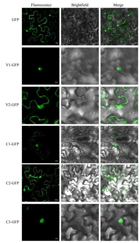
Viruses 2018, 10, 488                                                                             2 of 15

necrosis when expressed from a PVX-based vector; AGV C4 functions as a symptom determinant,
and the putative acylated residues Gly2 and Cys8 play important roles in its subcellular localization
and symptom development.

2. Materials and Methods

2.1. Construction of Plasmids
     To construct binary plasmids transiently expressing GFP fusion proteins for subcellular localization
assays in Nicotiana benthamiana, the coding sequences of AGV V1, V2, C1, C2, C3, and C4 were amplified
with the corresponding primer pairs, digested with XbaI and SmaI restriction enzymes and inserted into
the binary vector pCam35S-GFP to generate the recombinant vectors pCam35S-V1-GFP, pCam35S-V2-GFP,
pCam35S-C1-GFP, pCam35S-C2-GFP, pCam35S-C3-GFP, and pCam35S-C4-GFP expressing the fusion
proteins V1-GFP, V2-GFP, C1-GFP, C2-GFP, C3-GFP, and C4-GFP.
     To construct binary plasmids transiently expressing proteins for PTGS assays, the coding
sequences of AGV V1, V2, C1, C2, C3, and C4 were amplified and cloned into the restriction
sites on the binary vector pCHF to obtain pCHF-V1, pCHF-V2, pCHF-C1, pCHF-C2, pCHF-C3,
and pCHF-C4 recombinant plasmids for agroinfiltration.
     For PVX assays, the full-length coding sequence of AGV V1, V2, C1, C2, C3, and C4 were
PCR-amplified with the primer pairs listed in Table S1. The products were digested with the
corresponding restriction enzymes and ligated to the pGR106 vector to generate PVX-V1, PVX-V2,
PVX-C1, PVX-C2, PVX-C3, and PVX-C4.
     The mutants C4G2A , C4C8A , and C4G2AC8A were constructed using Fast Mutagenesis System
(Transgen, Beijing, China) with primers covering and flanking the mutation sites. To analyze the
subcellular localization of the C4 mutants, the coding sequences of C4G2A , C4C8A , and C4G2AC8A were
amplified using the primer pairs listed in Table S1, digested with the corresponding restriction enzymes
and inserted into the expression vector pCam35S-GFP. The recombinant vectors pCam35S-C4G2A -GFP,
pCam35S-C4C8A -GFP, and pCam35S-C4G2AC8A -GFP were designed to express fusion proteins
C4G2A -GFP, C4C8A -GFP, and C4G2AC8A -GFP. To investigate the pathogenicity of the C4 mutants,
the coding sequences of C4G2A , C4C8A , and C4G2AC8A were amplified and cloned into pGR106 resulting
in PVX-C4G2A , PVX-C4C8A , and PVX-C4G2AC8A , respectively.
     All primers and restriction enzyme sites used for plasmid construction are listed in supplemental
Table S1.
     All constructs were sequenced before use to make sure no amplification errors were induced
by PCR.

2.2. Agroinfiltration
      The recombinant PVX infectious clones were transformed into Agrobacterium tumefaciens strain
GV3101 and the binary expression vectors were transformed into EHA105. After incubating in
Luria–Bertani broth with the appropriate antibiotics at 28◦ C overnight, the cultures were centrifuged
and resuspended with infiltration buffer (10 mM MES, pH 5.7, 10 mM MgCl2 , 150 mM acetosyringone)
to a final OD600 of 0.5–1.0. After incubating at room temperature for 3 h, the suspensions were
infiltrated into leaves of 4-week-old N. benthamiana plants. The infiltrated plants were analyzed for
fluorescence images by confocal laser scanning microscopy (CLSM) at 3 d post infiltration.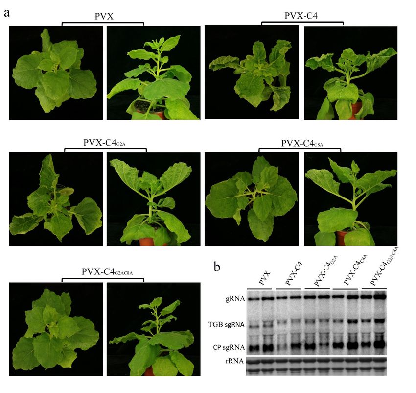
Viruses 2018, 10, 488                                                                         3 of 15

2.3. Fluorescence Observation
     To investigate the subcellular localization of GFP fusion proteins, fluorescence images of the
epidermal cells of N. benthamiana infiltrated with the transformed agrobacterium were captured with
a Zeiss LSM 880 CLSM using the preset settings for GFP (with 488 nm excitation and 500–550 nm
emission) and for chloroplast autofluorescence (with 561 nm excitation and 650–750 nm emission).

2.4. H2 O2 Detection in Plants
     Hydrogen peroxide detection in leaves of N. benthamiana was conducted with the 3,
3’-diaminobenzidine (DAB)-HCl uptake method as described previously with minor modifications [14].
Briefly, the leaves were cut off at the base of the stem and then immersed in 1mg/mL DAB solution in
Tris-HCl buffer (pH 3.8). After 12 h incubation at room temperature, the leaves were bleached with
96% ethanol in boiling water for 5 minutes and photographed. This method was used to decolorize
the leaves and detect the H2 O2 content with the dark brown precipitation produced by the reaction of
DAB with H2 O2 .

2.5. PTGS Assay
     Agrobacterium cultures carrying 35S-GFP, 35S-P19, pCHF-V1, pCHF-V2, pCHF-C1, pCHF-C2,
pCHF-C3, and pCHF-C4 were prepared as described before with a final OD600 of 1.0. The pCHF3 +
GFP agrobacterium culture mixture (harboring pCHF3 and 35S-GFP), V1 + GFP agrobacterium culture
mixture (harboring pCHF3-V1 and 35S-GFP), V2 + GFP agrobacterium culture mixture (harboring
pCHF3-V2 and 35S-GFP), C1 + GFP agrobacterium culture mixture (harboring pCHF3-C1 and
35S-GFP), C2 + GFP agrobacterium culture mixture (harboring pCHF3-C2 and 35S-GFP), C3 + GFP
agrobacterium culture mixture (harboring pCHF3-C3 and 35S-GFP), C4 + GFP agrobacterium culture
mixture (harboring pCHF3-C4 and 35S-GFP), and P19 + GFP agrobacterium culture mixture (harboring
35S-P19 and 35S-GFP) were infiltrated into the seedlings of GFP-transgenic N. benthamiana plant line
16c. GFP fluorescence was detected under a hand-held 100W, long-wave UV lamp (UV products,
Upland, CA, USA). The seedlings were photographed with a Nikon 80D digital camera (Nikon, Tokyo,
Japan) with a yellow filter.

2.6. RNA and Protein Analyses
     Total RNA was extracted from virus-infected leaves with TRIzol reagent (Invitrogen, Carlsbad,
CA, USA). For northern blotting analysis of PVX RNA, 5 µg of total RNA from each sample were
separated on a 1.5% denaturing agarose gel, transferred to a Hybond-N+ membrane (GE Healthcare,
Piscataway, NJ, USA), hybridized and detected with the DIG High Prime DNA Labeling and Detection
Starter Kit II (Roche Diagnostics, Mannheim, Germany). The probe was synthesized with the PCR DIG
Probe Synthesis Kit (Roche Diagnostics, Mannheim, Germany) with the primers listed in Table S1.
     Total proteins were extracted with extraction buffer (50 mM Tris-HCl, pH 7.5, 150 mM NaCl,
3 mM MgCl2 , 1 mM EDTA, 1 mM DTT) containing protease inhibitor cocktail (Roche Diagnostics,
Mannheim, Germany), which was followed by centrifugation at 3000 g for 20 min. The resulting
supernatant was used as total proteins. The nuclear and cytoplasmic proteins were extracted with
the Plant Nuclear and Cytoplasmic Proteins Extraction Kit (BestBio, Shanghai, China) and membrane
proteins were extracted with Plant Membrane Proteins Extraction Kit (BestBio, Shanghai, China) from
plant tissues. Western blot was conducted with the anti-GFP and anti-mCherry mouse monoclonal
antibodies (Transgen, Beijing, China).
Viruses 2018, 10, 488                                                                                4 of 15

3. Results

3.1. Subcellular Localization
     Determination of the subcellular localization of a viral protein is one of the crucial steps to unravel
its putative functions during virus infection. To understand the basic characteristics of AGV proteins,
subcellular localization of AGV encoded V1, V2, C1, C2, C3, and C4 was investigated using GFP tag
after agroinfiltration in epidermal cells of N. benthamiana. The full-length sequences were inserted
at the N-terminus of GFP in the vector pCam35S-GFP, obtaining fusion proteins V1-GFP, V2-GFP,
C1-GFP, C2-GFP, C3-GFP, and C4-GFP, respectively. The vector pCam35S-GFP expressing free GFP
was used as control. The agroinfiltrated leaves were examined by CLSM at 3 d post infiltration and
results are shown in Figures 1 and 2. As expected, free GFP was observed in the cytoplasm and
nucleus almost uniformly. The fusion V1-GFP accumulated exclusively in the nucleus, especially in
the nucleolus. V2-GFP and C1-GFP were distributed in the cytoplasm and nucleus, without nucleolus
accumulation. The fluorescence of C2-GFP shows nuclear-cytoplasmic distribution, with higher
relative concentration in the nucleus than free GFP. C3-GFP was targeted to the nucleus but not
to the nucleolus. The fluorescence of C4-GFP was detected in the cytoplasm, plasma membrane,
nucleus, and chloroplasts (Figure 2). The localization in chloroplasts was indicated by the overlap
with the auto-fluorescence of chlorophyll. The subcellular localization of C4 was also confirmed
through subcellular fraction assays. The total proteins, nuclear proteins, cytoplasmic proteins,
and membrane proteins were extracted from plant tissues expressing C4-GFP, GFP, and PIP2A-mCherry.
The GFP acted as markers of nuclear and cytoplasmic proteins, while PIP2A-mCherry acted as a
membrane-localization marker. Western blot analysis further indicated that C4-GFP was present in the
cytoplasm, nucleus, and membrane fraction (Figure S1).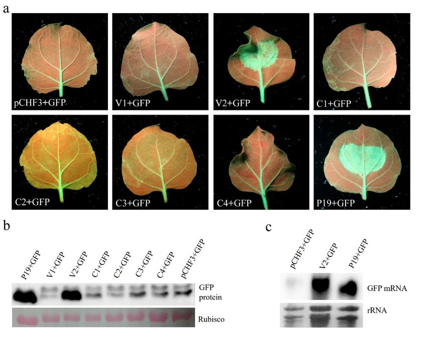
Viruses 2018, 10, 488                                                                                                         5 of 15
  Viruses 2018, 10, x                                                                                                     5 of 15

        Figure1.1.Subcellular
      Figure         Subcellular localization
                                  localization ofof AGV
                                                    AGVV1, V1,V2,
                                                                V2,C1,
                                                                     C1,C2,   andC3C3ininN.N.
                                                                         C2,and               benthamiana
                                                                                            benthamiana      leaves.
                                                                                                         leaves.  TheTheGFPGFP
      expressed     from pCam35S-GFP        is  distributed   evenly  in the cytoplasm    and  nucleus.
        expressed from pCam35S-GFP is distributed evenly in the cytoplasm and nucleus. The V1-GFP         The  V1-GFP     fusion
      protein   from   pCam35S-V1-GFP          is localized   to the nucleus   especially   the nucleolus
        fusion protein from pCam35S-V1-GFP is localized to the nucleus especially the nucleolus in the       in the  infiltrated
        infiltrated
      leaves   of N. leaves    of N. The
                      benthamiana.      benthamiana.
                                            V2-GFP from The pCam35S-V2-GFP
                                                               V2-GFP from pCam35S-V2-GFP
                                                                                    and C1-GFP from   andpCam35S-C1-GFP
                                                                                                            C1-GFP from
        pCam35S-C1-GFP
      are distributed in theare       distributed and
                                 cell cytoplasm       in nucleus,
                                                          the cellwithout
                                                                      cytoplasm    and accumulation.
                                                                             nucleolus     nucleus, without      nucleolus of
                                                                                                           The localization
        accumulation.
      C2-GFP             The localization ofisC2-GFP
                 from pCam35S-C2-GFP               mainlyfrom    pCam35S-C2-GFP
                                                            in the  nucleus with bits is mainly  in the nucleus
                                                                                         of fluorescence    in thewith   bits
                                                                                                                    cytoplasm.
        of fluorescence
      The                  in the cytoplasm.
           subcellular localization              The from
                                         of C3-GFP     subcellular  localization of
                                                             pCam35S-C3-GFP       is C3-GFP
                                                                                     targetedfrom
                                                                                               to thepCam35S-C3-GFP
                                                                                                       nucleus but not to  is the
        targeted to the nucleus but not to the nucleolus. Confocal laser scanning images were takenbars
      nucleolus.     Confocal   laser  scanning    images   were   taken  approximately     3 d post  infiltration.  Scale
        approximately
      indicated    in the 3photos
                            d postrepresent
                                    infiltration.
                                                10Scale
                                                    or 20bars
                                                          µm.indicated in the photos represent 10 or 20 μm.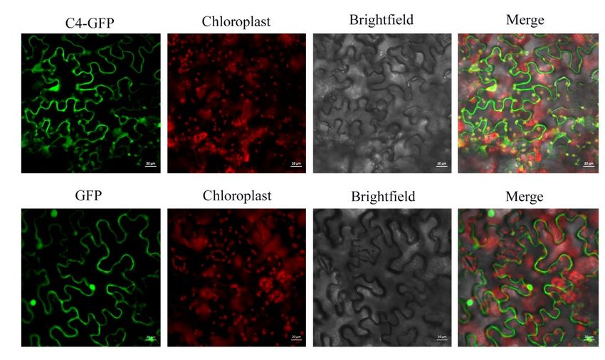
Viruses 2018, 10, 488
Viruses 2018, 10, x 6 6ofof15
15
Figure 2. Subcellular localization of AGV C4 in N. benthamiana leaves. The recombinant fluorescent
Figure
protein2. C4-GFP
Subcellular localization
expressed fromofpCam35S-C4-GFP
AGV C4 in N. benthamiana leaves.
is localized The recombinant
to cytoplasm, plasmafluorescent
membrane,
protein C4-GFP expressed from pCam35S-C4-GFP is localized to cytoplasm, plasma membrane,
nucleus, and chloroplasts, which is different from the free GFP from pCam35S–GFP distributed evenly
nucleus, and chloroplasts,
in the cytoplasm whichChloroplasts
and nucleus. is different are
from the free by
identified GFPthefrom pCam35S–GFP
auto-fluorescence of distributed
chlorophyll
evenly in the cytoplasm and nucleus. Chloroplasts are identified by the auto-fluorescence
through laser excitation of 561 nm. Confocal laser scanning images were taken approximately 3 d post of
chlorophyll through laser excitation of 561 nm. Confocal
infiltration. Scale bars indicated in the photos represent 20 µm. laser scanning images were taken
approximately 3 d post infiltration. Scale bars indicated in the photos represent 20 μm.
3.2. AGV V2 is a Suppressor of PTGS
3.2. AGV V2 is a Suppressor of PTGS
Recently, V2 (AV2), C2, and C4 of several geminivirus species have been shown to be RNA
Recently,
silencing V2 (AV2),
suppressors C2,ofand
(RSSs) PTGS, C4 blocking
of several geminivirus
local or systemicspecies have been
RNA silencing. Toshown
identifytowhich
be RNAviral
silencing
protein(s)suppressors
can suppress (RSSs)
PTGSofinPTGS,
AGV, we blocking local or systemic
used co-infiltration assaysRNA silencing. To identify
in GFP-transgenic which
N. benthamiana
viral protein(s)
16c plants. The can suppress PTGS
Agrobacterium in AGV,
cultures we used
harboring co-infiltration
one of the constructs assays in GFP-transgenic
capable of expressing AGV N.
benthamiana
proteins or GFP16c plants.
from the The Agrobacterium
CaMV 35S promoter cultures harboring
(V1 + GFP, one C1
V2 + GFP, of +the
GFP,constructs
C2 + GFP, capable of
C3 + GFP,
expressing
and C4 + GFP) AGVwere
proteins or GFPinto
infiltrated from 16cthe CaMV
plants. The35SpCHF3
promoter (V1Agrobacterium
+ GFP + GFP, V2 + GFP, C1 +harboring
culture GFP, C2
+empty
GFP, C3 + GFP,
vector wasand C4 + GFP)
infiltrated intowere infiltrated
16c plants into 16c
as negative plants.and
control TheP19+GFP
pCHF3 +Agrobacterium
GFP Agrobacteriumculture
culture harboring
expressing the P19empty
silencingvector was infiltrated
suppressor of tomatointo 16cstunt
bushy plants as was
virus negative control
infiltrated and P19+GFP
as positive control.
Agrobacterium culture expressing
At 5 d post infiltration, the intensity the ofP19 silencing
green suppressor
fluorescence in theof leaves
tomatoexpressing
bushy stunt C1 virus
+ GFP, wasC2
infiltrated
+ GFP, C3 as positive
+ GFP, C4 +control.
GFP, and At V15 d+ post
GFP infiltration, the intensity
declined substantially, of green
similarly to fluorescence
what is observedin thein
leaves expressing
the leaf infiltratedC1with+ GFP, C2 + GFP,
the empty C3 pCHF3
vector + GFP, C4 + GFP,
+ GFP. and V1 +the
In contrast, GFP declined
leaf substantially,
expressing V2 + GFP
similarly to what and
showed obvious is observed
strongerin the leaf
green infiltrated
fluorescence withUV
under thelight
empty vector
similar to pCHF3 + GFP.by
that produced In P19
contrast,
+ GFP
the leaf 3a),
(Figure expressing V2 + GFP
which correlates withshowed obviousaccumulation
the enhanced and stronger of green fluorescence
GFP proteins under
by Western UV
blot light
analysis
similar
(Figure to3b).that produced
Northern by also
blotting P19 showed
+ GFP that (Figure 3a), which correlates
the concentration of GFP mRNA withwas thesignificantly
enhanced
accumulation
higher in the ofleafGFP proteins
patches by Western
co-infiltrated withblot
V2analysis
+ GFP or (Figure 3b). Northern
P19 + GFP than in thoseblotting also showed
co-infiltrated with
that
pCHF3the concentration
+ GFP (Figureof GFP
3c). mRNA
These wassuggest
results significantly
that AGVhigher inan
V2 is theefficient
leaf patches co-infiltrated
and strong RSS. with
V2 + GFP or P19 + GFP than in those co-infiltrated with pCHF3 + GFP (Figure 3c). These results
suggest that AGV V2 is an efficient and strong RSS.Viruses 2018, 10, 488 7 of 15
Viruses 2018, 10, x 7 of 15
Figure 3. Suppression of local PTGS by AGV V2. (a) The leaves of GFP-transgenic 16c line were
co-infiltrated with agrobacterium suspension harboring 35S-GFP expressing GFP and one of the
Figure 3. Suppression of local PTGS by AGV V2. (a) The leaves of GFP-transgenic 16c line were
recombinant vectors expressing AGV proteins as indicated below the images. The leaves expressing
co-infiltrated with agrobacterium suspension harboring 35S-GFP expressing GFP and one of the
pCHF3 + GFP were used as negative control and leaves expressing P19 + GFP were used as positive
recombinant vectors expressing AGV proteins as indicated below the images. The leaves expressing
control. The photographs were taken under UV light at 5 d post infiltration. (b) Western blot analysis
pCHF3 + GFP were used as negative control and leaves expressing P19 + GFP were used as positive
of GFP accumulation in the co-infiltrated leaf patches at 5 d post infiltration. The Ponceau-stained
control. The photographs were taken under UV light at 5 d post infiltration. (b) Western blot
rubisco indicates the equal loading of total proteins. (c) Northern blotting analysis of GFP mRNA
analysis of GFP accumulation in the co-infiltrated leaf patches at 5 d post infiltration. The
accumulation from the co-infiltrated leaf patches at 5 d post infiltration. The rRNAs below indicate the
Ponceau-stained rubisco indicates the equal loading of total proteins. (c) Northern blotting analysis
equal loading of total RNAs.
of GFP mRNA accumulation from the co-infiltrated leaf patches at 5 d post infiltration. The rRNAs
3.3. V2below
and C1indicate the equal
Potentially loading
Enhance theof total RNAs. of PVX and C4 is a Putative Symptom Determinant
Pathogenicity
3.3.To
V2investigate the pathogenicity
and C1 Potentially of AGV proteins,
Enhance the Pathogenicity of PVXtheand
sixC4AGV proteinsSymptom
is a Putative were expressed from
Determinant
a PVX vector. The full-length V1, V2, C1, C2, C3, and C4 genes were cloned into a PVX vector
To investigate
and inoculated into N. thebenthamiana
pathogenicity of AGV proteins,The
by agroinfiltration. the inoculated
six AGV proteins were maintained
plants were expressed fromin a a
PVX vector. The ◦ full-length ◦ V1, V2, C1, C2, C3, and C4 genes were cloned
growth room (24 C day/22 C night, 16 h light/8 h dark) and symptoms were recorded periodically. into a PVX vector and
inoculated
Six plants were into N. benthamiana
inoculated with eachby agroinfiltration.
construct The inoculated
in at least three independentplants were maintained
experiments. in a
At 5 days post
growth room (24 °C day/22 °C night, 16 h light/8 h dark) and symptoms
inoculation (dpi), mild mosaic symptom appeared on the control PVX-inoculated plants. The plants were recorded periodically.
Six plants with
inoculated were PVX-V1,
inoculated with each
PVX-C2, andconstruct
PVX-C3 in at least three
produced independent
PVX-like symptoms experiments.
without anyAt other
5 days
post inoculation
discernible phenotype (dpi),through
mild mosaic symptom
continuous appeared(Figure
observation on theS2).
control PVX-inoculated
However, plants.and
obvious mosaic The
plants inoculated with PVX-V1, PVX-C2, and PVX-C3 produced PVX-like
downward curling of newly emerging leaves were observed in the PVX-V2-infected plants at 5 dpi. symptoms without any
other discernible phenotype through continuous observation (Figure S2). However,
Furthermore, the upper leaves became crinkled and necrotic lesions appeared on the inoculated leaves obvious mosaic
atand downward
9 dpi, which was curling
followedof newly emerging
by apical necrosisleaves
at 13were observed
dpi that in the
ultimately ledPVX-V2-infected plants
to the death of the plantsat 5
dpi. Furthermore, the upper leaves became crinkled and necrotic lesions
(Figure 4a). To determine whether severe symptoms are the consequence of higher virus accumulation, appeared on the
inoculated
Northern leaveswas
blotting at 9employed
dpi, which to was followed
examine the RNAby apical necrosis of
accumulation at PVX-V2.
13 dpi that ultimately
There was no led to the
obvious
difference in the accumulation of PVX genomic RNA (gRNA) between PVX-V2-infected plants andof
death of the plants (Figure 4a). To determine whether severe symptoms are the consequence
higher virus plants
PVX-infected accumulation,
at 5 dpi Northern
and 9 dpi, blotting
while was employed to examine
the accumulation of triple thegeneRNA accumulation
block subgenomicof
PVX-V2.
RNA (TGB There
sgRNA) was andnoCPobvious
sgRNA difference in theupregulated
were apparently accumulation at 9ofdpi PVX genomic RNA (gRNA)
in PVX-V2-infected plants
compared to PVX-infected plants (Figure 4b), which suggests that V2 can enhance 9thedpi,
between PVX-V2-infected plants and PVX-infected plants at 5 dpi and while of
expression the
accumulation of triple gene block subgenomic RNA (TGB sgRNA) and CP
TGB sgRNA and CP sgRNA. These results indicate that V2 is a virulence determinant inducing severe sgRNA were apparently
upregulated
crinkling at 9 dpi when
and necrosis in PVX-V2-infected
expressed from plants compared
a PVX-based to PVX-infected plants (Figure 4b), which
vector.
suggests that V2 can enhance the expression of TGB sgRNA and CP sgRNA. These results indicateViruses 2018, 10, 488 8 of 15
Viruses 2018, 10, x 9 of 15
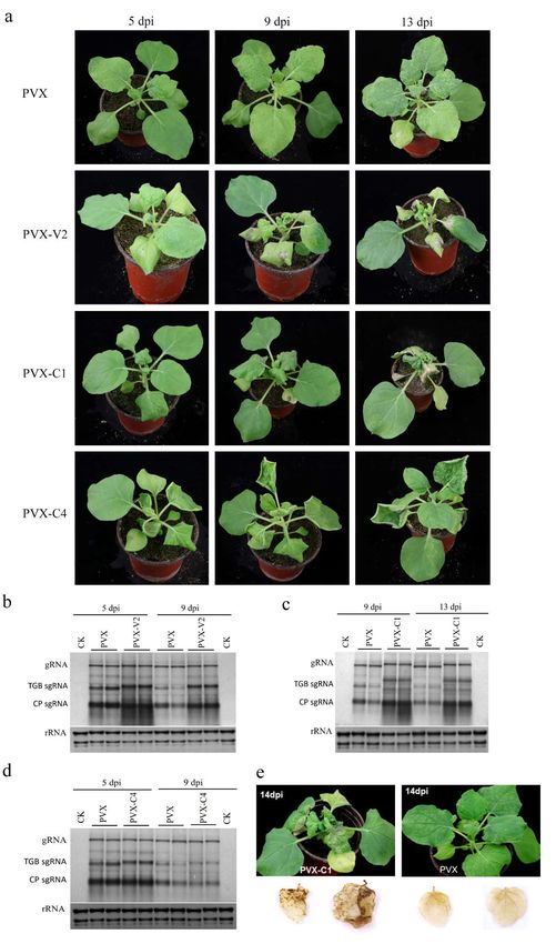
Figure4.4.AGV
Figure AGV V2V2
andand
C1 enhance pathogenicity
C1 enhance of chimeric
pathogenicity PVX and
of chimeric PVXAGVandC4AGV
is a putative
C4 is asymptom
putative
determinant. (a) Symptoms of N. benthamiana plants inoculated with PVX,
symptom determinant. (a) Symptoms of N. benthamiana plants inoculated with PVX, PVX-V2, PVX-C1 and
PVX-V2,
PVX-C4.
PVX-C1 and Symptoms
PVX-C4. were photographed
Symptoms at 5 dpi, 9atdpi,
were photographed and
5 dpi, 13 dpi.
9 dpi, and 13The
dpi.images shown
The images are
shown
representative plants out of six plants with each construct in at least three independent experiments.
are representative plants out of six plants with each construct in at least three independent
Northern blotting analyses of PVX-V2 (b), PVX-C1 (c), and PVX-C4 (d) accumulation compared
experiments. Northern blotting analyses of PVX-V2 (b), PVX-C1 (c), and PVX-C4 (d) accumulation
with PVX. The blot shown is a representative result of three independent experiments. The probe
compared with PVX. The blot shown is a representative result of three independent experiments.
used was specific for PVX RNA. (e) DAB staining for detection of H2 O2 accumulation in PVX-C1-
The probe used was specific for PVX RNA. (e) DAB staining for detection of H2O2 accumulation in
and PVX-inoculated N. benthamiana plants at 14 dpi, showing the higher accumulation of H2 O2 in
PVX-C1- and PVX-inoculated N. benthamiana plants at 14 dpi, showing the higher accumulation of
PVX-C1 inoculated leaves.
H2O2 in PVX-C1 inoculated leaves.Viruses 2018, 10, 488 9 of 15
Plants inoculated with PVX-C1 showed mild leaf curling at 5 dpi, which was followed by strong
downward curling and crinkling of the newly emerging leaves. The inoculated and systematic leaves,
but not the apical leaves, developed visible necrotic lesions at 13 dpi, while the plants inoculated with
PVX did not show any necrosis (Figure 4a). The cell death during pathogen infection is a typical feature
of necrotic tissue. Therefore, the production of H2 O2 in PVX-C1-infected leaves was investigated by in
situ detection with the DAB uptake method. In the presence of H2 O2 , DAB can polymerize to produce
deep brown products which can be visualized after decolorization by ethanol. Figure 4e shows that H2 O2
accumulated to a higher level in the systemically PVX-C1-infected leaves, especially at the base, at 14 dpi.
In contrast, there was no obvious accumulation of H2 O2 in PVX-infected leaves. Northern blotting
was conducted to quantify viral RNA accumulation, showing that the gRNA was not affected in the
presence of C1, while the TGB sgRNA and CP sgRNA increased substantially at 9 dpi and 13 dpi in
PVX-C1-infected plants compared to PVX-infected plants (Figure 4c). Taken together, these results show
that AGV C1 enhances the pathogenicity of PVX eliciting necrotic lesions in N. benthamiana.
The inoculation of PVX-C4 on N. benthamiana induced upward leaf curling at 5 dpi. The symptoms
aggravated progressively with time. Hyperplasia and strong leaf upward curling could be observed
in the upper leaves at 9 dpi and 13 dpi (Figure 4a). Northern blotting showed that there were
no differences in viral gRNA or sgRNA accumulation between PVX-C4- and PVX-infected plants
(Figure 4d), which revealed that C4 does not change the replication and expression of PVX.
However, AGV C4 can induce hyperplasia and severe upward leaf curling in N. benthamiana, which
implies that C4 could be a putative symptom determinant.
3.4. The N-terminal Acylated Sites of C4 are Associated with its Function as Symptom Determinant
In view of the fact that AGV C4 is a putative symptom determinant, the key amino acids
involved in establishing its subcellular localization or symptom determinant function were further
investigated through several point mutations. N-myristoylation is a common modification in some
C4 proteins of geminiviruses, so we predicted the potential post-translational modifications of AGV
C4 with several online programs (http://lipid.biocuckoo.org/, http://lishuyan.lzu.edu.cn/seqpalm/,
http://bioinfo.ncu.edu.cn/WAP-Palm.aspx). The results showed that the Gly residue at position
2 and Cys residue at position 8 were predicted to be acylated by N-myristoylation and S-palmitoylation,
respectively. The residues Gly2 and Cys8 were substituted with Ala, independently and simultaneously,
resulting in C4 mutants C4G2A , C4C8A , and C4G2AC8A . To identify whether the putative acylated
modifications affect the localization of C4, we fused the C4 mutants with GFP obtaining C4G2A -GFP,
C4C8A -GFP, and C4G2AC8A -GFP. Figure 5 shows that the green fluorescence of C4G2A -GFP mainly
colocalized with the red auto-fluorescence of chloroplasts with low fluorescence in the cytoplasm,
plasma membrane, and nucleus, which indicates that C4G2A -GFP is mainly targeted to chloroplasts.
In contrast, there was no chloroplast localization in the C4C8A -GFP. The fluorescence of C4G2AC8A -GFP
was detected in the cytoplasm, plasma membrane, nucleus, and chloroplasts, similar to that of C4-GFP
(Figure 5 and Figure S1).was conducted to analyze the viral RNA accumulation of PVX-C4, PVX-C4G2A, PVX-C4C8A, and
PVX-C4G2AC8A. The results demonstrate that there were no differences in viral gRNA and sgRNA
accumulation among PVX-C4- and C4 mutant variants-infected plants (Figure 6b). Taken together,
our results show that the residues G2 and C8 are essential for C4 localization and play important
roles 2018,
Viruses in AGV C4 function as a putative symptom determinant.
10, 488 10 of 15
Figure 5. Subcellular localization of AGV C4 mutant variants on N-terminal acylated sites in N. benthamiana
leaves. The recombinant fluorescent protein C4G2A -GFP expressed from pCam35S-C4G2A -GFP is mainly
localized to chloroplasts, C4C8A -GFP expressed from pCam35S-C4C8A -GFP is targeted to plasma membrane,
and nucleus and C4G2AC8A -GFP expressed from pCam35S-C4G2AC8A -GFP is localized in the cytoplasm,
nucleus, and chloroplasts. The chloroplasts are identified by the red auto-fluorescence of chlorophyll.
Confocal laser scanning images were taken approximately 3 d post infiltration. Scale bars indicated in the
photos represent 20 µm.
Furthermore, to investigate the effect of AGV C4 mutants on symptom formation, the PVX
vector was used to express the C4 mutant variants. Six plants for each construct were infected in at
least three independent experiments. At the early stage of infection (5 dpi), the plants infected with
PVX-C4 and its derivatives developed mild upward leaf curling. At the late stage of infection (21 dpi),
the plants infected with PVX-C4G2A showed PVX-C4-like phenotypes with severe upward leaf curling.
However, PVX-C4C8A induced very mild upward leaf curling and the PVX-C4G2AC8A -infected plants
showed no differences in phenotype compared with plants inoculated with PVX (Figure 6a). In order to
eliminate the effect of virus accumulation on symptom development, Northern blotting was conducted
to analyze the viral RNA accumulation of PVX-C4, PVX-C4G2A , PVX-C4C8A , and PVX-C4G2AC8A .
The results demonstrate that there were no differences in viral gRNA and sgRNA accumulation among
PVX-C4- and C4 mutant variants-infected plants (Figure 6b). Taken together, our results show that the
residues G2 and C8 are essential for C4 localization and play important roles in AGV C4 function as a
putative symptom determinant.pCam35S-C4G2A-GFP is mainly localized to chloroplasts, C4C8A-GFP expressed from
pCam35S-C4C8A-GFP is targeted to plasma membrane, and nucleus and C4G2AC8A-GFP expressed
from pCam35S-C4G2AC8A-GFP is localized in the cytoplasm, nucleus, and chloroplasts. The
chloroplasts are identified by the red auto-fluorescence of chlorophyll. Confocal laser scanning
images were taken approximately 3 d post infiltration. Scale bars indicated in the photos represent
Viruses 2018, 10, 488 11 of 15
20 μm.
Figure 6. The N-terminal acylated residues of C4 are associated with its symptom determinant
Figure 6.
function. (a)The N-terminal
Symptoms of N.acylated residues
benthamiana plantsofinoculated
C4 are associated
with PVX,with its symptom
PVX-C4, and PVX-C4 determinant
mutant
function.
variants (a) Symptoms
mutated of N. benthamiana
in its N-terminal plants
potentially inoculated
acylated with PVX,were
sites. Symptoms PVX-C4, and PVX-C4
photographed mutant
at 21 dpi.
variants
The imagesmutated
shown arein its N-terminal plants
representative potentially
out ofacylated
six plantssites. Symptoms
inoculated with were photographed
each construct at 21
in at least
dpi. The images shown are representative plants out of six plants inoculated with each construct in
three independent experiments. PVX-C4 G2A showed PVX-C4-like phenotype with severe upward
at curling,
leaf least three independent
PVX-C4 C8A inducedexperiments.
very mild PVX-C4
upwardG2A leafshowed
curling, PVX-C4-like phenotype
and PVX-C4G2AC8A withplants
-infected severe
upward leaf curling, PVX-C4C8A induced very mild upward leaf curling, and PVX-C4G2AC8A-infected
showed PVX-like symptoms. (b) Northern blotting analyses of viral RNA accumulation showed that
plants
there showed
were PVX-like
no obvious symptoms.
differences among(b) PVX-C4
Northern blotting
mutant analyses of viralplants
variants-inoculated RNA compared
accumulationto
showed that there were no obvious differences among PVX-C4 mutant variants-inoculated plants
PVX- and PVX-C4-inoculated plants. The blot shown is a representative one out of three independent
experiments.
compared toThe probe
PVX- andused was specific for PVX
PVX-C4-inoculated RNA.
plants. The blot shown is a representative one out of
three independent experiments. The probe used was specific for PVX RNA.
4. Discussion
4. Discussion
Viruses are obligate intracellular pathogens with relatively small genomes encoding very
few proteins, which means that they depend heavily on host factors to facilitate infection and
Viruses are obligate intracellular pathogens with relatively small genomes encoding very few
counter host defense responses [15]. The subcellular localization of viral proteins provides clues
proteins, which means that they depend heavily on host factors to facilitate infection and counter
to understand the interaction with host factors and their functions during virus infection. The AGV
host defense responses [15]. The subcellular localization of viral proteins provides clues to
C1 is the replication-associated protein (Rep) with conserved motifs with the Rep protein of other
understand the interaction with host factors and their functions during virus infection. The AGV C1
geminiviruses [12]. The cytoplasmic and nuclear localization of AGV C1 is in agreement with its
is the replication-associated protein (Rep) with conserved motifs with the Rep protein of other
function as Rep. The C2 and C3 proteins of monopartite geminiviruses function as transcriptional
activator and replication enhancer, respectively [16]. The localization of AGV C2 and C3 to the
nucleus implies that they are possibly involved in the transcription and replication of the AGV
genome. The chloroplast localization suggests that AGV C4 may perform some different functions
than other C4 homologs. V1 is the coat protein (CP) of monopartite geminiviruses and it exerts multiple
functions, including mediating vector transmission, shuttling of genome DNA into and out of the
nucleus, and facilitating cell-to-cell or systemic viral movement [16]. The AGV V1 displays the same
localization than CPs of many monopartite or bipartite geminiviruses, such as tomato yellow leaf curlViruses 2018, 10, 488 12 of 15
virus (TYLCV) [17] or tomato leaf curl Java virus [18]. The cytoplasmic and nuclear localization of
AGV V2 is in agreement with its function as RSS of PTGS.
It is widely accepted that the virus-derived small-interfering RNAs (vsiRNAs)-mediated RNA
silencing pathway is an important plant immune mechanism during virus infection [19]. In this
antiviral defense pathway, vsiRNAs are produced from viral dsRNA precursors by a set of Dicer-like
proteins, and then assembled with Argonaute proteins to form the RNA-induced silencing complex.
In this complex, the vsiRNAs are used to guide specific cleavage of the cognate viral RNAs. This process
is referred to as PTGS, which is a ubiquitous defense mechanism against RNA and DNA viruses.
Inhibition of RNA silencing by virus-encoded RSS proteins is an essential strategy adopted by viruses
in the arms race between plants and viruses. In geminiviruses, several proteins (i.e. AC2/C2, AC4/C4,
V2, and βC1) have been identified as RSSs targeting different steps of the PTGS pathway [20–24].
For example, the V2 protein of TYLCV was identified to suppress PTGS through its interaction with
SGS3 to prevent it from accessing substrate RNA [25,26]. SGS3 is a dsRNA-binding protein which
recognizes the 5´ overhang dsRNA for subsequent steps in PTGS [27]. In this study, we identified
AGV V2 as a RSS targeting PTGS using the co-agroinfiltration assays with 35S-GFP in 16c plants.
It cannot be excluded that there are other RSSs in AGV. Many RNA silencing suppressors have also
been identified as determinants of pathogenicity in plants. We have also found that AGV V2 expressed
from a PVX-based vector produced serious mosaic, crinkling and necrosis symptoms in N. benthamiana.
The hypersensitive response-like (HR-like) response was not observed when we expressed AGV V2 by
infiltration of agrobacterium cultures harboring the vector pCHF3-V2 under the control of cauliflower
mosaic virus 35S promoter (Figure 3a), which indicates that AGV V2 does not behave like a HR elicitor
in N. benthamiana. Therefore, we speculate that the severe symptoms of PVX-V2-infected N. benthamiana
plants were induced by the enhanced accumulation of PVX TGB sgRNA and CP sgRNA, which may
be attributed to the activity of V2 as an RSS.
Expression of C1 from the PVX-C1 construct induced crinkling and necrosis symptoms
in N. benthamiana, and H2 O2 accumulation can be detected in the systemically infected leaves.
However, there was no HR-like response in the leaves transiently expressing C1 protein by
agroinfiltration from the 35S promoter (Figure 3a). These results suggest that AGV C1 is not a
HR elicitor in N. benthamiana, but it potentially enhances the pathogenicity of PVX by increasing the
accumulation of PVX TGB sgRNA and CP sgRNA.
The C4 protein is the least conserved of all geminivirus proteins, and it has a divergent biological
function [16]. Many C4 proteins from different geminiviruses alter the host plant development and have
been proved to be symptom determinants [16,28]. For example, transgenic Arabidopsis plants expressing
beet curly top virus (BCTV) and beet severe curly top virus (BSCTV) C4 exhibit symptoms similar to
those cause by the viral infection, which are induced by ectopic cell division [29,30]; the C4 protein of
sweet potato leaf curl virus (SPLCV) alters plant development by regulating brassinosteroid signaling
through the interaction with AtBIN2 [31]; and tomato leaf curl Yunnan virus (TLCYnV) C4 induces cell
division through enhancing the stability of Cyclin D 1.1, which regulates the G1/S-phase transition in
plants [32]. We have proved that AGV C4 can induce severe upward leaf curling in N. benthamiana by
expressing it from a PVX-based vector. In addition, PVX-C4 did not change the PVX gRNA and sgRNA
accumulation, which implies that C4 can induce the symptom formation as a symptom determinant.
Acylation is a post-translational modification that has been reported in several geminivirus
C4 proteins. For example, the plasma membrane localization of BCTV C4 is dependent on its intact
N-terminal myristoylation motif [29], and the mutation of the N-myristoylation site disrupted its
plasma membrane localization and the induction of hyperplasia [33]. In addition, S-palmitoylation
also occurs in some geminivirus C4 proteins [34]. For example, the palmitoylation of African cassava
mosaic Zanzibar virus (EACMZV) AC4 contributed to its membrane localization [34]. In our research,
the subcellular localization studies showed that AGV C4 protein exhibits a complex localization with
strong fluorescent signal in the cytoplasm, plasma membrane, nucleus, and chloroplasts. The mutation
of the putative N-myristoylation site in C4 (C4G2A ) reduced its localization at the plasma membrane andViruses 2018, 10, 488 13 of 15
nucleus, so that this mutant version was mainly targeted at chloroplasts. Unlike the BCTV C4, the AGV
C4G2A mutant still induced PVX-C4-like symptoms. The mutation of the putative S-palmitoylation
site in C4 (C4C8A ) abolished its localization in chloroplasts. Furthermore, C4C8A displayed a reduced
induction of upward leaf curling at the late stage of infection. The simultaneous mutant on the
N-myristoylation and the S-palmitoylation sites (C4G2AC8A ) displayed a similar localization to that of
C4, while induced PVX-like symptom at the late stage of the virus infection. We speculate that
there are no obvious connections between localization and pathogenicity of AGV C4. In view
of the C4 localization, the putative S-palmitoylation modification is important for its chloroplast
localization; the putative N-myristoylation modification is important for its plasma membrane
localization; competitive balance may exist between the N-myristoylation and the S-palmitoylation
of C4. In consideration of pathogenicity, N-myristoylation and, especially, S-palmitoylation, play
essential roles in the function of C4 as a symptom determinant.
Taking into account all results obtained in this study, we conclude that: (I) AGV V2 is a suppressor
of PTGS; (II) AGV V2 and C1 enhance the pathogenicity of PVX inducing severe crinkling and necrosis;
and (III) AGV C4 is a putative symptom determinant and acylation is important for its localization
and symptom induction.
Supplementary Materials: The following are available online at http://www.mdpi.com/1999-4915/10/9/
488/s1, Figure S1: Subcellular fractionation of C4-GFP, C4G2A -GFP, C4C8A -GFP and C4G2AC8A -GFP. Figure S2:
The symptoms of PVX-V1-, PVX-C2- and PVX-C3-inoculated N. benthamiana plants show PVX-like symptoms.
Table S1: Primers used for PCR amplification and probe synthesis.
Author Contributions: B.Z., X.Y., and W.Z. performed the experiments and analyzed data. B.Z. wrote the
manuscript. X.Z., X.Y., and S.L. designed the experiment. X.Z. supervised the study and revised the manuscript.
Acknowledgments: This research was financially supported by the National Natural Science Foundation of China
(31701768 and 31390422) and National Key R&D Program of China (2017YFE0110900).
Conflicts of Interest: The authors declare no conflicts of interest.
References
1. Zerbini, F.M.; Briddon, R.W.; Idris, A.; Martin, D.P.; Moriones, E.; Navas-Castillo, J.; Rivera-Bustamante, R.;
Roumagnac, P.; Varsani, A. ICTV virus taxonomy profile: Geminiviridae. J. Gen. Virol. 2017, 98, 131–133.
[CrossRef] [PubMed]
2. Mansoor, S.; Zafar, Y.; Briddon, R.W. Geminivirus disease complexes: the threat is spreading. Trends Plant Sci.
2006, 11, 209–212. [CrossRef] [PubMed]
3. Varsani, A.; Roumagnac, P.; Fuchs, M.; Navas-Castillo, J.; Moriones, E.; Idris, A.; Briddon, R.W.;
Rivera-Bustamante, R.; Murilo, Z.F.; Martin, D.P. Capulavirus and Grablovirus: Two new genera in the
family Geminiviridae. Arch. Virol. 2017, 162, 1819–1831. [CrossRef] [PubMed]
4. Polston, J.E.; Londoño, M.A.; Capobianco, H. The complete genome sequence of new world jatropha mosaic
virus. Arch. Virol. 2014, 159, 3131–3136. [CrossRef] [PubMed]
5. Narayana, D.S.A.; Shankarappa, K.S.; Govindappa, M.R.; Prameela, H.A.; Rao, M.R.G.; Rangaswamy, K.T.
Natural occurrence of jatropha mosaic virus disease in India. Curr. Sci. 2006, 91, 584–586.
6. Srivastava, A.; Kumar, S.; Jaidi, M.; Raj, S.K. Molecular characterization of a new begomovirus associated
with leaf yellow mosaic disease of Jatropha curcas in India. Arch. Virol. 2015, 160, 1359–1362. [CrossRef]
[PubMed]
7. Rwahnih, M.A.; Dave, A.; Anderson, M.M.; Rowhani, A.; Uyemoto, J.K.; Sudarshana, M.R. Association of a
DNA virus with grapevines affected by red blotch disease in California. Phytopathology 2013, 103, 1069–1076.
[CrossRef] [PubMed]
8. Rwahnih, M.A.; Rowhani, A.; Golino, D.A.; Islas, C.M.; Preece, J.E.; Sudarshana, M.R. Detection and genetic
diversity of grapevine red blotch-associated virus isolates in table grape accessions in the National Clonal
Germplasm Repository in California. Can. J. Plant Pathol. 2015, 37, 130–135. [CrossRef]
9. Sudarshana, M.R.; Perry, K.L.; Fuchs, M.F. Grapevine red blotch-associated virus, an emerging threat to the
grapevine industry. Phytopathology 2015, 105, 1026–1032. [CrossRef] [PubMed]Viruses 2018, 10, 488 14 of 15
10. Loconsole, G.; Saldarelli, P.; Doddapaneni, H.; Savino, V.; Martelli, G.P.; Saponari, M. Identification of a
single-stranded DNA virus associated with citrus chlorotic dwarf disease, a new member in the family
Geminiviridae. Virology 2012, 432, 162–172. [CrossRef] [PubMed]
11. Ma, Y.; Navarro, B.; Zhang, Z.; Lu, M.; Zhou, X.; Chi, S.; Di, S.F.; Li, S. Identification and molecular
characterization of a novel monopartite geminivirus associated with mulberry mosaic dwarf disease.
J. Gen. Virol. 2015, 96, 2421–2434. [CrossRef] [PubMed]
12. Liang, P.; Navarro, B.; Zhang, Z.; Wang, H.; Lu, M.; Xiao, H.; Wu, Q.; Zhou, X.; Di, S.F.; Li, S. Identification and
characterization of a novel geminivirus with a monopartite genome infecting apple trees. J. Gen. Virol. 2015,
96, 2411–2420. [CrossRef] [PubMed]
13. Al, R.M.; Alabi, O.J.; Westrick, N.M.; Golino, D.; Rowhani, A. Description of a novel monopartite geminivirus
and its defective subviral genome in grapevine. Phytopathology 2017, 107, 240–251.
14. Sharma, P.; Ikegami, M. Tomato leaf curl Java virus V2 protein is a determinant of virulence, hypersensitive
response and suppression of posttranscriptional gene silencing. Virology 2010, 396, 85–93. [CrossRef]
[PubMed]
15. Ahlquist, P. Parallels among positive-strand RNA viruses, reverse-transcribing viruses and double-stranded
RNA viruses. Nat. Rev. Microbiol. 2006, 4, 371–382. [CrossRef] [PubMed]
16. Fondong, V.N. Geminivirus protein structure and function. Mol. Plant Pathol. 2013, 14, 635–649. [CrossRef]
[PubMed]
17. Rojas, M.R.; Jiang, H.; Salati, R.; Xoconostle-Cázares, B.; Sudarshana, M.R.; Lucas, W.J.; Gilbertson, R.L.
Functional analysis of proteins involved in movement of the monopartite begomovirus, tomato yellow leaf
curl virus. Virology 2001, 291, 110–125.
18. Sharma, P.; Ikegami, M. Characterization of signals that dictate nuclear/nucleolar and cytoplasmic shuttling
of the capsid protein of tomato leaf curl Java virus associated with DNA beta satellite. Virus Res. 2009, 144,
145–153. [CrossRef] [PubMed]
19. Li, F.; Ding, S.W. Virus counterdefense: diverse strategies for evading the RNA-silencing immunity.
Annu. Rev. Microbiol. 2006, 60, 503–531. [CrossRef] [PubMed]
20. Hanley-Bowdoin, L.; Bejarano, E.R.; Robertson, D.; Mansoor, S. Geminiviruses: masters at redirecting and
reprogramming plant processes. Nat. Rev. Microbiol. 2013, 11, 777–788. [CrossRef] [PubMed]
21. Vanitharani, R.; Chellappan, P.; Pita, J.S.; Fauquet, C.M. Differential roles of AC2 and AC4 of cassava
geminiviruses in mediating synergism and suppression of posttranscriptional gene silencing. J. Virol. 2004,
78, 9487–9498. [CrossRef] [PubMed]
22. Gopal, P.; Pravin, K.P.; Sinilal, B.; Jose, J.; Kasin, Y.A.; Usha, R. Differential roles of C4 and betaC1 in mediating
suppression of post-transcriptional gene silencing: evidence for transactivation by the C2 of bhendi yellow
vein mosaic virus, a monopartite begomovirus. Virus Res. 2007, 123, 9–18. [CrossRef] [PubMed]
23. Nawaz-Ul-Rehman, M.S.; Nahid, N.; Mansoor, S.; Briddon, R.W.; Fauquet, C.M. Post-transcriptional gene
silencing suppressor activity of two non-pathogenic alphasatellites associated with a begomovirus. Virology
2010, 405, 300–308. [CrossRef] [PubMed]
24. Luna, A.P.; Rodrígueznegrete, E.A.; Morilla, G.; Wang, L.; Lozanodurán, R.; Castillo, A.G.; Bejarano, E.R.
V2 from a curtovirus is a suppressor of post-transcriptional gene silencing. J. Gen. Virol. 2017, 98, 2607–2614.
[CrossRef] [PubMed]
25. Glick, E.; Zrachya, A.; Levy, Y.; Mett, A.; Gidoni, D.; Belausov, E.; Citovsky, V.; Gafni, Y. Interaction with host
SGS3 is required for suppression of RNA silencing by tomato yellow leaf curl virus V2 protein. Proc. Natl.
Acad. Sci. USA 2008, 105, 157–161. [CrossRef] [PubMed]
26. Zrachya, A.; Glick, E.; Levy, Y.; Arazi, T.; Citovsky, V.; Gafni, Y. Suppressor of RNA silencing encoded by
tomato yellow leaf curl virus-Israel. Virology 2007, 358, 159–165. [CrossRef] [PubMed]
27. Fukunaga, R.; Doudna, J.A. DsRNA with 50 overhangs contributes to endogenous and antiviral RNA
silencing pathways in plants. EMBO J. 2009, 28, 545–555. [CrossRef] [PubMed]
28. Michael, D.C.; Katherine, M.L. Toward understanding the molecular mechanism of a geminivirus C4 protein.
Plant Signal Behav. 2015, 10, e1109758.
29. Piroux, N.; Saunders, K.; Page, A.; Stanley, J. Geminivirus pathogenicity protein C4 interacts with Arabidopsis
thaliana shaggy-related protein kinase AtSKeta, a component of the brassinosteroid signalling pathway.
Virology 2007, 362, 428–440. [CrossRef] [PubMed]Viruses 2018, 10, 488 15 of 15
30. Lai, J.; Chen, H.; Teng, K.; Zhao, Q.; Zhang, Z.; Li, Y.; Liang, L.; Xia, R.; Wu, Y.; Guo, H. RKP, a RING finger
E3 ligase induced by BSCTV C4 protein, affects geminivirus infection by regulation of the plant cell cycle.
Plant J. 2009, 57, 905–917. [CrossRef] [PubMed]
31. Bi, H.; Fan, W.; Zhang, P. C4 protein of sweet potato leaf curl virus regulates brassinosteroid signaling
pathway through interaction with AtBIN2 and affects male fertility in Arabidopsis. Front. Plant Sci. 2017, 8,
1–15. [CrossRef] [PubMed]
32. Mei, Y.; Yang, X.; Huang, C.; Zhang, X.; Zhou, X. Tomato leaf curl Yunnan virus-encoded C4 induces cell
division through enhancing stability of Cyclin D 1.1 via impairing NbSKη -mediated phosphorylation in
Nicotiana benthamiana. PLoS Pathog. 2018, 14, e1006789. [CrossRef] [PubMed]
33. Millslujan, K.; Andrews, D.L.; Chou, C.; Deom, C.M. The roles of phosphorylation and SHAGGY-like protein
kinases in geminivirus C4 protein induced hyperplasia. Plos One 2015, 10, e0122356.
34. Fondong, V.N.; Reddy, R.V.; Lu, C.; Hankoua, B.; Felton, C.; Czymmek, K.; Achenjang, F. The consensus
N-myristoylation motif of a geminivirus AC4 protein is required for membrane binding and pathogenicity.
Mol. Plant Microbe Interact. 2007, 20, 380–391. [CrossRef] [PubMed]
© 2018 by the authors. Licensee MDPI, Basel, Switzerland. This article is an open access
article distributed under the terms and conditions of the Creative Commons Attribution
(CC BY) license (http://creativecommons.org/licenses/by/4.0/).You can also read