TIVITA Tissue FAQs - Diaspective Vision
←
→
Page content transcription
If your browser does not render page correctly, please read the page content below

TIVITA® Tissue FAQs
Below, you will find the most frequently asked questions of our clients. If you have a question and
cannot find the answer here, please send us an e-mail (office@diaspective-vision.com). We will be
glad to help you.

1   Why is it important to measure tissue oxygen saturation (StO 2), hemoglobin index
    (THI), NIR perfusion and other wound parameters? How does the TIVITA® Tissue
    system work?

The concentration of hemoglobin and the oxygenation in the tissue are two very important
physiological parameters, which indicate healthy conditions in the tissue and the smallest
capillaries respectively (keyword: micro circulation). Oxygen is imperative in all living cells and plays
an essential role in many wound healing processes, immune functions, the formation of new
tissue and blood vessels. Therefore, concerning normal and quick healing processes, a decrease
in tissue oxygenation has potentially severe and negative consequences; the development of ulcers
in patients with vascular disease or diabetes makes this clear.
The important role, which the measurement of the tissue oxygenation plays in many clinical
applications, such as the monitoring of vascular perfusion, the prognosis concerning amputation
level (e.g. in PAD, diabetes patients) or the assessment of reconstructive and plastic surgery, has
also been recognized for many years [1-5].
Other procedures for measuring perfusion and/or oxygenation of the blood/tissue are for example:
pulse oximetry, transcutaneous oxygen monitoring, Laser-Doppler-imaging, near-infrared
spectroscopy or ICG fluorescence imaging. A differentiation of these methods provides section 6.

2   How does the TIVITA® Tissue system work?

The hyperspectral camera TIVITA® Tissue records a complete spectrum in the visible (VIS) and near
infrared (NIR) spectral range for every single pixel of the image. The light spectrum in the range
between 500 nm and 1000 nm is dispersed into approx. 100 wavelengths. One recording usually
takes appr. 6 seconds. The parameters calculated from the data with specific physiological or
clinical significance are then made available to the user as scaled false color images. In addition,
the software can display the spectrum for each pixel. Furthermore, it is possible to export or save
the so-called hyperspectral data cube (3D-Cube) in different formats, thus providing the means
for further analysis of the data sets. In another step, different evaluations of the spectra can be
performed.

3   Which measuring procedure does the TIVITA® Tissue System use?

The TIVITA® Tissue is an imaging spectroscopic measurement system, which is based on the
physical principle of imaging remission spectroscopy. The measurement applies the laws of physics
according to which the light absorption depends on the concentration of chromophores while the
light dispersion depends on the structural characteristics of the tissue. The area measured is
illuminated with broadband white light and the penetrating light is diffused, absorbed and

www.diaspective-vision.com                                                                            |2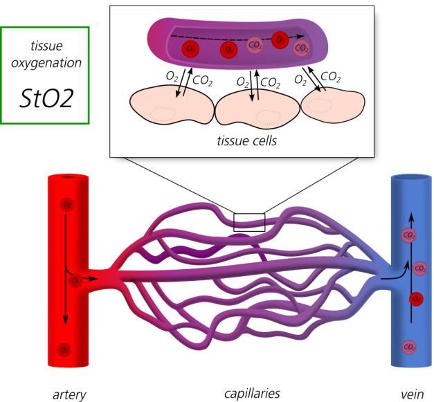
remitted. The TIVITA® Tissue system disperses the remitted light into its essential elements and
records it with a two-dimensional light sensor (camera sensor). Since the spectrum of the lighting
source is known, the analysis of the absorbed part of the light can be performed. The
measurement is done in the transflexion (remission), meaning lighting source and camera are
positioned in the same plane. The spectrum of the back scattered light is analyzed, which provides
the different medical parameters such as tissue oxygenation and hemoglobin or water index. Thus,
using contrast agents is no longer necessary.

The schematic assembly of the TIVITA® Tissue with the internal spectrometer and the entrance slit
is depicted in the following image.

    Figure 1: Schematic assembly of the TIVITA® Tissue with built-in transmission spectrometer and entrance slit.

4   Which parameters does the TIVITA® Tissue system measure?

The TIVITA® Tissue system measures tissue oxygenation in the micro circulation of the tissue (StO2),
the distribution of hemoglobin as an index (THI) [6], the perfusion of the underlying tissue parts
(NIR perfusion) as well as the distribution of water in the tissue as an index (TWI) and provides the
respective images. The NIR perfusion depends on the tissue oxygenation as well as the hemoglobin
distribution.

5   Why is the hemoglobin concentration given in arbitrary units (AU)?

On its way through the tissue, the light is deflected. This scattering of light is generally very hard
to quantify. Because of that, an absolute quantification of the hemoglobin concentration is not
possible. However, the relative concentration determined from measuring the oxygenated and
deoxygenated hemoglobin and other organic substances in the tissue is a reliable parameter. From
the parameter image and trend measurements valuable information can be retrieved. The fact
that this information is also provided as an image is a major enhancement and a big advantage
of the TIVITA® Tissue. As opposed to point-by-point or local measurements with a sensor, the
acquired parameter values can at least be assessed and analyzed in relation to the surrounding
area (whole measurement area) [7].

www.diaspective-vision.com                                                                                          |3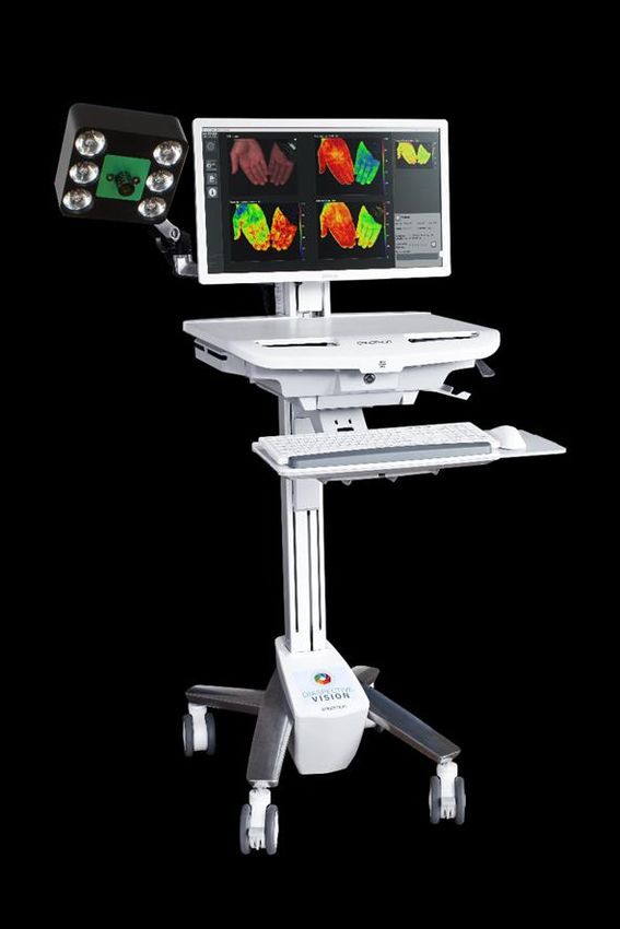
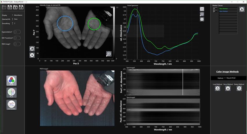
6   How does the tissue oxygenation measured by the TIVITA® Tissue system differ from
    other measurements concerning oxygen supply (SpO2, NIRS, tcpO2)?

The StO2-value measured with the TIVITA® Tissue reflects the percentage of hemoglobin
oxygenation in the capillary area of the tissue micro circulation and shows the changes in oxygen
supply and consumption directly in the tissue. The tissue oxygenation is generally lower than the
arterial oxygenation (SaO2) and the pulse oximetry oxygenation SpO2) respectively and lies
between the arterial and the venous oxygenation (SvO2). Models often use a relation of 70%
venous and 30% arterial oxygenation for the calculation of the current tissue oxygenation [8].

                       Figure 2: Principle-imaging of the micro circulation in the tissue

Many systems are calibrated applying hypoxia studies during which the test person’s supply with
oxygen is reduced in a controlled manner and the respective saturations are determined by taking
arterial and venous blood using blood gas analysis (BGA). The tissue oxygenation is then calculated
from a fixed ratio between arterial and venous blood (typically 25-30% SaO2 + 70-75% SvO2).
The size of the ratio is at the discretion of the manufacturer.

SpO2:
This value is measured using a pulse oximeter. It is the most common measurement unit for
hemoglobin-oxygenation. Unlike the StO2-value, the SpO2-value is calculated from the pulsing
(arterial) part of the quickly scanned measurement signal, which is why this value is the standard
value for arterial oxygenation. While it does provide useful information on pulmonary function, it
does not offer any information on tissue oxygenation or the oxygen absorption of the organs,
which is possible when using the TIVITA® Tissue system for measurements. Because measurements
with pulse oximeters depend on the hemoglobin pulse beat in the artery, SpO2-measuments – in
contrast to determining StO2 levels – also require a pulsing change via the heartbeat.

www.diaspective-vision.com                                                                      |4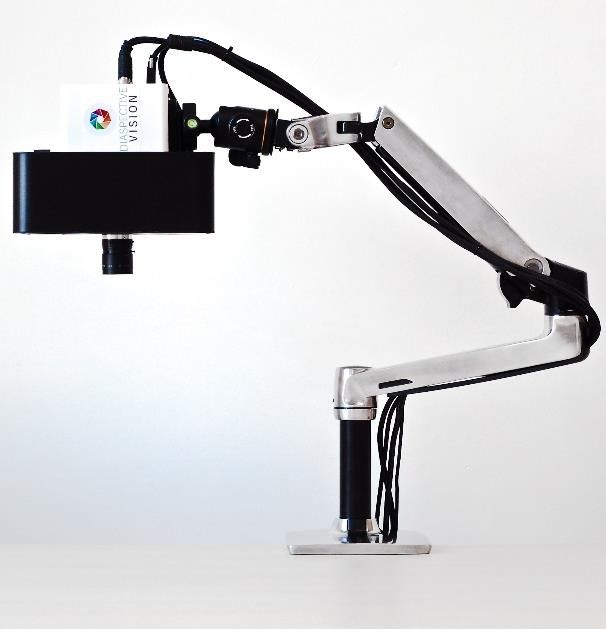
NIRS:
The tissue oxygenation measurement with near-infrared spectroscopy (NIRS) provides values for
local hemoglobin saturation at the measurement site itself. It differs from the StO2-measuring
technique of the TIVITA® Tissue because of the lighting source (white light vs. LEDs), the
wavelengths (visible range + near-infrared range vs. only near-infrared range) and the algorithms,
which are used for calculating tissue oxygenation. NIRS primarily was developed for the
assessment of deeper tissue layers, such as muscles and brain. Contrarily, the TIVITA® Tissue
system, which is based on spectroscopy with visible light (visible light spectroscopy, VLS) and NIR
spectroscopy (NIRS), measures the oxygenation in superficial tissue layers as well as in deeper
tissue layers. Furthermore, the hemoglobin absorption signal is much stronger in the visible range
than in the near-infrared range, which is why the VLS procedure has a much higher signal-to-
noise-ratio and much higher precision.

                             Visible light          Near-infrared wavelength range

                      Figure 3: Principle depiction of the interaction of light with tissue

NIRS-measurement technology can only be applied point by point and with direct contact to the
patient. The TIVITA® Tissue system on the other hand provides a contact free measurement
procedure and two-dimensional images.

tcpO2:
TcpO2-procedures transcutaneously measure partial oxygen pressure. An electrode is used in order
to warm up the tissue underneath and create a localized hyperemia. This means that the
transcutaneous measuring values reflect the maximum capacity of the local vascular system and
tissue to provide oxygen and remove carbon dioxide. Therefore, the tcpO2 value is more in the
range of arterial oxygen saturation and is not a measurement of tissue oxygen saturation under
normal conditions measured with the TIVITA® Tissue System. The tcpO2 method requires the skin
tissue to be heated to 40°C or higher for the measurement, so it takes several minutes to read
the first measurement. This procedure is also not suitable for long-term monitoring. In

www.diaspective-vision.com                                                                      |5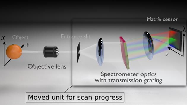
comparison, the TIVITA® Tissue system is a non-invasive, non-contact, imaging system and a much
faster and more convenient method for measuring tissue oxygenation.

Fluorescence Imaging:
One method that keeps resurfacing in angiography is fluorescence imaging with the color agent
indocyanine green (ICG). With this method, it is possible to depict the perfusion of tissues and the
lymph circulation. A big disadvantage of this method is the intravenous administration of the color
agent. On the whole, the information content of ICG imaging is limited and subject to scattering
effects, which increase with the depth of the tissue. The TIVITA® Tissue non-invasively provides
much more and more detailed information – without the application of color agents – thus making
it a far more suitable method for measuring tissue oxygenation.

7   What is the difference between tissue photometry with two wavelengths and the
    tissue spectrometry with over one hundred wavelengths?

Known procedures similar to the oxygenation measurements with the TIVITA® Tissue system are
the near-infrared tissue spectroscopy (NIRS), which, for example, is used by the INVOS of Covidien
or the multi-spectral camera system HyperVue by Hypermed. An important difference in the
methodology is the measured and evaluated wavelength range.
On the way through the tissue, the light is absorbed by different tissue pigments and partially
scattered. The absorption as well as the scattering changes the amount of light, which at a specific
wavelength comes back from the tissue to the sensor and can be measured there. Absorption,
which is mainly caused by the blood (hemoglobin), has certain forms over an entire wavelength
range and is changed due to scattering or other tissue pigments. At specific wavelengths, one or
the other factor has more influence. Looking only at a few wavelengths, it is difficult to determine,
what caused the change in the measured amount of light and other characteristics of the light
must be considered (e.g. runtime differences of the light). However, looking at the whole
wavelength spectrum, the influence of different factors can be determined by characteristic
changes in the forms of the spectra. The TIVITA® Tissue provides all wavelengths in the VIS and
NIR range, which can be influenced by blood. Accordingly, changes in the scattering behavior,
which influence specific points in the hemoglobin spectrum can be determined anew with every
single measurement and be taken into account for the calculation.

8   How does the tissue oxygen saturation measured by the TIVITA® Tissue System differ
    from the measurements by other tissue oximeters (NIRS)??

Even though often the term near-infrared spectroscopy is used for measuring tissue oxygenation,
they are in most cases photometric procedures with few wavelengths, similar to pulse oximeters.
For tissue measurements, numerous influences on the measurement signal must be taken into
account. These are mainly the skin pigment melanin, water and tissue scattering (cf. fig. 4)
With conventional photometric systems and the utilization of only a few wavelengths, this
problem can only be solved in a limited way.

www.diaspective-vision.com                                                                        |6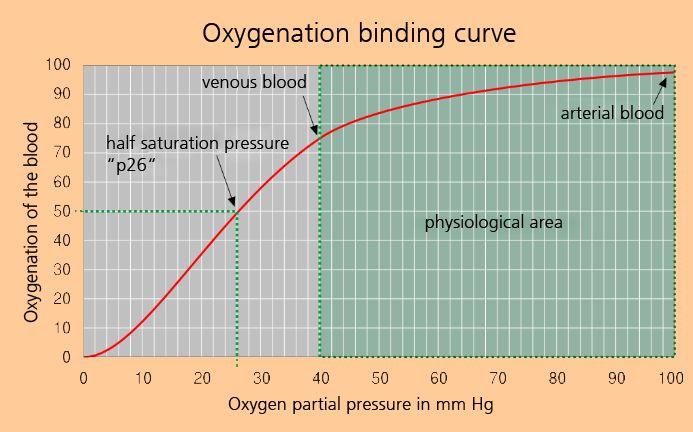
Because of the high quality of the spectra, the 2nd derivation of the tissue spectrum can be
calculated mathematically. Constant and linear influences are eliminated and an exact
chemometric analysis of the absorption band is created (cf. fig. 4).

Figure 4: Absorption curves of different tissue constituents. Next to the hemoglobin, it clearly shows the various other
            constituents which influence the measurement signal and whose concentration is unknown.

9    What is the difference between the TIVITA® Tissue System and laser Doppler or laser
     speckle contrast cameras?

The measuring principle and the parameters determined by Laser-Doppler or Laser-Speckle
cameras are very different from the measurement technology of the TIVITA® Tissue. Laser-Doppler-
(or Laser-Speckle-) procedures use the frequency shift of coherent irradiated laser light at a specific
wavelength, which is caused by light scattering on moving particles (Doppler effect). From this
frequency shift, conclusions about the movement of the scattering particles in the observed
measurement environment, i.e. essentially the blood, and in the measurement volume can be
drawn. This is used to calculate the so-called flux parameter from the measurement data, which
is intended to quantify the blood flow in the measurement volume.
Although this can be used to test the functioning of the perfusion, important parameters such as
the oxygenation of hemoglobin cannot be determined. By using the Speckle-Effect, the Laser-
Doppler-method can also be used relatively quickly for imaging.
Opposed to that, the TIVITA® Tissue system does not provide any direct information about the
movement of blood cells in the measurement area. The quality of perfusion, however, is
sufficiently determined by tissue oxygenation, the THI and other parameters. However, since less
well saturated blood also moves through the body or oxygen consumption in the tissue can be
greatly increased, it is more effective in many situations to determine the oxygen saturation in the
tissue..

10 How does the TIVITA® Tissue System differ from Pushbroom Imagers and other
     spectral technologies?

In technology, imaging spectrographs are often referred to as hyperspectral imaging systems.
These imaging spectrographs are also called Pushbroom Imagers. Generally, they can only record

www.diaspective-vision.com                                                                                           |7
one spectrally resolved line. In order to acquire a three-dimensional data cube, either the objects
or the Pushbroom Imagers and lighting units have to move. For the practical application in
hospitals this, of course, is rather inconvenient. Thus, Diaspective Vision has developed a
procedure which enables the recording of high-resolution hyperspectral images with a compact
camera system. It is important to note, that in the TIVITA® Tissue system, the spectrometer unit is
integrated. Thus, ensuring that the spectrum is not distorted or disturbed in any pixels. This is the
basis for a reliable chemometric analysis and equally reliable parameter images.

11 What is the measuring depth of the TIVITA® Tissue system?

Answering this question concerning the tissue penetration depth of the light is only possible in a
limited way. The penetration depth depends on the respective tissue, which varies individually.
The TIVITA® Tissue system uses a broad light spectrum in the VIS and NIR range for the analysis.
In the VIS range, tissue absorption is many times higher than in the NIR range. For this reason, the
penetration depth over the evaluated spectrum is not constant.

We do not consider it plausible to give absolute values for the wavelength-depending penetration
depths on living human tissue, since they are not verifiable in real terms. Additionally, the results
of theoretical model calculations are sometimes very different.

As rough guide values for the penetration depths, the range from 0.1 mm for blue light (450 nm)
and up to 6 mm for NIR light (> 750 nm) can be given (cf. fig. 3) [11].

12 How is the oxygenation determined?

The oxygenation is defined as the ratio of oxygenated and deoxygenated hemoglobin. Via the
different absorption spectra of both hemoglobin variations an analysis of the oxygenation is
generally possible (cf. fig. 4).
The TIVITA® Tissue system uses the spectral range from 500 nm to 1000 nm – thus the VIS light
as well as the NIR light – for analysis.
As already explained in sections 1 and 7, the measured spectra contain wavelength-dependent
contributions from different skin layers (penetration depths). Hence, for each range different,
adapted algorithms are used for parameter calculation.

13 How does skin pigmentation (melanin) influence the measurements with the TIVITA®
   Tissue system?

The distribution of melanin in the skin varies greatly from person to person and is mainly
concentrated in superficial skin layers. The consideration of the absorption spectrum of melanin,
which is nearly linear in the regarded spectral range, requires respective modulations in the
calculations (e.g. calculation of the hemoglobin levels).
The knowledge that the absorption curve of melanin in the spectral range under consideration is
almost linear can be partly taken into account for the calculation of the parameters. For example,
the influence of melanin on the calculation of oxygenation StO2 is very small. The parameters THI
and NIR Perfusion Index may be influenced by melanin. If the parameters StO2 and NIR perfusion

www.diaspective-vision.com                                                                        |8
differ strongly from each other, it should be checked whether this was caused by a strong
pigmentation of the skin. In the case of very dark skin, an influence on the oxygenation StO2
cannot be ruled out.
If the TIVITA® Tissue system is used to evaluate wounds in which no skin with melanin is present,
the parameters are unaffected and can be used to evaluate the wound.
The water index TWI is not influenced by melanin.

14 Can TIVITA® Tissue be used for dark skin or tattoos?

TIVITA® Tissue can be used on patients with dark skin or tattoos. However, the parameter images
may provide false information or may not be completely evaluated. With darker skin this is due to
the increased melanin content. An explanation can be found in question 13. The different
colorants of tattoos lead to an increased absorption, which can prevent a valid evaluation.

15 Are there normal/standard values for tissue oxygenation?

Unilateral norm or limiting values for tissue oxygenation are currently non-existent because there
is no gold standard and many devices are calibrated differently. Scientific studies establishing
standard limit values are still in progress. To this end, we work closely together with the doctors.
Tissue oxygenation in general is lower than the arterial oxygenation (SaO2) and is more commonly
located in the area of venous oxygenation (SvO2). The measuring values depend on the tissue, the
skin temperature and the patients overall status. The values for tissue oxygenation of healthy test
subjects usually lies between 50% - 70% [8].
Here there is a connection to the transcutaneous oxygen measurement [15], which is explained
by the oxygen binding curve. Further information is given in section 20.
Furthermore, there are some papers dealing with tissue oxygen saturationdetermined by NIRS
sensors [12 - 14].

16 How were the measuring values of the TIVITA® Tissue system calibrated?

For the calibration of the measuring values, different clinical and non-clinical studies were
conducted.
The TIVITA® Tissue StO2-value was calibrated using the blood flow model with a freely adjustable
oxygenation degree of the assessed blood volume. The calibration was done using reference
values determined by using a blood gas analyzer. Looking only at the absorption bands of the
hemoglobin for the analysis of the tissue oxygenation, this method is very well fitted for calibration
procedures. The consideration of further tissue components, can be achieved using phantom
systems for simulation and testing [9].
The results of the in vitro-calibration were verified by occlusion tests in combination with a NIRS
tissue oximeter sensor.

17 Do I have to calibrate the TIVITA® Tissue System prior to use?

No, it is not necessary to calibrate the TIVITA® Tissue. The TIVITA® Tissue system is calibrated during
production and the calibration data are saved within the camera.

www.diaspective-vision.com                                                                          |9
18 How do I choose the suitable lighting?

The TIVITA® Tissue system is delivered with a lighting unit optimized for the application.

                        Figure 5: TIVITA® Tissue system with integrated lighting unit

19 Will the measurement values be influenced by the general lighting in the room?

The TIVITA® Tissue system is an imaging spectrometer system and not a photometric multi-spectral
system. In photometric systems, the camera sensors are usually placed directly behind the entrance
lens. In spectrometers, incoming light must pass the optical entrance slit in the spectrometer unit.
Lighting sources for room illumination (e.g. in clinical operation theaters) have characteristic
spectra. These lighting sources are illuminating the measurement area in addition to the TIVITA®
Tissue lighting unit. The respective lighting component can also get into the TIVITA® Tissue system
and falsify the measurements because the TIVITA® Tissue system is not calibrated in respect to
these lighting conditions.
In order to determine whether the room lighting does influence the measurements, you can switch
the room lighting source on and off.
External light, which causes noticeable changes in the parameter values, must be avoided or the
measurement area must be shielded. Generally, the camera lighting is very intense in comparison
to the room’s lighting source. It is of advantage to darken the room in order to avoid wrong
measurements. Direct sunlight on the measuring region should be avoided, as this can cause an
error in the calculations which cannot be detected later.

20 Why is it so important to measure oxygenation, THI and NIR perfusion at the same
   time?

The oxygenation levels measured by the TIVITA® Tissue system show the percentage of oxygen
bound to the hemoglobin. This is important for determining tissue hypoxia, because through
measuring oxygenation, the amount of oxygen dissolved in the tissue is given. This is due to the
fact that the oxygen binding curve refers the oxygen saturation to a certain amount of dissolved

www.diaspective-vision.com                                                                      |10
oxygen. In order to determine how much oxygen is absolutely present, the blood flow is needed
in addition to the oxygenation. Only then can the absolute amount of inflow (which is determined
by the blood flow and the arterial oxygenation) and outflow (given by the blood flow and the
capillary-venous oxygenation) be used to determine the amount of oxygen delivered to the tissue.
Reasons for a decreased oxygenation, which are based on an increased release of oxygen or a
decreased blood flow, can be detected.
Figure 6: Oxygen saturation curve of the hemoglobin (source: Wikipedia)
Would only the blood flow be determined, assertions on the amount of oxygen delivered could
be made. However, no information on the amount of oxygen in the capillary-venous area can be
given.
21 Can the acquired hyperspectral data cubes be used for own research purposes?
The recording software of the TIVITA® Tissue enables the imaging of the spectral data for further
analysis. For external analyses, the hyperspectral data sets can be saved in binary format. The file
format can be opened and read using a standard development tool (e.g. LabView, Matlab or
Octave).
The standard resolution is 640 (Y) × 480 (X) × 100 (WL). The wavelength spectrum is measured
from 500 nm to 1000 nm. The Y-direction describes the scanning direction of the TIVITA® Tissue,
thus, the X-direction indicates the dimension along the entrance slit direction. One recording takes
approx. 6 sec with standard settings and the size of the respective raw data file is 120 MB.
For specific questions concerning the application of TIVITA® Tissue in particular scientific areas or
for external data analysis, please do not hesitate to contact us.
www.diaspective-vision.com |11Figure 7: Screenshot of software for the spectral analysis of the hyperspectral images 22 What is included in TIVITA® Tissue’s scope of delivery? The TIVITA® Tissue’s scope of delivery depends on the chosen TIVITA® Tissue package. The TIVITA® Tissue camera is available either with or without an objective lens. It is delivered with all the cables necessary for the operation of the TIVITA® Tissue and with an analyzing software. The TIVITA® Tissue camera with lighting unit includes all the accessories of the TIVITA® Tissue camera plus a lighting unit with all the necessary cables and power packs. The TIVITA® Tissue system is a complete package with all the necessary components for an immediate operation. It includes a cart with swivel arm and ball head for mounting the camera, a box-PC with a monitor, mouse and keyboard, a lighting unit, different lenses, all the cables and power packs necessary, and the software. 23 What do I need in addition to TIVITA® Tissue’s scope of delivery in order to operate and use it? When choosing the TIVITA® Tissue system, no further components and accessories are necessary. Only the power supply of the system must be ensured. For the TIVITA® Tissue camera with lighting unit, a save and rigid camera mount (e.g. a tripod) must be provided. Additionally, a PC or laptop is required in order to operate the camera. The same applies for the TIVITA® Tissue camera. Depending on the application, lighting may also be required. The TIVITA® Tissue camera is intended mainly for an application with microscopes/endoscopes, which generally do already provide an external illumination. www.diaspective-vision.com |12
24 What is the economic advantage of the TIVITA® Tissue?

Through a more targeted diagnostic with the support of the TIVITA® Tissue allowing an early
adaption of the therapy to the needs of the patient, a significant cost-saving potential can be
achieved – especially in the area of (chronic) wound care. Unnecessary long wound healing times
create high follow-up costs, which can be reduced considerably by a suitable treatment control.
Furthermore, with the technology, new wound treatment strategies can be validated scientifically
and implemented economically and thus made billable to health insurance companies.
In addition to the economic advantages, the focus is on shortening patients' time of suffering
through more efficient care. In plastic surgery, too, revisions can be prevented or optimized by
early detection of problems in the flap transplants. This can also reduce the time patients spend
in hospitals. A reduction of the lying times leads to considerable saving potentials.

25 Is a connection to the hospital information systems (HIS) ensured and if so via which
   interfaces?

The data of the TIVITA® Tissue can be fed into the HIS via a transport medium (USB stick, external
hard disk). After a recording, the evaluated parameter images are automatically saved in DICOM
and png format and can be transferred. We are working on the direct connection via LAN/WLAN.

26 Is there a billing number/regulated reimbursement for the use of TIVITA®?

There is currently no explicit accounting figure for the use of TIVITA® in the DRG system. At the
moment the use falls under the case lump sum.

27 In which indication areas can TIVITA® be used?

TIVITA® Tissue can be used for perfusion imaging. It is mainly used to assess flap transplants,
wounds, burns and to monitor the efficacy of therapies. Medical fields of application currently
include plastic reconstructive surgery, burn injuries, vascular surgery, dermatology, and wound
diagnostics. It can also be used in the field of oedema management.

28 Is the TIVITA® Tissue to be classified in risk class I or class IIa?

Medical devices, with the exception of in vitro diagnostic medical devices and active implantable
medical devices, are assigned to classes. Classification shall be carried out in accordance with the
classification rules set out in Annex IX to Directive 93/42/EEC. The products are divided into four
classes: I, IIa, IIb and III.
TIVITA® Tissue is a non-invasive, contact-free system, whose functionality does not require harmful
radiation or contrast agents and is therefore classified in Class I. In addition, no vital parameters
are determined. Two independent authorities have confirmed this classification (CEcert GmbH,
MedCert GmbH). The exact description of the classification is part of the product file and can be
viewed in document 0101001-GF-006_TIVITA Tissue Classification. Please contact us if you are
interested!

www.diaspective-vision.com                                                                       |13
29 Can therapeutic recommendations for action and therapy decisions be derived by the
   use of TIVITA® Tissue?

The aim of using TIVITA® Tissue is to objectively support the diagnosis of the attending physician.
Based on the output parameters, the physician receives a comprehensive overview of the tissue
area under consideration. By combining the various parameters it is possible to identify the causes
of a possible problem and to initiate therapeutic measures or to adapt the existing therapy. The
responsibility to interpret the images and to make a decision about the further procedure lies
solely with the physician.

30 Are there any recommendations for action for TIVITA® Tissue?

Clinical recommendations for action or guidelines do not yet exist. Together with our cooperating
physicians, we develop such recommendations for action.

31 Is there a health economic aspect to the use of TIVITA® Tissue?

By using TIVITA® Tissue to support diagnostics, physicians can gain a quick and comprehensive
overview of the tissue under consideration. First results show that the physicians are faster in
adapting therapies or making decisions regarding further interventions. With this gained speed,
lying times can be saved, interventions can be prevented and patient suffering times can be
reduced.

32 Are there side effects from the use of TIVITA® Tissue?

The use of TIVITA® Tissue does not result in any side effects for the patient.

www.diaspective-vision.com                                                                     |14
33 Literature

[1] Sowa MG1, Kuo WC2, Ko AC1, Armstrong DG3; Review of near-infrared methods for wound
assessment; J Biomed Opt. 2016 Sep;21(9):091304. doi: 10.1117/1.JBO.21.9.091304.

[2] Yudovsky D, Nouvong A, Schomacker K, Pilon L. Monitoring temporal development and
healing of diabetic foot ulceration using hyperspectral imaging. J Biophotonics 2011, 4, 565-576.

[3] Khaodhiar L, Thanh Dinh, Schomacker KT, Panasyuk SV et al; The Use of Medical Hyperspectral
Technology to Evaluate Microcirculatory Changes in Diabetic Foot Ulcers and to Predict Clinical
Outcomes; Diabetes Care, Vol 30, No 4, April 2007

[4] Perng CK, Recent advances in postoperative free microvascular flap monitoring, Formosan
Journal of Surgery (2013) 46, 145-148

[5] Marotz J, Siafliakis A, Holmer A, Kulcke A, Siemers F; First results of a new hyperspectral camera
system for chemical based wound analysis; Wound Medicine, Volumes 10–11, December 2015,
17–22

[6] Myers D, McGraw M, George M, Mulier C, Beilman G. Tissue hemoglobin index: a noninvasive
optical measure of total tissue hemoglobin; Crit Care. 2009, 13 Suppl 5:S2.

[7] Lu G, Fei B. Medical hyperspectral imaging: a review. J Biomed Opt 2014, 19, 1-23.

[8] Bickler PE1, Feiner JR, Rollins MD.; Factors affecting the performance of 5 cerebral oximeters
during hypoxia in healthy volunteers. Anesth Analg. 2013 Oct;117(4):813-23. doi:
10.1213/ANE.0b013e318297d763. Epub 2013 Sep 10.

[9] A N Bashkatov, E A Genina, V I Kochubey and V V Tuchin; Optical properties of human skin,
subcutaneous and mucous tissues in the wavelength range from 400 to 2000 nm; J. Phys. D:
Appl. Phys. 38 (2005) 2543–2555.

[10] Tetschke F., Markgraf W., Gransow M., Koch S., Thiele C., Kulcke A. and Malberg H.;
Hyperspectral imaging for monitoring oxygen saturation levels during normothermic kidney
perfusion; J. Sens. Sens. Syst., 5, 1–6, 2016 doi:10.5194/jsss-5-1-2016

[11] Eisenbeiß W., Marotz J., Schrade J. P.; Reflection-optical multispectral imaging method for
objective determination of burn depth; Burns 25(8):697-704, 2000; DOI: 10.1016/S0305-
4179(99)00078-9

[12] Seki, T., Fujioka, M., Fukushima, H., Matsumori, H., Maegawa, N., Norimoto, K., & Okuchi,
K. (2014). Regional tissue oxygen saturation measured by near-infrared spectroscopy to assess the
depth of burn injuries. International journal of burns and trauma, 4(1), 40.

www.diaspective-vision.com                                                                        |15
[13] Wong, J. K., Smith, T. N., Pitcher, H. T., Hirose, H., & Cavarocchi, N. C. (2012). Cerebral and
lower limb near‐infrared spectroscopy in adults on extracorporeal membrane oxygenation.
Artificial organs, 36 (8), 659-667.

[14] Hampton, D. A., & Schreiber, M. A. (2013). Near infrared spectroscopy: clinical and research
uses. Transfusion, 53, 52S-58S.

[15] Wright, L. P., Makhratchev, M., Yarbrough, A., Elmandjra, M., & Mao, J. M. (2006, February).
Comparison of TcPO 2 and StO 2 using the blood oxygen dissociation curve. In Photonic
Therapeutics and Diagnostics II (Vol. 6078, p. 60781X). International Society for Optics and
Photonics.

www.diaspective-vision.com                                                                      |16
Responsible for content:

Diaspective Vision GmbH

Unternehmenssitz:
Diaspective Vision GmbH
Strandstraße 15
18233 Am Salzhaff/Pepelow / Germany
Telefon:              +49 38294 166760
E-Mail:               info@diaspective-vision.com
Web:                  www.diaspective-vision.com

Document:              0101001-MD-012 TIVITA Tissue FAQ_EN
Revision:              B (DC-19-078)
Effective Date:        04.02.2019

www.diaspective-vision.com                                   |17
You can also read