Original Article Biological effects of electrical stimulation on pelvic floor muscle strength and neuropeptide Y expression
←
→
Page content transcription
If your browser does not render page correctly, please read the page content below
Int J Clin Exp Med 2019;12(3):2427-2434
www.ijcem.com /ISSN:1940-5901/IJCEM0088906

Original Article
Biological effects of electrical stimulation on pelvic floor
muscle strength and neuropeptide Y expression
Jie Zeng*, Juan Liu*, Chunyan Zeng, Dunjin Chen

The Third Affiliated Hospital, Guangzhou Medical University, Guangzhou 510150, Guangdong, China. *Equal
contributors.
Received November 25, 2018; Accepted January 8, 2019; Epub March 15, 2019; Published March 30, 2019

Abstract: Objective: This study aimed to evaluate neuropeptide Y (NPY) expression and pelvic floor muscle strength
of stress urinary incontinence (SUI) and diabetes rats before and after ES (electrical stimulation), and to compare
the neuromuscular morphology and pathological features. Materials and Methods: Animal models of SUI or diabe-
tes were induced by vaginal balloon dilation, or high-fat high-energy diet plus STZ injection respectively. The strength
of pubococcygeal muscles were measured utilizing a biomechanical system, NPY expression was detected by ELSIA
assay and western blotting. The histology of pelvic floor muscles was analyzed by H&E staining. Results: Stationary
contractility and forced contractility of pelvic floor muscles in rats of SUI or diabetes were significantly increased
after ES treatment. The NPY concentration in plasm of SUI or diabetes groups were largely promoted, and increased
expression of NPY amount was correlated with enhanced muscle contractility before and after ES treatment. The
pubococcygeal muscle morphology of SUI or diabetes animal models has appeared similar myogenic neurogenic
performance, which may be good simulated the etiology of pelvic floor dysfunction. Conclusions: The present study
has shown ES increases the release of NPY as a neurotransmitter, which reflects the recovery of the damaged nerve
to increase the muscle strength. ES may be an ideal physiotherapy for SUI patients in clinical.

Kewords: Stress urinary incontinence, SUI, electrical stimulation, ES, pelvic floor muscles, neuropeptide Y, NPY,
muscle strength

Introduction                                                    Therefore, physiotherapies targeted to the re-
                                                                covery of pelvic muscles have become part
Pelvic floor dysfunction (PFD) is a position and                of the clinical management of SUI and have
function disorder of pelvic organs that has                     achieved outstanding effects [5]. Specifically,
many different causes [1]. Patients who have                    electrical stimulation (ES), one of the most com-
PFD usually exhibit various clinical symptoms,                  mon interventions used by physiotherapists,
such as pelvic organ prolapse (POP), chronic                    manages to produce muscle hypertrophy, nor-
pelvic pain, and stress urinary incontinence                    malize the reflex activity of the lower urinary
(SUI) [2]. SUI is the complaint of involuntary                  tract, and increase circulation to muscles or the
urine loss on effort or exertion such as cough-                 capillary system [6].
ing or sneezing, and it is frequently diagnosed
and recognized as the most common type of UI                    Previously, an animal model of SUI was estab-
in women [3]. It has been reported that nearly                  lished by using vaginal balloon dilation, and
30 billion US dollars are spent each year on the                dysregulation of transforming growth factor
clinical therapy of SUI, which is much more than                (TGF)/Smad signaling pathway was found to
total cost for breast, ovarian, and cervical can-               possibly play an important role in the patho-
cer [4].                                                        genesis of SUI. Expression of TGF-β receptor II
                                                                (TβR-2) and Smad7 protein was significantly in-
Although reasons for SUI are currently unknown,                 creased [7]. As for the current study, postpar-
more and more research has been focused on                      tum rats with SUI as well as rats with diabetes
the supporting system changes of the pelvic                     were used, because both of these groups have
floor, especially dysfunction of pelvic muscles.                typical SUI symptoms. ES treatment was used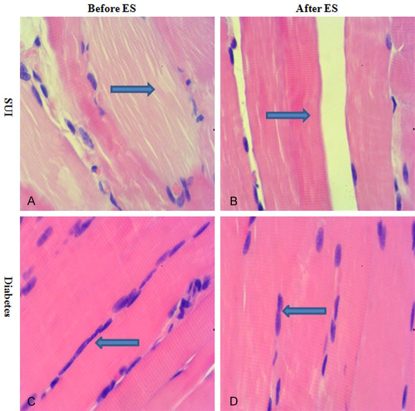
ES increased pelvic floor muscle strengh and NPY expression

Table 1. Results of the sneezing experiment         emptied again and filled with a volume of me-
in the SUI animal model                             thylene blue solution equaling half of the ma-
                         Sneezing Experiment        ximum bladder capacity. The sneezing reflex
Groups      Number                                  was induced by inserting a piece of severed
                       Positive (+) Negative (-)
                                                    rat’s beard into the nostril to increase abdo-
Control        5            0            5
                                                    men pressure. If any amount of methylene bl-
SUI           10           10            0
                                                    ue outflow was observed from the external
                                                    meatus, the sneezing experiment was consid-
to test whether it can increase pelvic muscle       ered positive. For evaluating the diabetes ani-
strength and resolve the dysfunction. To assess     mal model, the body weight and blood glucose
its effects, a neurotransmitter of neuropeptide     were examined after 1 month on the high-fat,
Y (NPY) amount was detected by enzyme-linked        high-energy diet and injections. Only rats with
immunosorbent assay (ELISA) and Western             typical symptoms of diabetes were considered
blotting and the histological characteristics of    to be induced with type 2 diabetes. Glucose
pelvic muscle tissue were analyzed.                 standards of type 2 diabetes in the present
                                                    study were set as fasting blood-glucose >11.1
Materials and methods                               mmol/L or postprandial blood sugar >16.7
                                                    mmol/L.
Establishment of SUI and diabetes in animal
models                                              Electrical stimulation as the normal treatment

Eight-week-old healthy female Wistar rats were      All rats in the control, SUI, and diabetes grou-
provided by the experimental animal center of       ps were given ES for the pubococcygeal mu-
Guangdong province, and randomized into             scles. ES was performed with the MyoBravo
three groups: the control group (n=5), the SUI      electro stimulation instrument (MTR+Vertiebs
group (n=10), and the diabetes group (n=10).        GmbH, Berlin). A vaginal probe was inserted,
Rats in the control group were non-pregnant         and a medium-frequency (25 Hz) alternating
and without any treatment, rats in the SUI          current was administered for stimulation with
group underwent normal parturition plus po-         a duty cycle of 1, 3, 6, 12, and 30 times. The
st-partum vaginal balloon dilation for 6 hours,     interval was 60 seconds, and total cycle time
and rats in the type 2 diabetes group were          was 343 second. Each rat received a 30 min-
developed with high-fat high-energy diets plus      ute session every 2 days, and the strength of
Streptozocin (STZ, Sigma, USA) injections.          pelvic floor muscles was assessed at baseline
Vaginal balloon dilation and STZ injections were    and after the completion of the experimental
performed as previously described [7]. Ethics       sessions (7 days).
were approved by the Institutional Animal Care
and Ethics Committee of Guangzhou Medical           Strength measurement of the pubococcygeal
University.                                         muscles

Evaluation of SUI and diabetes in animal mod-       The pubococcygeal muscles of all rats were dis-
els                                                 sected on day 7 and attached to a tension
                                                    transducer by using the non-plastic thread (10
The SUI and diabetes animal models were eval-       cm). Muscle strength was measured by using
uated by sneezing experiment and glucose            a biomechanical test system (BL-420, Cheng-
test, respectively. For the sneezing experiment,    du, China). Briefly, initial force was set as 1
rats of the SUI group were intraperitoneally        gram and stabilized for 3 seconds, and then ES
anesthetized with 2% pentobarbital sodium           (25 Hz, 50 ms, 3.6 V) of the pubococcygeal
(0.2 mL/100 g), and the bladder was emptied         muscles was performed for 3 seconds. Chang-
by an epidural catheter. The maximum bladder        es of muscle strength were recorded as resul-
capacity was measured by filling the bladder         ts of stationary contractility. After 3 min of rest,
with methylene blue (Jili, Jiangsu, China), dis-    the thread was extended to 10.5 cm, and ES of
solved in sterile saline, until the first drop of   pubococcygeal muscles was performed imme-
urine leakage outside the external orifice of        diately. Changes of muscle strength were re-
urethra was observed. Then, the bladder was         corded as results of forced contractility.

2428                                                     Int J Clin Exp Med 2019;12(3):2427-2434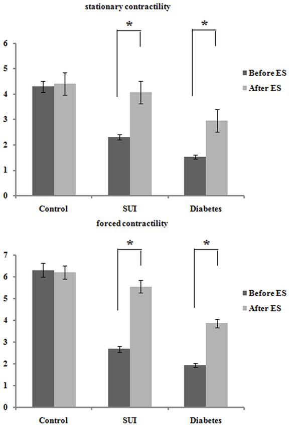
ES increased pelvic floor muscle strengh and NPY expression

Table 2. Results of the glucose test in the diabetes animal model            (3000 rpm) before and aft-
                                              After (mmol/L)                 er ES treatment. Supernatant
Groups     Number Before (mmol/L)                                            was obtained and used for
                                      1 month 2 months 3 months
                                                                             NPY measurement. Ninety-six
Control       5        3.58±0.54     3.60±0.67 3.72±0.48 3.84±0.57
                                                                             well plates were used, and
Diabetes      6        3.47±0.59     21.3±1.82 16.8±1.26 23.3±0.79
                                                                             NPY production was detected
                                                                             by using an enzyme-linked
                                                                             immunosorbent assay (ELISA)
                                                                             kits (CSB-E13431r, Wuhan,
                                                                             China) following the manufac-
                                                                             turer’s protocol.

                                                                             Western blot analysis

                                                                               The pubococcygeal muscle ti-
                                                                               ssue was collected from rats
                                                                               in the diabetes group before
                                                                               and after ES treatment, hom-
                                                                               ogenized, and lysed in rad-
                                                                               io immune-precipitation ass-
                                                                               ay (RIPA, Bio-teke Corporati-
                                                                               on, China) buffer with 1 mM
                                                                               phenylmethanesulfonyl fluori-
                                                                               de (PMSF, Bio-teke Corpora-
                                                                               tion, China). The protein con-
                                                                               centration was measured us-
                                                                               ing the Bio-Rad protein ass-
                                                                               ay kit (Bio-Rad, Hercules, CA).
                                                                               Forty micrograms of each pr-
                                                                               otein was separated on 10%
                                                                               sodium dodecyl sulfate pol-
                                                                               yacrylamide gels (SDS-PAGE)
                                                                               and then transferred to cellu-
                                                                               lose acetate membranes (Mi-
                                                                               llipore, MA, USA). The mem-
                                                                               branes were blocked with 5%
                                                                               non-fat dry milk in Tween/Tr-
                                                                               is-buffered solution (TTBS) for
                                                                               2 hours at room temperature,
                                                                               followed by incubation with ra-
                                                                               bbit anti-NPY (1:500, Abcam,
                                                                               USA) and rabbit anti-glycer-
                                                                               aldehyde 3-phosphate dehy-
                                                                               drogenase (GAPDH, 1:5000,
Figure 1. ES enhanced the strength of pelvic floor muscles in rats of the SUI  Abcam, USA) antibodies ov-
and diabetes groups. Both stationary contractility and forced contractility of
pelvic floor muscles in rats of the SUI and diabetes groups were significantly
                                                                               ernight at 4°C. The membra-
enhanced after ES treatment (*, p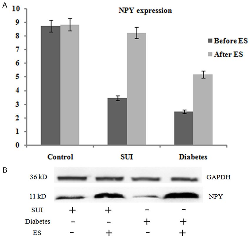
ES increased pelvic floor muscle strengh and NPY expression

                                                                               Statistical analysis

                                                                               Data analysis was perform-
                                                                               ed using SAS program (SAS
                                                                               version 8.1, USA). Student’s t
                                                                               test was used to compare the
                                                                               two means. One-way analysis
                                                                               of variances (ANOVA) was us-
                                                                               ed to compare more than two
                                                                               means. A p value of less than
                                                                               0.05 was regarded as signi-
                                                                               ficant.

                                                                               Results

                                                                               Establishment of SUI and dia-
                                                                               betes in animal models

                                                                                 According to the results of the
                                                                                 sneezing experiment, all 10
                                                                                 rats in the SUI group were
                                                                                 verified as having SUI (Table
                                                                                 1). As for the diabetes gro-
Figure 2. ES increased the amount of NPY expression in rats of SUI and dia-      up, 6 of 10 rats were recog-
betes groups. The NPY concentration in the supernatant of peripheral blood       nized as having type 2 diab-
from rats in the SUI or diabetes groups was significantly increased after ES     etes based on the 3 months
(A, *, p
ES increased pelvic floor muscle strengh and NPY expression Figure 3. Increased NPY was correlated with enhanced strength of pelvic floor muscles before and after ES treat- ment (Pearson r=0.98, p
ES increased pelvic floor muscle strengh and NPY expression
trunk, such as the abdomin-
als, quadrates lumborum, sp-
inal muscles of multifidus, hip
muscles, the diaphragm, and
pubococcygeal muscles [9]. In
essence, the pelvic floor mu-
scles are the floor of the core.
In the present study, contrac-
tility of pelvic floor muscles
was found in rats of the SUI
group and the diabetes gro-
up was significantly enhanc-
ed after ES treatment, which
indicates its prominent effe-
cts. The physiological objecti-
ves of ES are to produce mu-
scle hypertrophy and increa-
se circulation to muscles. It
may increase conscious aw-
areness of muscles to prod-
uce improved ability to perf-
orm voluntary muscle contr-
action [10].
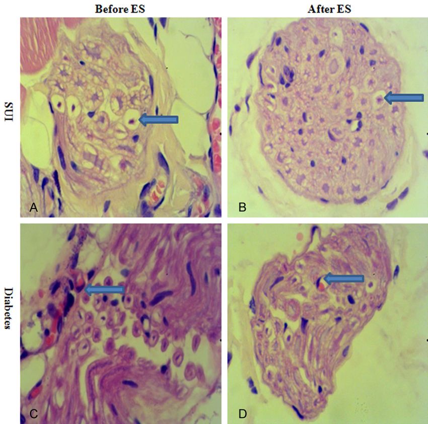
Interestingly, the enhanced co-
Figure 4. Histological characteristics of pelvic floor muscles before and after
ES treatment. For SUI rats before ES, muscle fibers were swollen and the ntractility of pelvic floor mus-
cytoplasm was lightly stained (arrow, A). The striations were not prominent. cles follows with the increas-
After ES, no pathological changes were observed in the target fibers. Muscle ed expression of NPY, which
bundles had integrated structures with a pink appearance (arrow, B). For suggests its potential value
diabetes rats, muscle fibers showed chain changes, similar to myogenic to assess the effects of ES
neurogenic changes (arrow, C). After ES, the nucleus numbers under sarco-
lemma had significantly increased in partial muscle fibers (arrow, D). treatment. NPY, a 36-amino-
acid neuropeptide, is involved
in the regulation of blood flow
cells (Figure 5). After ES, the nucleus numbers and is commonly found among the nerve fibers
under sarcolemma had significantly increased [11, 12]. NPY is always synthesized in the neu-
in partial muscle fibers. Slightly swelling endo- ron and then transported to the vaginal wall
thelial cells were found to be embedded in the through the nerve axon; therefore, NPY is one
endo-neural membrane, and the disarrange- of the most abundant neuropeptides observ-
ment of neuron fibers was observed less ed underlying the vaginal epithelium [13]. For
frequently. example, the staining of NPY protein was found
in the anterior vaginal wall tissue of healthy
Discussion women by Hu et al [14]. In menopausal pati-
ents with POP, as the symptoms intensify, NPY
There is a growing need to understand how expression decreases progressively. Results of
physiotherapies impact SUI symptoms, impair- ELISA and Western blotting in the present study
ments, and functional limitations. The target of showed not only the NPY concentration but
physiotherapies is the pelvic floor muscles. The also its protein amount from rats of SUI and
pelvic floor consists of striated muscles arran- diabetes groups significantly increased after
ged in a dome-shaped sheet, which is usually ES, and expression of NPY was correlated with
described as a sling [8]. These muscles are muscle contractility. Based on these results,
referred to as lying within either the deep or the increased amount of NPY in the pelvic floor
superficial pelvic floor. Although it is infrequent- muscles is proposed to be related to nerve
ly discussed, the pelvic floor has an important recovery or regeneration, resulting in a change
role in the function of core muscle stabilization. in blood flow, atrophy, and pelvic floor laxity
The core muscles are known as muscles of the [15].
2432 Int J Clin Exp Med 2019;12(3):2427-2434ES increased pelvic floor muscle strengh and NPY expression
the pubococcygeal muscle
morphology in both SUI and
diabetes rat models have sh-
own similar myogenic chan-
ges. Neurogenic myopatholo-
gy may provide the answer to
the etiology of pelvic floor dy-
sfunction, and clearly explain
the pathological mechanis-
ms of ES treatment on SUI
symptoms.
Conclusion
In summary, the present stu-
dy found that, the increased
concentration of NPY in SUI
and diabetes rats after ES
treatment was related to the
recovery of the pubococcygeal
muscle strength, which may
be related to NPY in promoting
the generation of the pelvic
floor muscles and blood ves-
Figure 5. Neural changes of pelvic floor muscles before and after ES treat-
sels. ES treatment has shown
ment. Swollen neuronal cells were observed in SUI rats before ES. Con- considerable therapeutic eff-
densed nuclei with marked hyperchromatism and dissolution of Nissl bodies ects, and would be an ideal
(arrow, A) were also observed. After ES, few neuron cells showed swelling physiotherapy to solve pati-
and partial dissolution of Nissl bodies (arrow, B). For diabetes rats before ents’ SUI symptoms.
ES, neural changes included the congestion of blood vessels and swelling of
endothelial cells (arrow, C). After ES, slightly swollen endothelial cells were
Acknowledgements
found to be embedded in the endoneural membrane, and disarrangement
of neuron fibers was observed less frequently (arrow, D).
This study was supported by
funds from National Natural
To further explore the histological characteris- Science Foundation of China (Grant No.
tics of pubococcygeal muscles, H&E staining 8167060599).
and histological analysis were performed. The
neurons and muscle fibers showed ischemic Disclosure of conflict of interest
changes in the SUI group before ES. Muscle
fibers were swollen, and the cytoplasm was None.
lightly stained. Neural changes included swe-
lling neuronal cells, condensed nuclei with Address correspondence to: Dunjin Chen, The Third
marked hyperchromatism, and the dissolution Affiliated Hospital, Guangzhou Medical University,
of Nissl bodies. The diabetes group showed Guangzhou 510150, Guangdong, China. Tel: 86-20-
typical pathological changes, muscle fibers dis- 81292183; Fax: 86-20-81292407; E-mail: djch-
played chain changes, the congestion of blood engz@126.com; gzdrchen@gzhmu.edu.cn
vessels, and swelling of endothelial cells. After
ES, muscle bundles had integrated structures References
with a pink appearance in the SUI group, and
[1] Ahmad AN, Hainsworth A, Williams AB, Schizas
few neurons showed swelling and partial disso-
AM. A review of functional pelvic floor imaging
lution of Nissl bodies. In the diabetes group, modalities and their effectiveness. Clin Imag-
the nucleus numbers under sarcolemma had ing 2015; 39: 559-65.
significantly increased in partial muscle fibers. [2] Van Geluwe B, Wolthuis A, D’Hoore A. Laparos-
Slightly swelling endothelial cells were embe- copy for pelvic floor disorders. Best Pract Res
dded in the endoneural membrane. Results of Clin Gastroenterol 2014; 28: 69-80.
2433 Int J Clin Exp Med 2019;12(3):2427-2434ES increased pelvic floor muscle strengh and NPY expression
[3] Garely AD, Noor N. Diagnosis and surgical [10] Correia GN, Pereira VS, Hirakawa HS, Driusso
treatment of stress urinary incontinence. Ob- P. Effects of surface and intravaginal electrical
stet Gynecol 2014; 124: 1011-27. stimulation in the treatment of women with
[4] Anderson KM, Davis K, Flynn BJ. Urinary incon- stress urinary incontinence: randomized con-
tinence and pelvic organ prolapse. Med Clin trolled trial. Eur J Obstet Gynecol Reprod Biol
North Am 2015; 99: 405-16. 2014; 173: 113-8.
[5] Qaseem A, Dallas P, Forciea MA, Starkey M, [11] Glipin SA, Gosling JA, Sminth AR, Warrell DW.
Denberg TD, Shekelle P; Clinical Guidelines The pathogenesis of genitourinary prolapsed
Committee of the American College of Phy- and stress incontinence of urine. A histological
sicians. Nonsurgical management of urinary and hisochemical study. Br J Obstet Gynaecol
incontinence in women: a clinical practice 1989; 96: 15-23.
guideline from the American College of Physi- [12] Geloso MC, Corvino V, Di Maria V, Marchese E,
cians. Ann Intern Med Sep 2014; 161: 429-40. Michetti F. Cellular targets for neuropeptide Y-
[6] Ghaderi F, Oskouei AE. Physiotherapy for wom- mediated control of adult neurogenesis. Front
en with stress urinary incontinence: a review Cell Neurosci 2015; 9: 85.
article. J Phys Ther Sci 2014; 26: 1493-9. [13] Zhu L, Lang J, Jiang X, Jiang F, Chen J, Wong F.
[7] Wang H, Liu J, Zeng J, Zeng C, Zhou Y. Expres- Neuropeptide Y expression in vaginal epitheli-
sion of TβR-2, Smad3 and Smad7 in the vagi- um of women with pelvic organ prolapse and
nal anterior wall of postpartum rats with stress stress urinary incontinence. Int J Gynaecol Ob-
urinary incontinence. Arch Gynecol Obstet stet 2008; 102: 65-8.
2015; 291: 869-76. [14] Hu JM, Wang L, Cheng X, Zhou LH, Li ZG. Neu-
[8] Pregazzi R, Sartore A, Troiano L, Grimaldi E, ropeptide Y innervation in the vaginal mucosa
Bortoli P, Siracusano S, Guaschino S. Postp- among patients with pelvic organ prolapse.
artum urinary symptoms: prevalence and risk Mol Med Rep 2012; 5: 444-8.
factors. Eur J Obstet Gynecol Reprod Biol [15] Guralnick ML, Kelly H, Engelke H, Koduri S,
2002; 103: 179-82. O’Connor RC. InTone: a novel pelvic floor reha-
[9] Wei JT, De Lancey JO. Functional anatomy of bilitation device for urinary incontinence. Int
the pelvic floor and lower urinary tract. Clin Ob- Urogynecol J 2015; 26: 99-106.
stet Gynecol 2004; 47: 3-17.
2434 Int J Clin Exp Med 2019;12(3):2427-2434You can also read