Moxibustion Protects Dopaminergic Neurons in Parkinson's Disease through Antiferroptosis - Hindawi.com
←
→
Page content transcription
If your browser does not render page correctly, please read the page content below
Hindawi
Evidence-Based Complementary and Alternative Medicine
Volume 2021, Article ID 6668249, 11 pages
https://doi.org/10.1155/2021/6668249

Research Article
Moxibustion Protects Dopaminergic Neurons in Parkinson’s
Disease through Antiferroptosis

          Zifeng Huang,1,2 Wenwen Si,1 Xinrong Li,1,2 Shanyu Ye,3 Xuelei Liu,1 Yichun Ji,4
          Xiaoqian Hao,1,2 Dongfeng Chen ,3 and Meiling Zhu 1
          1
            Traditional Chinese Medicine Innovation Research Center, Shenzhen Hospital of Integrated Traditional
            Chinese and Western Medicine, Shenzhen, Guangdong 518104, China
          2
            Graduate School, Guangzhou University of Chinese Medicine, Guangzhou, Guangdong 510405, China
          3
            Department of Anatomy, The Research Center of Basic Integrative Medicine, Guangzhou University of Chinese Medicine,
            Guangzhou, China
          4
            Shenzhen Bao An Traditional Chinese Medicine Hospital, Guangzhou University of Chinese Medicine, Shenzhen, China

          Correspondence should be addressed to Dongfeng Chen; cdf27212@21cn.com and Meiling Zhu; meilingzhu2020@126.com

          Received 4 November 2020; Revised 20 March 2021; Accepted 29 March 2021; Published 16 April 2021

          Academic Editor: Mohammad Fattahi

          Copyright © 2021 Zifeng Huang et al. This is an open access article distributed under the Creative Commons Attribution License,
          which permits unrestricted use, distribution, and reproduction in any medium, provided the original work is properly cited.

          Ferroptosis is associated with neural degeneration of dopaminergic neurons in Parkinson’s disease (PD). However, how to control
          the level of ferroptosis in PD remains unclear. Clinically, moxibustion has been used to treat PD and has an apparent therapeutic
          effect on improving the motor symptoms of PD. In the present study, the PD rat model was constructed by two-point stereotactic
          6-hydroxydopamine injection. Then, moxibustion was used to treat the PD rats. The expression of glutathione peroxidase 4
          (GPX4) and Ferritin Heavy Chain 1 (FTH1), the level of reactive oxygen species (ROS), and the morphology of mitochondrial
          were detected to evaluate the level of ferroptosis. The results showed that moxibustion treatment of Shi’s moxa sticks could reduce
          the behavioral score, alleviate the level of ferroptosis, decrease mitochondrial damage, and improve dopaminergic neuron
          survival. In conclusion, the present study results indicated that Shi’s moxa sticks could effectively suppress the level of ferroptosis,
          thereby improving the survival of dopaminergic neurons in the SNpc of PD rats, which may provide a promising complementary
          and alternative therapy for PD patients.

1. Introduction                                                           symptoms [6]. However, motor complications caused by
                                                                          levodopa’s long-term use are a significant drawback of
Parkinson’s disease (PD) is a degenerative disease of the                 levodopa in PD treatment [7]. Thus, there is an urgent need
aging people’s nervous system. It has become the second                   to search for effective treatment of PD.
primary neurodegenerative disease worldwide [1]. It is es-                    Moxibustion is a form of external therapy in traditional
timated that the prevalence rate of PD is about 2% in the                 Chinese medicine [8]. Recent research suggested that
population over 50 years of age and 2.5% in the population                moxibustion treatment can postpone PD progression and
over 70 years of age world [2]. PD is primarily characterized             has an apparent therapeutic effect on motor symptom im-
by the degeneration and death of the dopaminergic neurons                 provement in PD patients [9, 10]. Moreover, numerous
in the SNpc [3]. PD’s motor symptoms, such as resting                     animal experiments have confirmed that moxibustion can
tremor, bradykinesia, and rigidity, can severely affect the                alleviate PD injury. The mechanism may be related to the
patient’s daytime function and quality of life [4]. Further-              protection of dopaminergic neurons in SNpc [11, 12]. In the
more, patients’ gradual deterioration leads to higher dis-                present study, we investigated the protective effects of Shi’s
ability rates, which will pose a sizeable economic burden to              moxa stick on PD rat’s dopaminergic neuron injury. Shi’s
the family and society [5]. At present, the primary treatment             moxa stick was developed by Shi Xuemin, an academician of
for PD is levodopa, which can significantly reduce PD                      the Chinese Academy of Engineering and a master of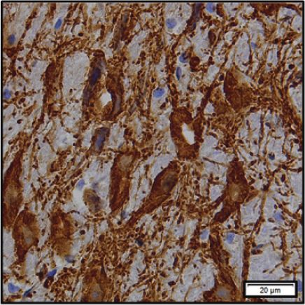
2                                                                  Evidence-Based Complementary and Alternative Medicine

traditional Chinese medicine, and has been used in our            the rats (n � 18/group) were anesthetized and sacrificed, with
hospital to relieve PD symptoms.                                  the fresh right side of the brains being quickly removed to
    There is increasing evidence that ferroptosis is associated   −80°C (n � 6/group). At the same time, the other was fixed
with the occurrence of PD [13]. Ferroptosis is a novel form of    using 4% paraformaldehyde solution (n � 6/group) and 2.5%
nonapoptotic programmed cell death closely related to iron        glutaraldehyde (n � 6/group).
metabolism disturbance. It is closely associated with various
neurodegenerative diseases, including PD [14–16]. The oc-
                                                                  2.3. PD Rat Model Preparation. The PD rat model was
currence of ferroptosis is related to a decrease of glutathione
                                                                  established using a two-point 6-hydroxydopamine injection
peroxidase 4 (GPX4) and Ferritin Heavy Chain 1 (FTH1)
                                                                  in the right SN and ventral tegmental area [24]. The rats used
[17], an increase of reactive oxygen species (ROS) [18], and
                                                                  in the experiments were under anesthesia (100 mg/kg ket-
the damage of mitochondrial [18–20]. GPX4 is a type of
                                                                  amine and 10 mg/kg xylazine, intraperitoneal injection). In
antioxidase that can reduce intracellular lipid peroxide levels
                                                                  the PD model rats, the rat’s head was shaved to expose the
and avoid oxidative damage. The decrease of GPX4 activity
                                                                  scalp and fixed on the stereotactic frame. 4 μl 6-OHDA (cat.
will lead to ROS accumulation, which will eventually induce
                                                                  no. H116-5 mg; Sigma-Aldrich; use 0.02% ascorbate solution
ferroptosis [21]. FTH1 is a major iron storage protein and
                                                                  to dissolve to 8 μg/μl) was intracerebroventricularly injected
maintains intracellular iron balance [22]. The excess iron
                                                                  into two sites (AP: −4.9 mm, L: −1.9 mm, and DV: −7.5 mm
may lead to ferroptosis in the cells [23]. Our previous studies
                                                                  and AP: −4.9 mm, L: −1.1 mm, DV: 8.0 mm relative to the
have shown that the pathology mechanisms of PD were
                                                                  bregma and dural surface) by 5 μl microsyringe. After in-
related to the level of ferroptosis, which can be regulated by
                                                                  jection, we left the needle in the brain for 10 minutes and
FTH1 [17, 24]. Observation of mitochondrial morphology
                                                                  then withdrew the needle at a rate of 1 mm/min. Finally, we
and size by transmission electron microscope is an essential
                                                                  put a piece of gelatin sponge into the burr holes to prevent
indicator of ferroptosis. The morphological changes of
                                                                  bleeding and cerebrospinal fluid leakage and suture the skin
ferroptosis include reducing mitochondrial volume, an in-
                                                                  wound. The rat’s body temperature was kept at 36.5°C with a
crease of double-layer membrane density, and reduction or
                                                                  heating pad during the procedure until the rat recovers from
disappearance of mitochondrial cristae [18, 25]. These in-
                                                                  anesthesia. The same procedures were performed in the
dicators will be used to detect the level of ferroptosis and
                                                                  sham group, but 4 μl of saline was administered that con-
explore how to suppress ferroptosis in PD.
                                                                  tained 0.02% ascorbic acid instead of 6-OHDA.
    In this study, we investigated the specific mechanism of
moxibustion in improving symptoms of PD. Our results
confirmed that moxibustion could effectively suppress the           2.4. Moxibustion Treatment of Shi’s Moxa Stick. After four
level of ferroptosis, thereby improving the survival of do-       weeks of 6-OHDA lesioning, rats in the moxibustion group
paminergic neurons in the SNpc of PD rats, which may              were treated with Shi’s moxa sticks after model establish-
provide a promising complementary and alternative therapy         ment. The current study chose Baihui (GV20) acupoint. For
for PD patients.                                                  the moxa stick to reach the acupoint, the area of Baihui on
                                                                  the rat’s head was shaved to expose the skin at the beginning
2. Materials and Methods                                          of treatment. The moxibustion treatment of Shi’s moxa
                                                                  sticks lasts for 30 minutes per day, six times a week, for four
2.1. Animals. A total of 108 male Sprague-Dawley (SD;             weeks. The rats in the control group, the sham group, and the
180–200 g; 8-9 weeks old) rats were purchased from                model group received normal feeding and did not undergo
Guangzhou Dean Gene Technology Co., Ltd., and were                any treatment for four weeks.
placed under standard laboratory conditions (21–25°C,
50–70% relative humidity, 12-hour light-dark cycle, and
                                                                  2.5. Behavioral Test. Before collecting tissue, all rats were
food and water ad libitum). All rats were allowed to accli-
                                                                  injected with intraperitoneal apomorphine (APO, cat. no.
mate for one week before PD surgery. All animal procedures
                                                                  017–18321; Wako; 0.5 mg/kg) for behavioral test. Ten
followed the rules of the National Institute of Health Guide
                                                                  minutes after APO injection, the rat started to spin and the
for the Care and Use of Laboratory Animals.
                                                                  number of complete turns (360°) to the left side for 30 min
                                                                  was recorded.
2.2. Experiment Protocol

2.2.1. Experiment 1. To observe the degeneration of dopa-         2.6. Immunohistochemical Staining. To observe the ex-
minergic neurons and the occurrence of ferroptosis in the         pression of TH in the substantia nigra, six rats in each group
PD rat model, the rats were randomly divided into three           were in under anesthesia. Firstly, we used normal cold saline
groups: control group, sham group, and 6-OHDA group.              for blood washing through the cardiac aorta injection and
                                                                  then fixed the brain tissue with 4% paraformaldehyde so-
                                                                  lution. After fixation, the brain tissue was gently removed,
2.2.2. Experiment 2. To validate the efficacy of moxibustion        placed in a 4% paraformaldehyde solution, and sectioned
treatments in PD, the rats were randomly divided into four        into 10 μm thick coronal slices. Frozen sections (10 μm
groups: control group, sham group, 6-OHDA group, and              thickness) of the brain were permeabilized in 0.3% Triton
moxibustion group. When the behavioral test was complete,         X-100 solution for 30 min. Then, we performed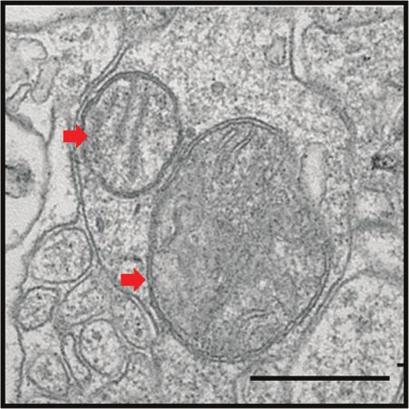
Evidence-Based Complementary and Alternative Medicine                                                                            3

immunohistochemical staining with Histostain-Plus Kits            secondary antibodies (horseradish peroxidase- (HRP-)
(cat. no. SP-0022; Bioss) according to the manufacturer’s         conjugated goat anti-rabbit, 1 : 5000, cat. no ab6721, Abcam;
protocol. Primary antibody against TH (1 : 1000; cat. no.         HRP-conjugated goat anti-mouse, 1 : 5000, cat. no. ab6789,
ab152; Millipore) was incubated at 4°C overnight. After           Abcam) for 1 hour at room temperature. The ECL substrate
washing with PBS, diaminobenzidine (DAB) working so-              (Pierce, Thermo Fisher Scientific, Inc. USA) was used to
lution (cat. no. C02-04001; Bioss) was added for color            visualize the bands. The data were analyzed by image j.
rendering. Each section was restained by hematoxylin (cat.
no. DH0005; Mayer), dehydrated, and fixed with ethanol
                                                                  2.11. Extraction of Total RNA and RT-qPCR. Total RNA from
according to the concentration gradient. The histopatho-
                                                                  the SNpc was extracted and purified using the Direct-zol
logical observations were documented by light microscopy
                                                                  RNA Kit (cat. no. R2070; ZYMO research) according to the
at 400x magnification, and images were captured. We re-
                                                                  manufacturer’s protocol, and the RNA was stored at −80°C.
peated each experiment three times, and data were expressed
                                                                  Reverse transcription and qPCR assays were performed
as the mean ± SEM.
                                                                  using the PrimeScript RT reagent kit (Cat.no. RR600 A;
                                                                  Takara) and the TB Green Premix Ex Taq II (Cat. no.
2.7. Hematoxylin/Eosin (HE) Staining. To observe neurons’         RR820 A; Takara) on Light Cycler 480 SYBR Green I Master
morphology in the substantia nigra, frozen sections (10 μm        (Roche Diagnostics, GmbH, Mannheim, Germany). Each
thickness) were used for HE staining. HE staining was             experiment was replicated three times, and data are pre-
performed according to the standard protocol, and histo-          sented as the mean ± SEM. Primers are shown as follows:
pathological observation results were recorded by light
                                                                       TH forward: 5′-ATTGCCTTCCAGTACAAGCAC-3′;
microscopy at 400x magnification, and images were
captured.                                                              TH reverse: 5′-CCTTCAGCGTGACATATACCTCC-
                                                                       3′;
                                                                       GPX4 forward: 5′-ATAAGAACGGCTGCGTGGTGA
2.8. ROS Detection of Articular Cartilage. To observe the level
                                                                       AG-3′;
of ROS, fresh SN tissue from each group was made into
homogenate with buffer. Then, we performed ROS detection                GPX4 reverse: 5′-TAGAGATAGCACGGCAGGTCCT
with BBoxiProbe O13 (cat. no. BB-470512; BestBio)                      TC-3′;
according to the manufacturer’s protocol. We repeated each             FTH1 forward: 5′-TTCAGGGCCACATCATCCCG-3’;
experiment three times, and data were expressed as the                 FTH1 reverse: 5′-GCAAGTGCGCCAGAACTACC-3′;
mean ± SEM.
                                                                       β-acting forward: 5′-CTCAGGAGAGGAGCCATTTA
                                                                       TT-3′;
2.9. Transmission Electron Microscope. A transmission                  β-acting reverse: 5′-CCCGATCAGAGTGAAGCTAT
electron microscope was used to observe the morphology of              T-3′.
mitochondria. Six rats in each group were anesthetized.
Through the injection of the cardiac aorta, we used normal
cold saline for blood washing at first and then fixed the brain     2.12. Statistical Analyses. The experimental data are presented
tissue with 2.5% glutaraldehyde solution (cat. no. DF0156;        as the mean ± SEM. SPSS 22.0 software (IBM Corp., Armonk,
Leagene) at 4°C. We cut out the SN tissue, fixed it with 1%        NY, USA) was used for statistical analysis. The figures were all
osmium acid, and washed it 3 times with 0.1 M phosphate           produced using GraphPad Prism (Version 6.0; GraphPad
buffer. The tissue was then fixed with 1% osmium tetroxide          Software, Inc., La Jolla, CA, USA). A two-tailed unpaired t-test
and dehydrated through an alcohol gradient before being           was used to analyze the difference between the two groups. The
embedded in resin. The sample was detected by transmission        comparison between multiple groups was analyzed using a
electron microscopy.                                              one-way analysis of variance followed by Tukey’s multiple
                                                                  comparisons test as the post hoc test. P < 0.05 was considered
                                                                  to indicate a statistically significant difference.
2.10. Western Blot Analysis. The protein was extracted from
the SNpc tissue by RIPA lysis buffer (cat. no. R0278; Sigma-       3. Result
Aldrich). After centrifugation at 12,000 rpm for 20 min at
4°C, total proteins were harvested. The bicinchoninic acid        3.1. The PD Rats Model Exhibits Degeneration of Dopami-
assay (BCA; cat. no. 23225; Thermo Fisher Scientific) was          nergic Neurons. Behavioral test was applied to explore
used to determine the concentration of total proteins. The        whether the PD rats model was successfully constructed after
total protein (30 ug) was separated with 10% SDS-PAGE gels        four weeks of two-point stereotactic 6-hydroxydopamine in-
and transferred to polyvinylidene difluoride membranes             jection [26] (Figure 1(a)). The behavioral test revealed that the
(PVDF; Millipore). After blocking with 5% BSA for 2 h,            behavioral score increased significantly after stereotactic in-
the membrane was incubated with primary antibodies (TH,           jection, and no animal in the normal group or sham group
1 : 1000, cat. no. ab152, Millipore; GPX4, 1 : 1000, cat. no.     exhibited rotating behavior (Figure 1(b); sham versus 6-
ab125066, Abcam; FTH1, 1 : 1000, cat. no. ab183781, Abcam;        OHDA; P < 0.001). The expression of TH is a marker of
GAPDH, 1 : 5000, cat. no. ab8245, Abcam) at 4°C overnight.        dopaminergic neurons [27] and reflects the activity of dopa-
The membrane was then incubated with HRP-labelled                 minergic       neurons.      The     Western        blot     and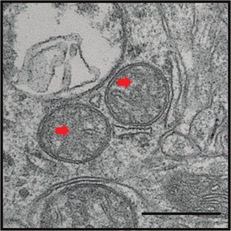
4                                                                                                                   Evidence-Based Complementary and Alternative Medicine

                                                                                   6-OHDA lesioning
                                                                                      for 4 weeks
                                                                                                                                                 End

                                       D0                             D7                                                           D35
                                                                                                                               Behavioral test
                                                                                                (a)
                                                              15
                                                                                                                         ∗∗∗

                                        Number of rotations   10

                                                              5

                                                              0
                                                                            Control                               Sham         6-OHDA
                                                                                               (b)
                      Control      Sham                            6-OHDA                                         1.5

                TH                                                            62 kDa
                                                                                                                                                                                    ∗∗∗
                                                                                          Expression of protein

           GAPDH                                                              36 kDa                              1.0

                                                                                                                  0.5

                                                                                                                  0.0
                                                                                                                                Control            Sham                                   6-OHDA
                                                                                                (c)
                                                                                                                                                                                    1.5

                                                                                                                                                                                                            ∗∗∗
                                                                                                                                                          Average optical density

                                                                                                                                                                                    1.0

                                                                                                                                                                                    0.5

                                                                                                                                                                                    0.0
                                                                                                                                                                                           Control   Sham     6-OHDA
                      Control                                               Sham                                               6-OHDA
                                                                                               (d)

Figure 1: The behavioral score of PD rats increased, and the protein expression level of TH decreased. (a) The timeline of animal experiment
1. (b) The behavioral score of the control group, the sham group, and the 6-OHDA group. (c) TH level of total protein in SNpc of the control
group, the sham group, and the 6-OHDA group. (d) IHC showed the expression of TH in the substantia nigra of the control group, the sham
group, and the 6-OHDA group.

immunohistochemistry showed that the expression of TH was                                                          3.2. The PD Rats Model Exhibits an Increased Level of
significantly decreased in the 6-OHDA group compared with                                                           Ferroptosis. To evaluate the level of ferroptosis in the PD rats
the sham group (Figure 1(c); sham versus 6-OHDA; P < 0.001;                                                        model, we detected the expression of GPX4, FTH1, and ROS.
Figure 1(d); sham versus 6-OHDA; P < 0.001). These results                                                         The Western blot showed that the expression of GPX4 and
showed that the PD rat model was successfully constructed,                                                         FTH1 was significantly decreasing in the 6-OHDA group
and the survival of dopaminergic neurons was decreased.                                                            compared with the sham group (Figure 2(a); sham versus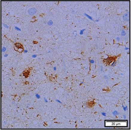
Evidence-Based Complementary and Alternative Medicine                                                                                                                            5

                                     Control         Sham   6-OHDA                                      1.5

                              GPX4                                   21 kDa
                                                                                                                                          ∗∗∗                          ∗∗∗

                                                                              Expression of protein
                              FTH1                                   17 kDa                             1.0

                            GAPDH                                    36 kDa

                                                                                                        0.5

                                                                                                        0.0
                                                                                                                                      GPX4                       FTH1
                                                                                                                                Control
                                                                                                                                Sham
                                                                                                                                6-OHDA
                                                                                      (a)
                                                                                                                                                                 ∗∗∗
                            1.5                                                                                              10000
                                               ∗∗∗                   ∗∗∗
                                                                                                                              8000
       Expression of mRNA

                                                                                                      Tissue ROS intensity
                            1.0
                                                                                                                              6000

                                                                                                                              4000
                            0.5

                                                                                                                              2000

                            0.0                                                                                                  0
                                        GPX4                     FTH1                                                                     Control         Sham         6-OHDA
                                  Control
                                  Sham
                                  6-OHDA
                                                     (b)                                                                                            (c)

Figure 2: The level of ferroptosis in PD rats is increased. (a) GPX4 and FTH1 level of total protein in the control group, the sham group, and
the 6-OHDA group. (b) GPX4 and FTH1 levels of total mRNA in the control group, the sham group, and the 6-OHDA group. (c) The level of
ROS in the control group, the sham group, and the 6-OHDA group.

6-OHDA; P < 0.001). A similar trend was observed for the                                                       (Figure 3(a)). To evaluate whether the moxibustion treatment
GPX4 and FTH1 expression levels of mRNA (Figure 2(b);                                                          of Shi’s moxa stick could improve the survival of dopaminergic
sham versus 6-OHDA; P < 0.001). The occurrence of fer-                                                         neurons, we detected the behavioral test, the expression of TH,
roptosis is associated with the accumulate of ROS. The result                                                  and the morphology of neurons. As a show in Figure 3(b), we
showed that the level of ROS was increased obviously in the                                                    detected that the score of behavioral test was still increased after
6-OHDA group (Figure 2(c); sham versus 6-OHDA;                                                                 D35 in the 6-OHDA group, which demonstrates that also the
P < 0.001). These data suggested that the level of ferroptosis                                                 PD model was established and the 6-OHDA was still prompt
in PD rats is increased.                                                                                       neuronal death. In contrast, the behavioral score was signifi-
                                                                                                               cantly decreased after moxibustion treatment of Shi’s moxa
                                                                                                               sticks in the moxibustion group. And the moxibustion group’s
3.3. Moxibustion Treatment of Shi’s Moxa Stick Has a                                                           behavioral score was decreased significantly compared with the
Positive Effect on Suppressing the Death of Dopaminergic                                                        6-OHDA group (Figure 3(c); 6-OHDA versus moxibustion;
Neurons in the PD Rats Model. To assess whether mox-                                                           P < 0.001). According to the result of western blotting, the
ibustion could alleviate the injury of dopamine neurons in PD,                                                 expression of TH in the moxibustion group was significantly
we performed two-point stereotactic 6-hydroxydopamine in-                                                      increased compared with the 6-OHDA group (Figure 3(d); 6-
jection on rats again. After the PD rats’ model was established,                                               OHDA versus moxibustion; P < 0.001). A similar trend was
moxibustion was performed on the PD rats with Shi’s moxa                                                       confirmed by the results of immunohistochemistry
stick for 30 minutes per day, six times a week, for four weeks                                                 (Figure 3(e); 6-OHDA versus moxibustion; P < 0.001). The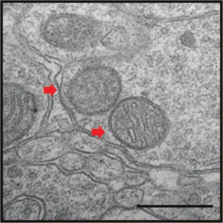
6                                                                                                                                      Evidence-Based Complementary and Alternative Medicine

                                                               6-OHDA lesioning                                                                  Moxibustion treatment
                                                                  for 4 weeks                                                                         for 4 weeks
                                                                                                                                                                                           End

                                       D0       D7                                                   D35                                                                     D63
                                                                                                 Behavioral test                                                         Behavioral test
                                                                                                       (a)
                           20                                                                                                               20
                                                                                                                                                                                           ∗∗∗

                                                                                                                     Numbers of rotations
    Numbers of rotations

                           15                                                                                                               15

                           10                                                                                                               10

                           5                                                                                                                5

                           0                                                                                                                0
                                       D7                    D35                           D63                                                         Control     Sham          6-OHDA Moxibustion

                                     Control                            6-OHDA
                                     Sham                               Moxibustion
                                                       (b)                                                                                                               (c)
                                                                                                                                   1.5
                                                                             Moxibustion
                                                               6-OHDA
                                      Control

                                                                                                        Expression of protein
                                                Sham

                                                                                                                                                                                           ∗∗∗
                                                                                                                                   1.0
                                TH                                                           62 kDa

           GAPDH                                                                             36 kDa                                0.5

                                                                                                                                   0.0
                                                                                                                                                      Control     Sham          6-OHDA Moxibustion
                                                                                                       (d)
                                                                                           Figure 3: Continued.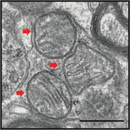
Evidence-Based Complementary and Alternative Medicine 7
20 µm 20 µm 20 µm 20 µm
Control Sham 6-OHDA Moxibustion
1.5
Average optical density ∗∗∗
1.0
0.5
0.0
Control 6-OHDA
Sham Moxibustion
(e)
Figure 3: Moxibustion treatment of Shi’s moxa stick has a positive effect on suppressing the death of dopaminergic neurons in the PD rats
model. (a) The timeline of animal experiment 2. (b) The change of behavioral score in D35 and D63 of the control group, the sham group, the
6-OHDA group, and the moxibustion group. (c) The behavioral score in D63 of the control group, the sham group, the 6-OHDA group, and
the moxibustion group. (d) TH level of total protein in SNpc of the control group, the sham group, the 6-OHDA group, and the moxibustion
group. (e) IHC showed the expression of TH in the substantia nigra of the control group, the sham group, the 6-OHDA group, and the
moxibustion group.
result showed that, after moxibustion treatment of Shi's moxa mitochondrial morphology damage is reduced. This proved
stick, the activity of dopaminergic neurons was increased that the protective effect of moxibustion treatment of Shi’s
According to HE staining (Figure 4), the shape of the nerve moxa stick on dopaminergic neurons may be related to ef-
cells in the sham group was normal. However, in the 6-OHDA fectively suppressing the level of ferroptosis.
group, the shape of neurons was rounded, and the nucleus was
concentrated or swollen. After moxibustion treatment of Shi’s 4. Discussion
moxa sticks, although some never cells still have abnormal
morphology, the number of nerve cells with healthy mor- As a progressive neurodegenerative movement disorder,
phology was increased. This proved that moxibustion treat- there is an urgent need for an effective treatment for PD [1].
ment of Shi’s moxa stick could alleviate the injury of dopamine In this study, we hope to provide promising therapies for
neurons in PD. PD. The present study constructed the PD rat model by a
two-point stereotactic injection of 6-hydroxydopamine and
investigated moxibustion treatment’s functional roles using
3.4. Moxibustion Treatment of Shi’s Moxa Stick Suppresses Shi’s moxa stick in PD. The present study’s significant results
the Level of Ferroptosis in the PD Rats Model. To investigate were as follows: (i) the level of ferroptosis is associated with
whether the moxibustion treatment of Shi’s moxa stick can the injury of dopamine neurons in PD models; (ii) mox-
suppress the level of ferroptosis in the PD rats model, the ibustion treatment of Shi’s moxa stick can alleviate the
expression of GPX4, FTH1, and ROS was used. The Western damage of dopamine neurons by suppressing ferroptosis.
blot and Rt-PCR analysis results showed that GPX4 and FTH1 Our research provides a promising treatment and thera-
expressions were increased significantly after moxibustion peutic target to improve the clinical outcome of PD
treatment of Shi’s moxa stick (Figure 5(a); 6-OHDA versus treatment.
moxibustion; P < 0.001; Figure 5(b); 6-OHDA versus mox- Research shows that abnormal iron metabolism and iron
ibustion; P < 0.001), whereas ROS was decreased obviously homeostasis are involved in PD’s pathological process
after moxibustion treatment of Shi’s moxa stick (Figure 5(c); [14, 28]. Previous studies have shown that FTH1, GPX4, and
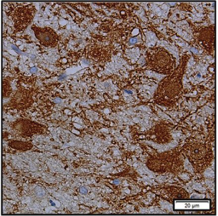
6-OHDA versus moxibustion; P < 0.05). The morphological ROS can be used as a marker to monitor the level of fer-
changes of ferroptosis are mainly the damage of mitochondria. roptosis [22, 29]. In contrast, the morphology of ferroptosis
As shown in Figure 5(d), in the 6-OHDA group, mitochondrial mainly manifested the reduced mitochondrial volume, the
cristae ruptured or disappeared, and the structure was fuzzy. increase of double-layer membrane density, and the re-
After moxibustion treatment of Shi’s moxa stick, duction or disappearance of mitochondrial cristae [18, 25].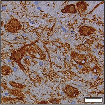
8                                                                                                                                                                   Evidence-Based Complementary and Alternative Medicine

                                                                       20 µm                                     20 µm                                                                    20 µm                         20 µm

                                                    (a)                                              (b)                                                                        (c)                            (d)

Figure 4: Moxibustion treatment of Shi’s moxa stick positively affects neurons’ morphology in the PD rats model. HE staining
was performed in the SN of the control group, the sham group, the 6-OHDA group, and the moxibustion group. (a) Control. (b) Sham.
(c) 6-OHDA. (d) Moxibustion.

                                                                                           Moxibustion
                                                                                                                                                           1.5
                                                                               6-OHDA
                                        Control

                                                          Sham

                                                                                                                                                                                                  ∗∗∗                           ∗∗∗

                                                                                                                         Expression of protein
                                                                                                                                                           1.0
                             GPX4                                                                           21 kDa

                             FTH1                                                                           17 kDa
                                                                                                                                                           0.5
                        GAPDH                                                                               36 kDa

                                                                                                                                                           0.0
                                                                                                                                                                                       GPX4                           FTH1
                                                                                                                                                                            Control                            6-OHDA
                                                                                                                                                                            Sham                               Moxibustion
                                                                                                                         (a)
                                                                                                                                                                                                                        ∗∗∗
                            1.5                                                                                                                                         15000

                                                          ∗∗∗                                              ∗∗∗
       Expression of mRNA

                                                                                                                                                 Tissue ROS intensity

                            1.0                                                                                                                                         10000

                            0.5                                                                                                                                          5000

                            0.0                                                                                                                                             0
                                                  GPX4                                    FTH1                                                                                        Control           Sham     6-OHDA Moxibustion

                                    Control                                        6-OHDA
                                    Sham                                           Moxibustion
                                                                 (b)                                                                                                                                     (c)
                                                                                                           Figure 5: Continued.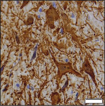
Evidence-Based Complementary and Alternative Medicine                                                                                      9

                                                                                                                             500 nm

                Control                          Sham                           6-OHDA                         Moxibustion
                                                                   (d)

Figure 5: After moxibustion treatment of Shi’s moxa stick, the level of ferroptosis is decreased. (a) GPX4 and FTH1 level of total protein in
the control group, the sham group, the 6-OHDA group, and the moxibustion group. (b) GPX4 and FTH1 levels of total mRNA in the control
group, the sham group, the 6-OHDA group, and the moxibustion group. (c) The level of ROS in the control group, the sham group, the 6-
OHDA group, and the moxibustion group. (d) Observe the morphology of mitochondrial in the control group, the sham group, the 6-
OHDA group, and the moxibustion group. The red arrow refers to mitochondria.

An essential finding of the present study is that GPX4 and                the 6-OHDA still promotes neuronal death. The protective
FTH1 decrease significantly, and the level of ROS was in-                 effect of moxibustion treatment may be related to the effective
creased obviously in the PD rats model. Simultaneously,                  suppression of ferroptosis levels.
directly observing the mitochondrial morphology of dif-                      The limitation of this study is that the specific molecular
ferent groups through transmission electron microscopy,                  mechanism of how moxibustion treatment of Shi’s moxa
mitochondrial cristae ruptured or disappeared, and the                   sticks suppresses ferroptosis level by increasing the ex-
structure was fuzzy in the PD rats model, representing high              pression of FTH1 has not been explained in this study, which
ferroptosis levels in the PD rat model. These results are                needs further research. In conclusion, we found that the
consistent with a recent study, which found that ferroptosis             moxibustion treatment of Shi’s moxa sticks improves DA
plays a vital role in PD development [30]. This study shows              neurons’ survival by suppressing the ferroptosis level.
that antiferroptosis of moxibustion treatment can be used as             Moxibustion treatment of Shi’s moxa stick is an economical,
a protection pathway for PD patients.                                    safe, and convenient way for PD treatment and a promising
    Another important finding of this study is that mox-                  complementary and alternative therapy for PD patients.
ibustion treatment of Shi’s moxa stick can effectively suppress
dopaminergic neurons’ death. As a form of external therapy               Data Availability
in traditional Chinese medicine, moxibustion treatment re-
ported can ameliorate motor symptoms in PD patients [9, 10].             The data and materials produced during the study can be
Shi’s moxa sticks were developed by Shi Xuemin, an aca-                  obtained from the corresponding authors upon reasonable
demician of the Chinese Academy of Engineering and a                     request.
master of traditional Chinese medicine. Shi’s moxa sticks have
been used in hospitals to relieve PD symptoms. Baihui                    Ethical Approval
(GV20) is located at the midpoint of the two ears above the
head. Because the head is the meeting point of Governor                  All animal experiments were approved by the Experimental
Vessel and all Yang meridians, Baihui (GV20) can regulate the            Animal Care and Use Committee of Guangzhou University
yang qi of all Yang meridians [24]. Existing research proves             of Traditional Chinese Medicine and conducted following
that Baihui improves blood circulation in the brain and                  the guidelines for the care and use of experimental animals
enhances memory [31]. Therefore, this study chose Baihui as              of the National Institutes of the Health of the United States.
an acupoint. The expression of TH is a crucial link of the DA
neuron [32]. Our results show that, after moxibustion                    Disclosure
treatment of Shi’s moxa stick, the rotational behavioral score           Zifeng Huang and Wenwen Si are co-first authors.
of PD rats was decreased, the expression of TH, GPX4, and
FTH1 was increased, the level of ROS was decreasing, and
                                                                         Conflicts of Interest
mitochondrial damage was relieved. However, in the 6-
OHDA group, the symptoms of PD rats continued to worsen.                 The authors declare no conflicts of interest.
As shown in the behavioral score, the behavioral score slightly
increased in the D63 compared to the D35, which demon-                   Authors’ Contributions
strates that also PD model has been established in D35, and
the 6-OHDA was still promoting neuronal death. This finding               Zifeng Huang and Wenwen Si contributed equally to this
may indicate that moxibustion treatment of Shi’s moxa stick              work. Zifeng Huang performed the experiments and wrote
can effectively suppress dopaminergic neurons’ death, while               the manuscript. Wenwen Si designed the experiments and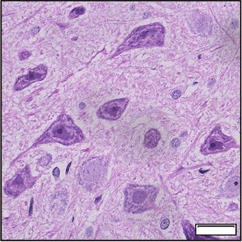
10                                                                       Evidence-Based Complementary and Alternative Medicine

analyzed the data. Xinrong Li, Shanyu Ye, Xuelei Liu, Yichun                   neuronal cell death in rats,” Neuroscience Letters, vol. 384,
Ji, and Xiaoqian Hao performed animal experiments.                             no. 1-2, pp. 133–138, 2005.
Meiling Zhu and Dongfeng Chen conceived and designed                    [13]   E. C. Hirsch and B. A. Faucheux, “Iron metabolism and
the experiments. All authors have read and approved the                        Parkinson’s disease,” Movement Disorders Official Journal of
final manuscript.                                                               the Movement Disorder Society, vol. 13, no. Suppl 1, pp. 39–45,
                                                                               1998.
                                                                        [14]   A. Scott and L. Peng, “Nigral iron elevation is an invariable
Acknowledgments                                                                feature of Parkinson’s disease and is a sufficient cause of
                                                                               neurodegeneration,” BioMed Research International,
This study was supported by the Traditional Chinese                            vol. 2014, Article ID 581256, 9 pages, 2014.
Medicine Bureau of Guangdong Province, China (grant no.                 [15]   H. Nishizawa, M. Matsumoto, T. Shindo et al., “Ferroptosis is
20203010), the Natural Science Foundation of Guangdong                         controlled by the coordinated transcriptional regulation of
Province (grant no. 2020A151501325), the Shenzhen Science                      glutathione and labile iron metabolism by the transcription
and Technology Innovation Committee Subject (grant no.                         factor BACH1,” Journal of Biological Chemistry, vol. 295,
                                                                               no. 1, pp. 69–82, 2020.
JCYJ20190807112405520), the Bao'an TCM Development
                                                                        [16]   A. Weiland, Y. Wang, W. Wu et al., “Ferroptosis and its role in
Foundation (grant nos. 2020KJCX-KTYJ-130 and
                                                                               diverse brain diseases,” Molecular Neurobiology, vol. 56, no. 7,
2020KJCX-KTYJ-131), and the Joint Research Project of                          pp. 4880–4893, 2019.
Guangdong Academy of Traditional Chinese Medicine                       [17]   Y. Tian, J. Lu, X. Hao et al., “FTH1 inhibits ferroptosis through
(grant no. 2017A020213036).                                                    ferritinophagy in the 6-OHDA model of parkinson’s disease,”
                                                                               Neurotherapeutics, vol. 17, no. 4, pp. 1796–1812, 2020.
                                                                        [18]   S. J. Dixon, K. M. Lemberg, M. R. Lamprecht et al., “Fer-
References                                                                     roptosis: an iron-dependent form of nonapoptotic cell death,”
 [1] S. V. Campenhausen, B. Bornschein, R. Wick et al., “Preva-                Cell, vol. 149, no. 5, pp. 1060–1072, 2012.
     lence and incidence of parkinson’s disease in Europe,” Eu-         [19]   M. Abdalkader, R. Lampinen, K. Kanninen, and T. Malm,
     ropean Neuropsychopharmacology, vol. 15, no. 4, pp. 473–490,              “Targeting Nrf2 to suppress ferroptosis and mitochondrial
     2005.                                                                     dysfunction in neurodegeneration,” Frontiers in Neuroence,
 [2] L. ML de Lau’s and M. M. B. Breteler, “Epidemiology of                    vol. 12, p. 466, 2018.
     Parkinson’s disease,” The Lancet Neurology, vol. 5, no. 6,         [20]   S. Neitemeier, A. Jelinek, V. Laino et al., BID links ferroptosis
     pp. 525–535, 2006.                                                        to mitochondrial cell death pathways. RedoxBiology, vol. 12,
 [3] J. M. Beitz, “Parkinson’s disease: a review,” Frontiers in                pp. 558–570, 2017.
     Bioscience, vol. 6, no. 1, pp. 65–74, 2014.                        [21]   I. Ingold, C. Berndt, S. Schmitt et al., “Selenium utilization by
 [4] J. A. Obeso, M. C. Rodriguez-Oroz, M. Rodriguez et al.,                   GPX4 Is required to prevent hydroperoxide-induced fer-
     “Pathophysiology of the basal ganglia in Parkinson’s disease,”            roptosis,” Cell, vol. 172, no. 3, pp. 409–422, 2018.
     Trends in Neurosciences, vol. 23, no. 1, pp. S8–S19, 2000.         [22]   Z. Zhang, Z. Yao, L. Wang et al., “Activation of ferritinophagy
 [5] O. Rascol, A. Lozano, M. Stern, and W. Poewa, “Milestones in              is required for the RNA-binding protein ELAVL1/HuR to
     Parkinson’s disease therapeutics,” Movement Disorders,                    regulate ferroptosis in hepatic stellate cells,” Autophagy,
     vol. 26, no. 6, pp. 1072–1082, 2011.                                      vol. 14, no. 12, pp. 2083–2103, 2018.
 [6] Z. Lily Mao and N. B. Modi, “Dose-response analysis of the         [23]   X. Sun, Z. Ou, R. Chen et al., “Activation of the p62-Keap1-
     effect of carbidopa-levodopa extended-release capsules                     NRF2 pathway protects against ferroptosis in hepatocellular
     (IPX066) in levodopa-naive patients with Parkinson disease,”              carcinoma cells,” Hepatology, vol. 63, no. 1, pp. 173–184, 2016.
     Journal of Clinical Pharmacology, vol. 56, no. 8, pp. 974–982,     [24]   J. Lu, X. Liu, Y. Tian et al., “Moxibustion exerts a neuro-
     2016.                                                                     protective effect through antiferroptosis in parkinson’s dis-
 [7] Y. C. Lam, W. Kum, F. Xiao et al., “Efficacy and safety of                  ease,” Evidence-based Complementary and Alternative
     acupuncture for idiopathic Parkinson’s disease: a systematic              Medicine, vol. 2019, Article ID 2735492, 10 pages, 2019.
     review,” Journal of Alternative & Complementary Medicine,          [25]   W. S. Yang and B. R. Stockwell, “Synthetic lethal screening
     vol. 14, no. 6, pp. 663–671, 2008.                                        identifies compounds activating iron-dependent, non-
 [8] T. Yi, L. Qi, J. Li et al., “Moxibustion upregulates hippocampal          apoptotic cell death in oncogenic-RAS-harboring cancer
     progranulin expression,” Neural Regeneration Research,                    cells,” Chemistry & Biology, vol. 15, no. 3, pp. 234–245, 2008.
     vol. 11, no. 4, pp. 610–616, 2016.                                 [26]   R. K. Schwarting and J. P. Huston, “The unilateral 6-
 [9] X. Zhang, Y. Wu, and L. Ding, “Moxibustion for parkison                   hydroxydopamine lesion model in behavioral brain research.
     disorder:a systematic review and meta-analysis,” Asia-Pacific              Analysis of functional deficits, recovery and treatments,”
     Traditional Medicine, vol. 12, no. 14, pp. 96–100, 2016.                  Progress in Neurobiology, vol. 50, no. 2-3, pp. 275–331, 1996.
[10] Z. Li, Y.-Y. Hu, C. Y. Zheng et al., “Rules of meridians and       [27]   M. T. Hayes, “Parkinson’s disease and parkinsonism,” The
     acupoints selection in treatment of Parkinson’s disease based             American Journal of Medicine, vol. 132, no. 7, pp. 802–807,
     on data mining techniques,” Chinese Journal of Integrative                2019.
     Medicine, vol. 26, no. 8, pp. 624–628, 2018.                       [28]   R. J. Ward, F. A. Zucca, J. H. Duyn, R. R. Crichton, and
[11] S.-J. Wang, Q. Wang, J. Ma, P. H. Yu, Z.-M. Wang, and                     L. Zecca, “The role of iron in brain ageing and neurode-
     B. Wang, “Effect of moxibustion on mTOR-mediated auto-                     generative disorders,” Lancet Neurology, vol. 13, no. 10,
     phagy in rotenone-induced Parkinson’s disease model rats,”                pp. 1045–1060, 2014.
     Neural Regeneration Research, vol. 13, no. 1, pp. 112–118,         [29]   A. Massie, A. Schallier, S. W. Kim et al., “Dopaminergic
     2018.                                                                     neurons of system x (c)-deficient mice are highly protected
[12] Y. K. Kim, H.-H. Lim, Y.-K. Song et al., “Effect of acupuncture            against 6-hydroxydopamine-induced toxicity,” The FASEB
     on 6-hydroxydopamine-induced nigrostratal dopaminergic                    Journal, vol. 25, no. 4, pp. 1359–1369, 2011.
Evidence-Based Complementary and Alternative Medicine 11
[30] C.-F. Zhu, L.-D. Zhang, X.-G. Song et al., “Moxibustion
improves learning-memory ability by promoting cellular
autophagy and regulating autophagy-related proteins in
hippocampus and cerebral cortex in APP/PS1 transgenic
alzheimer’s disease mice,” Acupuncture Research, vol. 44,
no. 4, pp. 235–241, 2019.
[31] S. B. Rangasamy, S. Dasarathi, P. Pahan, M. Jana, and
M. Pahan, “Low-dose aspirin upregulates tyrosine hydroxy-
lase and increases dopamine production in dopaminergic
neurons: implications for Parkinson’s disease,” Journal of
Neuroimmune Pharmacology the Official Journal of the Society
on Neuroimmune Pharmacology, vol. 14, no. 2, pp. 173–187,
2019.
[32] D. S. Rothblat, E. Rubin, and J. S. Schneider, “Effects of
chronic alcohol ingestion on the mesostriatal dopamine
system in the rat,” Neuroscience Letters, vol. 300, no. 2,
pp. 63–66, 2001.You can also read