Klotho promoter methylation status and its prognostic value in ovarian cancer
←
→
Page content transcription
If your browser does not render page correctly, please read the page content below
MOLECULAR AND CLINICAL ONCOLOGY 15: 181, 2021

                          Klotho promoter methylation status and its
                              prognostic value in ovarian cancer
                          MARYAM H. AL‑ZAHRANI1*, FATIMAH M. YAHYA1*, MOURAD ASSIDI2,3,
                                 ASHRAF DALLOL2,3 and ABDELBASET BUHMEIDA2

                1
                 Biochemistry Department, Faculty of Science, 2Center of Excellence in Genomic Medicine Research,
                              3
                                Medical Technology Department, Faculty of Applied Medical Sciences,
                                      King Abdulaziz University, Jeddah 21589, Saudi Arabia

                                       Received February 14, 2021; Accepted May 21, 2021

                                                  DOI: 10.3892/mco.2021.2343

Abstract. Among all gynecological cancers, ovarian                 Introduction
cancer (OC) is one of the deadliest types of cancer worldwide.
Epigenetic silencing of some genes has been reported to be asso‑   Among the different gynecologic cancers, ovarian cancer (OC)
ciated with OC. In this context, Klotho (KL) gene methylation      is the leading cause of death (1). In comparison to the breast
is a promising biomarker for OC. The present study aimed to        cancer, OC mortality rate is approximately three times higher
investigate the methylation profiles of KL and assess its prog‑    despite its lower incidence rate (2). This is attributed to its
nostic value. A total of 63 formalin‑fixed paraffin‑embedded       asymptomatic initial stages and late diagnosis at advanced
tissue samples from patients with primary OC were collected        stages (3,4). The incidence of OC in Saudi Arabia has increased
and analyzed in the present study. The methylation profiles        4‑fold between 1990 and 2016 (5). It affects more than 3% of
of KL were assessed by performing DNA bisulfate treatment          Saudi women in their lifetime (6,7). There are different
followed by DNA promoter methylation analysis using the            types of ovarian malignancy; however, the epithelial ovarian
MethyLight assay. The results revealed KL promoter hyper‑          cancer (EOC) is the most common malignancy (90%) (4,8‑10).
methylation in 62% of the OC cohort. Additionally, significant     This EOC is a heterogeneous disease with several histologic
associations were observed between KL methylation profiles         subtypes that exhibited distinct cytogenetic features, molec‑
and tumor subtype (P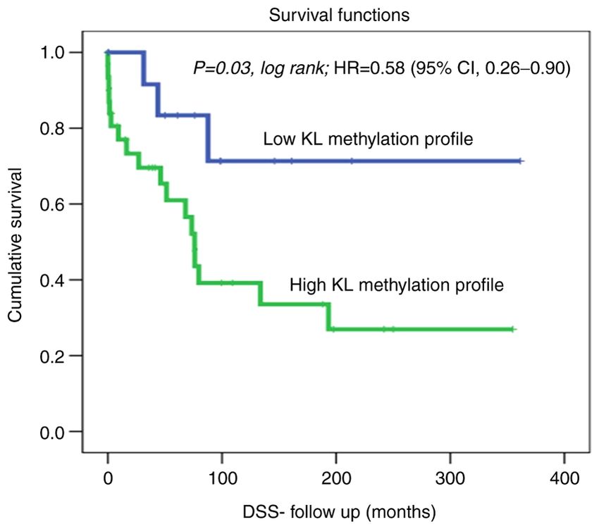
2 AL-ZAHRANI et al: KLOTHO METHYLATION STATUS AS PROGNOSTIC DETERMINANT OF OVARIAN CANCER
Genomic instability is one of the main causes of cancer Patients and methods
onset attributed to the interactions among a patient's
susceptible genome, environment, and lifestyle; hence, some Patients and samples. This retrospective study comprised
DNA alterations occur at the epigenomic level. Epigenetic 63 formalin‑fixed paraffin‑embedded (FFPE) tissues that
alterations encompass histone acetylation, noncoding were surgically resected after obtaining written consent
RNAs, and CpG island methylation, are among the most from female patients who were diagnosed with OC and were
frequent epigenetic alterations observed in OC (20‑22). selected based on the availability of both tissues and patient
Currently, there is an increasing interest in examining the clinicopathological follow‑up data. The FFPE samples
specific patterns of hypermethylation of CpG islands in were collected between 1995 and 2014 at the Departments
OC. Both targeted and whole‑genome analyses for DNA of Pathology & Gynecology, King Abdulaziz University
methylation patterns have shown potential in identifying Hospital (KAUH).
methylation biomarkers and their prognostic value in The cohort of patients was selected from a total of
OC (23‑26). Moreover, epigenetic silencing of some genes, 100 accessible samples based on the availability of patients'
including hMLH1, RASSF1A, and FANCF due to methyla‑ annotated clinicopathological follow‑up data, and the
tion is associated with ovarian tumor drug resistance (27). quality/quantity of extracted DNA. Patients' cohort was classi‑
Consequently, some key epigenetic events are likely to fied based on histopathological features using the tumor node
provide cancer cells an advantage to proliferate and invade, metastasis (TNM) classification system.
thereby promoting metastasis (28,29). Thus, further studies The main clinicopathological features (such as age,
are required to analyze the methylation profiles of several menopausal status, stage, grade, and lymph node status)
genes to gain deeper insights into the epigenetic alterations and follow‑up and survival data are summarized in Table I.
in OC, especially in the Arabian Peninsula, where only a few The samples and data were retrieved from the patients after
studies have been reported (25,26,30‑32). In this context, obtaining all the relevant ethical approvals according to the
klotho (KL) gene is an interesting candidate aging marker guidelines of the Ethical Committee of King Abdulaziz
gene that could be associated with aging‑related diseases University Hospital (Ref. number: KAU‑189‑14).
e.g. cancer. In fact, KL gene was reported with an aberrantly
methylated promoter reported in several malignancies (33). DNA extraction. Before DNA extraction, 10 µm thin FFPE
Initially discovered as an anti‑aging factor (34), KL is slices were deparaffinized using xylol (Sigma‑Aldrich)
located in chromosome 13q12. It is 50 kb in length and is and then washed with ethanol 99‑100% (Sigma‑Aldrich).
composed of five exons and four introns (35). It is highly DNA extraction was performed using the QIAamp DNA
expressed in the kidneys and brain but can also be expressed FFPE Tissue Kit (Qiagen) according to the manufacturer's
in the placenta, breast, ovaries, uterus, and fallopian tube protocol. The quality and concentration of all DNA samples
tissues (36). In the female reproductive system, particularly were assessed using NanoDrop 2000 Spectrophotometer
in the ovary, the level of abnormal expression of KL in gran‑ (Thermo Fisher Scientific, Inc.).
ulosa cells is tightly linked to the severity of ovary‑related
diseases (37). KL expression levels decrease in the brain KL methylation analysis. Bisulfite treatment of the KL promoter
with advancing age in mammals. This decrease could be due region was performed using the Qiagen EpiTect® Bisulfite
to hypermethylation of the promoter region of KL (38,39). Conversion kit as detailed in the manufacturer's handbook and
Furthermore, aberrant methylation in the promoter region reported by Dallol et al (47). Briefly, 0.5 µg extracted DNA
of KL decreases gene transcription (40). Recently, KL has was incubated with NaH2SO4 (3.12 M). Only the unmethylated
been reported as a potential tumor suppressor gene that cytosine (C) residues were converted into uracil (U) residues.
inhibits the IGF‑1 pathway and is a novel target gene for The MethyLight assay for the candidate gene was performed
epigenetic silencing in cancers of breast (41), cervix (34), using the EpiTect® MethyLight PCR Kit (Qiagen, Inc.) and
stomach (42), bladder, and ovaries (43). However, low EpiTect® Control DNA Set (Qiagen, Inc.) according to the
expression levels of KL are associated with weakness in protocol guidelines, fluorescent dual‑labeled TaqMan Probe
skeletal muscle, difficulty in executing daily activities, 6FAM‑CGGTTGGGTTAATCGCGTTTT‑BHQ1, and specif‑
and increased mortality in the elderly. Deficiency in IGF‑1 ically designed primers: Forward primer 5'‑AGCGTTTGT
expression level is a reported feature of aging, and is likely AGGACGT TTAC‑3', and reverse primer 5'‑TAACGA A AA
affected by methylated KL (40). The 1439‑bp long promoter CAAA ACTCCG C‑3') (48); qPCR was performed using the
region of KL lacks a TATA box and instead, contains four StepOnePlus™ real‑time PCR system (Applied Biosystems).
potential Sp1 binding sites (35,44). Moreover, KL expres‑
sion can be restored using demethylation reagents (35). So Statistical analysis. Statistical analyses were performed using
far, only few studies have assessed the methylation profiles SPSS® version 19 (IBM Corp.). Differentially hypo/hyper‑
of KL in OC and showed potential prognosis value. In that, methylated genes' profiles were assessed compared to the
higher KL promoter methylation profiles were associated EpiTect® Control DNA Set using a t‑test. The association
with cancer progression (45,46). Therefore, additional between KL methylation profile and clinicopathological
studies are required to further understand the prognosis features of patients was analyzed using Fisher‑Freeman‑Halton
value of KL methylation status in OC, particularly, in the Exact test. Furthermore, disease‑specific survival (DSS) and
Arabian Peninsula population. In this study, we analyzed disease‑free survival (DFS) based on KL methylation profiles
KL gene promoter methylation profiles in OC and assessed were calculated by univariate Kaplan‑Meier analysis along
its prognostic value. with hazard ration calculation at 95% CI, and equality of theMOLECULAR AND CLINICAL ONCOLOGY 15: 181, 2021 3
Table I. Association between KL methylation profiles and clinicopathological features of patients with ovarian cancer.
KL methylation profile
‑‑‑‑‑‑‑‑‑‑‑‑‑‑‑‑‑‑‑‑‑‑‑‑‑‑‑‑‑‑‑‑‑‑‑‑‑‑‑‑‑‑‑‑‑‑‑‑‑‑‑‑‑‑‑‑‑‑‑‑‑‑‑‑‑‑‑‑‑‑‑‑‑‑‑‑‑‑‑‑‑‑‑‑‑‑‑‑‑‑‑‑‑‑‑‑‑‑‑‑‑‑‑‑‑‑‑‑‑
Clinicopathological features N (%) 0 unmethylated, n (%) 1 hypermethylated, n (%) P‑valuea
Age, years
50 22 (34.9) 5 (20.8) 17 (44.7)
NA 1 (1.6)
Diagnosis method
Surgery 58 (92.1) 22 (91.7) 36 (94.7) 0.637
Biopsy 4 (6.3) 2 (8.3) 2 (5.3)
NA 1 (1.6)
Tumor site
Right 11 (17.5) 4 (16.7) 7 (18.4) 0.039b
Left 11 (17.5) 8 (33.3) 3 (7.9)
Bilateral 40 (63.5) 12 (50.0) 28 (73.7)
NA 1 (1.6)
Tumor size, cm
1‑5 18 (28.6) 9 (39.1) 9 (23.7) 0.370
6‑10 16 (25.4) 6 (26.1) 10 (26.3)
>10 27 (42.9) 8 (34.8) 19 (50.0)
NA 2 (3.2)
Tumor subtype
Serous 30 (47.6) 12 (50.0) 18 (47.4)4 AL-ZAHRANI et al: KLOTHO METHYLATION STATUS AS PROGNOSTIC DETERMINANT OF OVARIAN CANCER
Table I. Continued.
KL methylation profile
‑‑‑‑‑‑‑‑‑‑‑‑‑‑‑‑‑‑‑‑‑‑‑‑‑‑‑‑‑‑‑‑‑‑‑‑‑‑‑‑‑‑‑‑‑‑‑‑‑‑‑‑‑‑‑‑‑‑‑‑‑‑‑‑‑‑‑‑‑‑‑‑‑‑‑‑‑‑‑‑‑‑‑‑‑‑‑‑‑‑‑‑‑‑‑‑‑‑‑‑‑‑‑‑‑‑‑‑‑‑‑
Clinicopathological features N (%) 0 unmethylated, n (%) 1 hypermethylated, n (%) P‑valuea
Tumor stage
Stage I 16 (25.4) 8 (38.1) 8 (22.9) 0.660
Stage II 4 (6.3) 1 (4.8) 3 (8.6)
Stage III 31 (49.2) 10 (47.6) 21 (60.0)
Stage IV 5 (7.9) 2 (9.5) 3 (8.6)
NA 7 (11.1)
Endpoint status
Living 24 (38.1) 11 (73.3) 13 (41.9) 0.063
Deceased 22 (34.9) 4 (26.7) 18 (58.1)
NA 17 (27.0)
Recurrence status
No 20 (31.7) 7 (41.2) 13 (48.1) 0.760
Yes 24 (83.1) 10 (58.8) 14 (51.9)
NA 19 (30.2)
P‑values were calculated using Fisher‑Freeman‑Halton Exact test. bPMOLECULAR AND CLINICAL ONCOLOGY 15: 181, 2021 5
Table II. Cox regression analysis of the prognostic values of KL methylation, age at diagnosis, grade and histological subtypes.
Feature P‑value Standard error value Relative risk 95% CI
KL methylation (low vs. high methylation) 0.029 0.716 4.758 1.169‑19.366
Age at diagnosis (50 years) 0.843 0.540 1.113 0.386‑3.205
Histological subtypes (serous vs. mucinous) 0.470 0.296 1.238 0.693‑2.213
Tumor grade (low vs. high grade) 0.140 0.186 1.317 0.914‑1.898
KL, Klotho; CI, confidence interval.
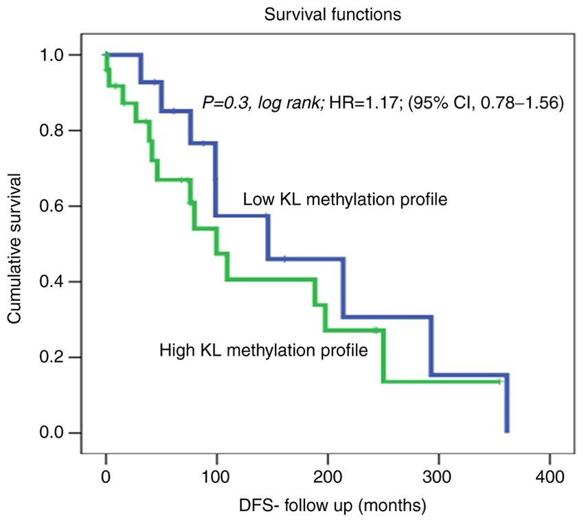
Figure 1. KL methylation profile status in the ovarian cancer cohort using
Figure 2. KL methylation profile status in the ovarian cancer cohort using
the cut‑off [unmethylated (0; 15 cases) vs. hypermethylated (1; 31 cases)] as a
the cut‑off [unmethylated (0; 17 cases) vs. hypermethylated (1; 27 cases)] as a
determinant of DSS in univariate (Kaplan‑Meier) analysis. DSS, disease‑spe‑
determinant of DFS in univariate (Kaplan‑Meier) analysis. DFS, disease‑free
cific survival; KL, Klotho; HR, hazard ratio.
survival; KL, Klotho; HR, hazard ratio.
Discussion
in the Arabian Peninsula to assess this biomarker in patients
OC is one of the deadliest gynecological cancers mainly with OC and particular clinicopathological features. Unlike
because of delayed diagnosis and asymptomatic early‑stage previous studies, most of our cohort was diagnosed with OC
progression. Moreover, the deleterious effects of OC can be before menopause (66.1%) and was younger than 50 years
attributed to the anatomical position of the ovaries within of age (64.5%). In contrast, other studies reported OC as an
the deep pelvic cavity which may hide the disease expan‑ age‑related disease, and as mainly postmenopausal (57).
sion and delay the diagnosis (49‑51). Scientists are seeking to Moreover, the incidence increased more noticeably in women
improve the detection, prognosis, management, and treatment over 65 years of age (58), and the median age at diagnosis
approaches toward precision oncology to alleviate the burden was between 50 and 79 years (59,60). According to the data
of this disease on patients, their families, and the health‑ released by Cancer Research UK, 53% of British women diag‑
care system (52). With the development of high‑throughput nosed with OC were 65 years and older. Moreover, according
sequencing technologies, it is possible to assess changes in the to the American Cancer Society report, 50% of the diagnosed
genetic and epigenetic profiles of the target genes/genomes and OC patients were aged 63 years or above (61). These findings
identify potential molecular biomarkers for OC (21,53‑56). highlight an interesting 15‑year shift in the onset of OC in the
KL is located at chromosome 13q12, with a length of 50 kb Arabian Peninsula, and therefore, warrant additional studies
including a promoter region of 1,439 bp. It is a known tumor to demystify such an early onset. Parity has been reported to
suppressor gene that has been reported to undergo aber‑ reduce the risk of OC in contrast to nulliparous women (62).
rant promoter methylation in many tumors (35). The level However, the majority of our patient cohort was parous
of abnormal expression of KL in granulosa cells of the women (69%), which may be attributed to the genomic blue‑
ovary is tightly linked with the acuteness of ovary‑related print, environmental factors, and/or lifestyle choices; however,
diseases (37). Thus, in this study, the methylation pattern of this needs further investigation.
KL were determined to evaluate its potential as a promising Till date, only Wiley et al have assessed the KL methyla‑
molecular biomarker in OC and assess its prognostic value. tion profiles in 215 epithelial OC patients. They found that KL
To the best of our knowledge, this is the first study conducted was hypermethylated in 14% of the OC patients compared with6 AL-ZAHRANI et al: KLOTHO METHYLATION STATUS AS PROGNOSTIC DETERMINANT OF OVARIAN CANCER the controls and the hypermethylation status was associated Authors' contributions with a significant reduction in DFS (46). Strikingly, approxi‑ mately 62% of our cohort had hypermethylated KL, which MHAZ and AB contributed to study design, statistical is 4‑fold higher than the results reported by Wiley et al (46). analysis and contributed in both drafting and critical revision Our results showed significant associations between KL meth‑ of the manuscript. FMY and MA contributed to data collec‑ ylation profile and tumor subtype (P
MOLECULAR AND CLINICAL ONCOLOGY 15: 181, 2021                                                      7

13. Norquist BM, Brady MF, Harrell MI, Walsh T, Lee MK,                 33. Xie B, Zhou J, Yuan L, Ren F, Liu DC, Li Q and Shu G: Epigenetic
    Gulsuner S, Bernards SS, Casadei S, Burger RA, Tewari KS, et al:        silencing of Klotho expression correlates with poor prognosis of
    Mutations in homologous recombination genes and outcomes in             human hepatocellular carcinoma. Hum Pathol 44: 795‑801, 2013.
    ovarian carcinoma patients in GOG 218: An NRG oncology/gyne‑        34. Lee J, Jeong DJ, Kim J, Lee S, Park JH, Chang B, Jung SI, Yi L,
    cologic oncology group study. Clin Cancer Res 24: 777‑783, 2018.        Han Y, Yang Y, et al: The anti‑aging gene Klotho is a novel
14. Maradeo ME and Cairns P: Translational application of                   target for epigenetic silencing in human cervical carcinoma. Mol
    epigenetic alterations: Ovarian cancer as a model. FEBS Lett 585:       Cancer 9: 109, 2010.
    2112‑2120, 2011.                                                    35. Rubinek T and Wolf I: Klotho tumor suppressor. In: Encyclopedia
15. Sa leh i F, Dun f ield L, Ph illips K P, K rewsk i D a nd               of Cancer. Schwab M (ed). Springer, Berlin, Heidelberg, pp1‑5,
    Vanderhyden BC: Risk factors for ovarian cancer: An overview            2016.
    with emphasis on hormonal factors. J Toxicol Environ Health B       36. Fan CF, Tan C and Wang SQ: α‑Klotho: A novel regulator in
    Crit Rev 11: 301‑321, 2008.                                             female reproductive outcomes and hormone‑related cancer. Int J
16. Irodi A, Rye T, Herbert K, Churchman M, Bartos C, Mackean M,            Clin Exp Med 10: 8511‑8521, 2017.
    Nussey F, Herrington CS, Gourley C and Hollis RL: Patterns of       37. Xie T, Ye W, Liu J, Zhou L and Song Y: The emerging key role
    clinicopathological features and outcome in epithelial ovarian          of Klotho in the hypothalamus‑pituitary‑ovarian axis. Reprod
    cancer patients: 35 years of prospectively collected data.              Sci 28: 322‑331, 2021.
    BJOG 127: 1409‑1420, 2020.                                          38. Duce JA, Podvin S, Hollander W, Kipling D, Rosene DL and
17. Sapiezynski J, Taratula O, Rodriguez‑Rodriguez L and                    Abraham CR: Gene profile analysis implicates Klotho as an
    Minko T: Precision targeted therapy of ovarian cancer. J Control        important contributor to aging changes in brain white matter of
    Release 243: 250‑268, 2016.                                             the rhesus monkey. Glia 56: 106‑117, 2008.
18. Currie G and Delles C: Precision medicine and personalized          39. King GD, Rosene DL and Abraham CR: Promoter methylation
    medicine in cardiovascular disease. In: Sex‑Specific Analysis           and age‑related downregulation of Klotho in rhesus monkey.
    of Cardiovascular Function. Vol. 1065. Kerkhof PLM and                  Age 34: 1405‑1419, 2012.
    Miller VM (eds). Springer International Publishing AG, Cham,        40. Semba RD, Moghekar AR, Hu J, Sun K, Turner R, Ferrucci L
    pp589‑605, 2018.                                                        and O'Brien R: Klotho in the cerebrospinal fluid of adults with
19. Schmid BC and Oehler MK: New perspectives in ovarian cancer             and without Alzheimer's disease. Neurosci Lett 558: 37‑40, 2014.
    treatment. Maturitas 77: 128‑136, 2014.                             41. Rubinek T, Shulman M, Israeli S, Bose S, Avraham A,
20. Keita M, Wang ZQ, Pelletier JF, Bachvarova M, Plante M,                 Zundelevich A, Evron E, Gal‑Yam EN, Kaufman B and Wolf I:
    Gregoire J, Renaud MC, Mes‑Masson AM, Paquet ÉR and                     Epigenetic silencing of the tumor suppressor Klotho in human
    Bachvarov D: Global methylation profiling in serous ovarian             breast cancer. Breast Cancer Res Treat 133: 649‑657, 2012.
    cancer is indicative for distinct aberrant DNA methylation          42. Wang LJ, Wang X, Wang XJ, Jie P, Lu H, Zhang S, Lin X,
    signatures associated with tumor aggressiveness and disease             Lam EK, Cui Y, Yu J and Jin H: Klotho is silenced through
    progression. Gynecol Oncol 128: 356‑363, 2013.                          promoter hypermethylation in gastric cancer. Am J Cancer
21. Ganzfried BF, Riester M, Haibe‑Kains B, Risch T, Tyekucheva S,          Res 1: 111‑119, 2011.
    Jazic I, Wang XV, Ahmadifar M, Birrer MJ, Parmigiani G, et al:      43. Yan Y, Wang Y, Xiong Y, Lin X, Zhou P and Chen Z: Reduced
    curatedOvarianData: Clinically annotated data for the ovarian           Klotho expression contributes to poor survival rates in human
    cancer transcriptome. Database (Oxford) 2013: bat013, 2013.             patients with ovarian cancer, and overexpression of Klotho
22. Rajendram R, Rajendram R, Patel VB, Preedy VR: Biomarkers               inhibits the progression of ovarian cancer partly via the inhi‑
    in health and disease: Cancer further knowledge. In: Biomarkers         bition of systemic inflammation in nude mice. Mol Med Rep 15:
    in Cancer. Biomarkers in Disease: Methods, Discoveries and              1777‑1785, 2017.
    Applications. Preedy V, Patel V (eds). Springer, Dordrecht,         44. Matsumura Y, Aizawa H, Shiraki‑Iida T, Nagai R, Kuro‑o M and
    pp981-986, 2015.                                                        Nabeshima Y: Identification of the human klotho gene and its
23. Strathdee G, Appleton K, Illand M, Millan DW, Sargent J, Paul J         two transcripts encoding membrane and secreted klotho protein.
    and Brown R: Primary ovarian carcinomas display multiple                Biochem Biophys Res Commun 242: 626‑630, 1998.
    methylator phenotypes involving known tumor suppressor genes.       45. Lu L, Katsaros D, Wiley A, de la Longrais IA, Puopolo M and
    Am J Pathol 158: 1121‑1127, 2001.                                       Yu H: Klotho expression in epithelial ovarian cancer and its asso‑
24. Wu JH, Liang XA, Wu YM, Li FS and Dai YM: Identification                ciation with insulin‑like growth factors and disease progression.
    of DNA methylation of SOX9 in cervical cancer using meth‑               Cancer Invest 26: 185‑192, 2008.
    ylated‑CpG island recovery assay. Oncol Rep 29: 125‑132, 2013.      46. Wiley A, Katsaros D, Lu L, de La Longrais IAR, Puopolo M,
25. Lee MP and Dunn BK: Influence of genetic inheritance on global          Si J and Yu H: DNA methylation of the human Klotho Gene:
    epigenetic states and cancer risk prediction with DNA meth‑             Associations with IGF‑I, IGF‑II, and IGFBP‑3 expression and
    ylation signature: Challenges in technology and data analysis.          ovarian cancer survival. Cancer Res 66 (Suppl 8): S1204, 2006.
    Nutr Rev 66 (Suppl 1): S69‑S72, 2008.                               47. Dallol A, Al‑Ali W, Al‑Shaibani A and Al‑Mulla F: Analysis
26. Shen SC, Liao CH, Lo YF, Tsai HP, Kuo WL, Yu CC, Chao TC,               of DNA methylation in FFPE tissues using the MethyLight
    Chen MF, Chang HK, Lin YC, et al: Favorable outcome of                  technology. In: Formalin‑Fixed Paraffin‑Embedded Tissues:
    secondary axillary dissection in breast cancer patients with            Methods and Protocols. Al‑Mulla F (ed). Humana Press, Totowa,
    axillary nodal relapse. Ann Surg Oncol 19: 1122‑1128, 2012.             NJ, pp191‑204, 2011.
27. Balch C, Huang TH, Brown R and Nephew KP: The epigenetics           48. Dallol A, Buhmeida A, Merdad A, Al‑Maghrabi J, Gari MA,
    of ovarian cancer drug resistance and resensitization. Am J             Abu‑Elmagd MM, Elaimi A, Assidi M, Chaudhary AG,
    Obstet Gynecol 191: 1552‑1572, 2004.                                    Abuzenadah AM, et al: Frequent methylation of the Klotho gene
28. Esteller M: Epigenetics in cancer. N Engl J Med 358: 1148‑1159,         and overexpression of the FGFR4 receptor in invasive ductal
    2008.                                                                   carcinoma of the breast. Tumour Biol 36: 9677‑9683, 2015.
29. Herman JG and Baylin SB: Gene silencing in cancer in asso‑          49. Losi L, Fonda S, Saponaro S, Chelbi ST, Lancellotti C, Gozzi G,
    ciation with promoter hypermethylation. N Engl J Med 349:               Alberti L, Fabbiani L, Botticelli L and Benhattar J: Distinct DNA
    2042‑2054, 2003.                                                        methylation profiles in ovarian tumors: Opportunities for novel
30. Ahluwalia A, Yan P, Hurteau JA, Bigsby RM, Jung SH,                     biomarkers. Int J Mol Sci 19: 1559, 2018.
    Huang TH and Nephew KP: DNA methylation and ovarian                 50. Ebell MH, Culp MB and Radke TJ: A systematic review of
    cancer. I. Analysis of CpG island hypermethylation in human             symptoms for the diagnosis of ovarian cancer. Am J Prev
    ovarian cancer using differential methylation hybridization.            Med 50: 384‑394, 2016.
    Gynecol Oncol 82: 261‑268, 2001.                                    51. Rooth C: Ovarian cancer: Risk factors, treatment and
31. Zeller C, Dai W, Curry E, Siddiq A, Walley A, Masrour N,                management. Br J Nurs 22: S23‑S30, 2013.
    Kitsou‑Mylona I, Anderson G, Ghaem‑Maghami S, Brown R               52. Ramaswami R, Bayer R and Galea S: Precision medicine from
    and El‑Bahrawy M: The DNA methylomes of serous borderline               a public health perspective. In: Annual Review of Public Health.
    tumors reveal subgroups with malignant‑ or benign‑like profiles.        Vol. 39. Fielding JE, Brownson RC and Green LW (eds). Annual
    Am J Pathol 182: 668‑677, 2013.                                         Reviews, Palo Alto, CA, pp153‑168, 2018.
32. Shi H, Wei SH, Leu YW, Rahmatpanah F, Liu JC, Yan PS,               53. Bachvarov D, L'Esperance S, Popa I, Bachvarova M, Plante M
    Nephew KP and Huang TH: Triple analysis of the cancer                   and Têtu B: Gene expression patterns of chemoresistant and
    epigenome: An integrated microarray system for assessing gene           chemosensitive serous epithelial ovarian tumors with possible
    expression, DNA methylation, and histone acetylation. Cancer            predictive value in response to initial chemotherapy. Int J
    Res 63: 2164‑2171, 2003.                                                Oncol 29: 919‑933, 2006.
8 AL-ZAHRANI et al: KLOTHO METHYLATION STATUS AS PROGNOSTIC DETERMINANT OF OVARIAN CANCER
54. L'Espérance S, Popa I, Bachvarova M, Plante M, Patten N, Wu L, 61. Smith RA, Cokkinides V and Brawley OW: Cancer screening in
Têtu B and Bachvarov D: Gene expression profiling of paired the United States, 2012: A review of current American Cancer
ovarian tumors obtained prior to and following adjuvant chemo‑ Society guidelines and current issues in cancer screening. CA
therapy: Molecular signatures of chemoresistant tumors. Int J Cancer J Clin 62: 129-142, 2012.
Oncol 29: 5‑24, 2006. 62. Kim SJ, Rosen B, Fan I, Ivanova A, McLaughlin JR, Risch H,
55. Sabatier R, Finetti P, Cervera N, Birnbaum D and Bertucci F: Narod SA and Kotsopoulos J: Epidemiologic factors that predict
Gene expression profiling and prediction of clinical outcome in long‑term survival following a diagnosis of epithelial ovarian
ovarian cancer. Crit Rev Oncol Hematol 72: 98‑109, 2009. cancer. Br J Cancer 116: 964‑971, 2017.
56. García‑Sánchez A and Marqués‑García F: Review of methods to 63. Earp MA and Cunningham JM: DNA methylation changes in
study gene expression regulation applied to asthma. In: Molecular epithelial ovarian cancer histotypes. Genomics 106: 311‑321, 2015.
Genetics of Asthma. Isidoro García M (ed). Springer New York, 64. Köbel M, Bak J, Bertelsen BI, Carpen O, Grove A, Hansen ES,
NY, pp71‑89, 2016. Levin Jakobsen AM, Lidang M, Måsbäck A, Tolf A, et al: Ovarian
57. Chan JK, Urban R, Cheung MK, Osann K, Shin JY, Husain A, carcinoma histotype determination is highly reproducible,
Teng NN, Kapp DS, Berek JS and Leiserowitz GS: Ovarian and is improved through the use of immunohistochemistry.
cancer in younger vs older women: A population‑based analysis. Histopathology 64: 1004‑1013, 2014.
Br J Cancer 95: 1314‑1320, 2006.
58. Mohammadian M, Ghafari M, Khosravi B, Salehiniya H,
Aryaie M, Bakeshei FA and Mohammadian‑Hafshejani A: This work is licensed under a Creative Commons
Variations in the incidence and mortality of ovarian cancer and Attribution-NonCommercial-NoDerivatives 4.0
their relationship with the human development index in European International (CC BY-NC-ND 4.0) License.
countries in 2012. Biomed Res Ther 4: 1541‑1557, 2017.
59. Zheng G, Yu H, Kanerva A, Försti A, Sundquist K and
Hemminki K: Familial risks of ovarian cancer by age at
diagnosis, proband type and histology. PLoS One 13: e0205000,
2018.
60. Arora N, Talhouk A, McAlpine JN, Law MR and Hanley GE:
Long‑term mortality among women with epithelial ovarian
cancer: A population‑based study in British Columbia, Canada.
BMC Cancer 18: 1039, 2018.You can also read