Beyond the Visible: Managing heart disease and cancer with nuclear science
←
→
Page content transcription
If your browser does not render page correctly, please read the page content below

Atoms for Peace: The First Half Century
            1957–2007

          Beyond the Visible:
          Managing heart disease and cancer
          with nuclear science

                                                                      1

Beyond the visible.indd 1                                                 19/9/06 11:11:43
Nuclear Medicine at Work
              Around the World

              Table of Contents
              Introduction                                                    3

              Targeting the Heart                                             4

              Lung Cancer — Customizing Care                                  6

              Breast Cancer — Finding the Right Treatment for the Patient     8

              The Role of the IAEA                                          10

              Photo Captions and Credits                                    11

Beyond the visible.indd 2                                                   19/9/06 11:11:51
Introduction
                   THE ROLE OF NUCLEAR TECHNIQUES IN MANAGING HEART DISEASE AND CANCER.

          Heart disease and cancer are the world’s number one and two killers. According to the World Health
          Organization (WHO), heart disease kills 17 million people a year — almost one third of all deaths worldwide
          — and cancer causes 7 million deaths every year.

          Early and accurate diagnosis is vital for effective treatment of both heart disease and cancer. Nuclear medicine
          techniques are helping to provide the vital information that doctors need to make decisions about treatment
          and disease management for patients.

           What is nuclear medicine?
           Nuclear medicine techniques make use of radiation emitted by radioisotopes. Radiation is energy released, in
           the form of waves or particles, that results from changes in the nucleus of an atom. Detecting this release of
           energy and transforming it into images is the basis of nuclear medicine techniques.

                     Tailored to specific functions

           Every organ in our body responds differently from a chemical point of view. Scientists have identified a
           number of chemicals that are absorbed by specific organs. With this knowledge, scientists can attach various
           radioisotopes to biologically active substances.

                     Radiopharmaceuticals defined

           A radiopharmaceutical is a compound that has been tagged with radioisotopes for diagnostic or therapeutic
           purposes. The amount of the radiopharmaceutical given to a patient is just enough to obtain the required
           information. The patient experiences no discomfort during the test and typically after a few hours there is no
           trace that the test was ever done. Once a radiopharmaceutical enters the body, it is incorporated into natural
           biological processes and excreted normally.

           There are up to 200 radioisotopes routinely used as tracers in biological substances. Radioisotopes can be
           attached to over 350 biological substances to target functions in over 200 diseases. The non-invasive nature
           of this technology, together with the ability to observe an organ functioning from outside the body, makes this
           technique a powerful diagnostic tool.

                                                                                                                        3

Beyond the visible.indd 3                                                                                                    19/9/06 11:12:06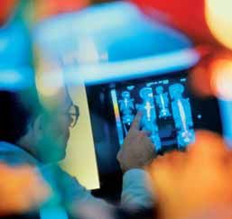
Targeting the Heart
                        HEART DISEASE WILL BE THE LEADING CAUSE OF DEATH IN THE DEVELOPING WORLD BY 2010.

              Every muscle in the body needs a constant flow of blood to remain healthy. The heart is a muscle too, so in
              addition to the blood that goes through the heart as it pumps, blood is carried into the heart muscle itself by a
              network of coronary arteries. This flow of blood supplies the heart muscle with oxygen and nutrients.

              In a person with heart disease, one or more of the coronary arteries has been narrowed or blocked by a build-
              up of fatty deposits. If the blood flow is reduced too much, starving the muscle of oxygen, part of the heart
              muscle is injured, causing a heart attack. This injured part of the heart muscle can be permanently damaged.

              Understanding heart disease risks
              Heart disease has several risk factors, including smoking, hypertension, or high cholesterol levels. But by far,
              diabetes remains the most serious risk factor. It is rising rapidly in developing countries, partly as a result of
              lifestyle changes, poor diet and rapid urbanization. When a patient has uncontrolled diabetes, glucose builds
              up in the bloodstream and heart tissue can be permanently damaged in only a few years.

                                                           Early diagnosis
                                                              Chongqing, China: Huáng Yunbiao* is a typical Chinese victim of heart
                                                              disease. Yunbiao was diagnosed with diabetes seven years ago. Last
                                                            year he was diagnosed with heart disease.

                                                When his doctor told him he had heart disease he was shocked. He’d never had a
                                                heart attack, and never even had any chest pain.

                                      Yunbiao’s doctor used nuclear medicine techniques to diagnose his heart disease,
                                 long before he began feeling symptoms. Yunbiao ran on a treadmill for about five or six
              minutes, and then doctors took images of his heart using a gamma camera.

              Yunbiao’s heart scans showed reduced blood flow when under stress that wasn’t seen while at rest. This
              reduced blood flow was jeopardizing the muscle tissue, and Yunbiao found that he needed treatment right away.

              What is a gamma camera?
              Radiopharmaceuticals injected into a patient produce                   Images of the heart taken with a SPECT gamma
              a signal which can be seen using a gamma camera —                      camera record how much blood is flowing into all parts
              a device that detects gamma radiation. Single-photon                   of the heart muscle.The results of nuclear cardiology
              emission computed tomography — or SPECT— uses a                        tests show the current status of blood flow to the heart
              rotating gamma camera to obtain images from multiple                   muscle and help doctors determine whether a patient
              angles of the organ being studied.                                     has heart disease, and if so, how severe it is.

              * To protect individual privacy, names and medical details have been changed in patient profiles.

                    4

Beyond the visible.indd 4                                                                                                               19/9/06 11:12:12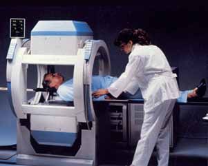
Making treatment decisions
          If the heart scans show that defects are minimal, medication for high blood pressure or removal of other risk
          factors might be an appropriate treatment. If, however, the image shows that the defects are large, the patient
          might be referred for angioplasty — a balloon that is inserted into a blood vessel to open up a blocked area in
          the vessel — or may be judged to be a candidate for bypass surgery. Nuclear medicine techniques help doctors
          decide whether a patient needs additional tests or treatment, and judge a patient’s risk of having a heart attack.

          Yunbiao’s SPECT tests were fast and painless. But more importantly, doctors found his condition
          early enough to take action. Since then, he has had three bypasses to keep his blood vessels
          open, and he is on medication. Thanks to SPECT, Yunbiao’s heart is healthier and now
          through treatment he is learning how to stay healthy.

          The Cost of SPECT                                              to experience symptoms — such as pain and
                                                                         shortness of breath — and ensure that the patient can
          The ability to obtain two measurements at the same             begin treatment early and continue living an active and
          time — one of blood flow to the heart and one of cardiac       productive life.
          pumping — is a value difficult to overlook. That is exactly
          what SPECT imaging offers.
                                                                                      In a study cited by the American Society of Nuclear
          Although SPECT is more expensive as an initial                              Cardiology, the use of SPECT for intermediate-risk
          diagnostic strategy, it is more cost effective because it is                              patients cut costs almost in
          more accurate in identifying the severity of the disease        without       with        half ($2 582) when compared with
                                                                          SPECT        SPECT
          and appropriately directing resources.                                                    direct cardiac catheterization without
                                                                          $4 854       $2 582
                                                                                                    SPECT ($4 854).
          For low-risk patients, SPECT can avoid unnecessary
          referrals for very expensive procedures, such as cardiac
          catheterization or coronary angiography, by filtering out
          patients who don’t need these procedures. SPECT can            The burden of heart disease
          also shorten the length of a patient’s hospital stay — also
          resulting in significant cost savings.                         Heart disease not only takes lives, but also causes
                                                                         an enormous economic burden. The World Health
                                                                         Organization estimates the total direct lifetime cost of
               Cutting the cost of treatment in half                     coronary heart disease, including primary care, clinical
                                                                         care and rehabilitation, at $26 billion, and the total
          Using a SPECT gamma camera, doctors can diagnose               indirect cost in loss of productivity, caused by disability
          coronary artery disease long before a patient begins           and death, at $48 billion.

                                                                                                                                       5

Beyond the visible.indd 5                                                                                                                    19/9/06 11:12:12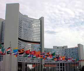
Lung Cancer — Customizing Care
                        DOCTORS NEED TO KNOW HOW FAR CANCER HAS SPREAD TO DETERMINE TREATMENT.

              Lung cancer is the uncontrolled growth of abnormal cells in one or both of the lungs. While normal cells
              reproduce and develop into healthy lung tissue, these abnormal cells reproduce rapidly and never grow into
              normal tissue. Lumps of cancer cells then form and disrupt the lung, making it difficult to function properly.

              Most lung cancers are smoking-related. Quitting smoking reduces risk significantly, although former smokers
              are still at greater risk of lung cancer than people who never smoked. Exposure to other carcinogens such as
              asbestos and radon gas also increases a person’s risk, especially when combined with smoking.

              Smoking on the rise
              While industrialized countries have seen a decline in the number of lung cancers after widespread anti-
              smoking campaigns, developing countries are experiencing the opposite trend. And while lung cancer is still
              more common in Europe and North America, this is changing as tobacco companies focus huge marketing
              campaigns on emerging markets. Of the ten million tobacco-related deaths expected by the World Health
              Organization for 2030, 70% of those will be in developing countries.

                                              Mumbai, India: Nadir Gupta is one of 1.2 million people diagnosed with lung
                                              cancer each year. He began smoking when he was fifteen, almost 35 years ago. But
                                            when he heard that he had lung cancer, quitting was easy.

                                 Nadir’s doctors used nuclear medicine techniques to understand the full extent of his lung cancer.
                             Determining the stage of the disease allows doctors to more accurately decide how to proceed with
                            care and treatment.

              What is staging?
              The term staging is used to describe the severity of the disease based on how far the cancer has spread and
              the size of the tumours. Staging helps doctors plan a patient’s treatment, and is used to assess the patient’s
              prognosis or the likely outcome or course of the disease.

              Staging is based on the knowledge of the way cancer develops. Cancer cells divide and grow without control or
              order to form a mass of tissue, called a tumour. As the tumour grows it can invade nearby organs and tissues.
              Cancer cells can also break away from the tumour and enter the bloodstream or lymphatic system. By moving
              through the bloodstream or lymphatic system, cancer can spread from the primary site to form new tumours in
              other organs. The spread of cancer is called metastasis.

                    6

Beyond the visible.indd 6                                                                                                       19/9/06 11:12:15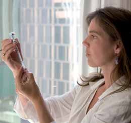
Refining the diagnosis
          Nadir’s doctor used Positron Emission Tomography — or PET — to determine the stage of the cancer and the
          best way to treat it. Doctors needed to see if surgery was still an option for Nadir, or if it had spread too far. For
          Nadir’s PET scan, doctors injected a radioactive sugar into his bloodstream to light up the tumours in his body.

          Doctors saw from Nadir’s PET scans that the cancer wasn’t limited to his lungs, but they could see that
          it hadn’t spread very far. Based on these images, his doctors knew that the best treatment for Nadir
          wasn’t surgery, but a more appropriate radiation treatment.

          How does PET contribute to staging?
          Positron Emission Tomography (PET) is a precise and sophisticated imaging technique using radioisotopes. PET
          makes it possible for the first time, to show both organ function and the development of the disease within it.

          Sugar is the fundamental source of energy for all body cells. Cancer cells, however, are hyperactive and therefore
          need more sugar than normal body cells. Simple sugars — glucose, for example — can be labelled with signal-
          emitting radioisotopes and injected into the patient. The PET scanner records the signals that these radioisotopes
          emit as they collect in organs targeted for examination. A computer then translates the signals into images.

                                                                                                                            7

Beyond the visible.indd 7                                                                                                       19/9/06 11:12:15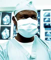
Breast Cancer — Finding the Right
              Treatment for the Patient
                        ONE OUT OF EIGHT WOMEN WILL DEVELOP BREAST CANCER.

              Less than two decades ago, breast cancer was not a significant health concern in Latin America. Today,
              breast cancer is the second leading cause of cancer deaths and there is an alarming increase in the number of
              women developing the disease.

              The risk factors for breast cancer cover a broad range of conditions, from age to inherited genes, reproductive
              history, alcohol consumption and high fat diets. In addition to age, family history is probably the most widely
              acknowledged and scientifically proven risk factor.

                                          Belém, Brazil: Juliana da Costa’s grandmother died of breast cancer. So she
                                          knew to begin examinations early. Because of her doctor’s vigilance, they were
                                         able to catch her breast cancer at an early stage.

                                Tracking the spread of cancer
                             One of the ways breast cancer spreads through the body is through the lymphatic system.
                        Breast cancer doesn’t spread randomly into the underarm lymph nodes, however, but tends to
                       spread in an orderly fashion through a ‘sentinel’ node before becoming more generally disseminated.
                     The sentinel node is simply the first node in a chain of nodes. If one imagines a string of pearls lying
              tangled on a dressing table, the cancer has to go through the first pearl to get to the rest.

              Until recently, standard surgical practice for breast cancer was to remove the underarm lymph nodes in order
              to assess the prognosis of the disease, which is related to the number of lymph nodes involved. There are 25 to
              30 underarm nodes which must be examined in a laboratory to see whether they are cancerous, and if so how many.

              Avoiding unnecessary pain and side effects
              Removing the underarm lymph nodes may cause swelling, arm pain, or motor deficiencies. For the vast
              majority of these removals, this surgery may not be necessary. A new nuclear medicine procedure, called
              sentinel lymph node detection, could spare patients this pain and yet yield the information needed to
              determine the best treatment.

              What is Sentinel Lymph Node Detection?
              Sentinel lymph node detection involves injecting a radiopharmaceutical near the breast tumour site. After the
              injection, the tracer is drained through the lymphatic vessels. In the operating theatre, a gamma probe — a hand-
              held device that detects gamma radiation — is used to locate the area of radioactivity and detect node location.

              Doctors find and remove the sentinel node, and then analyse it to detect even the smallest deposit of metastatic
              cancer cells. This procedure allows for detection of lymph nodes that are free of cancer thus identifying patients
              who don’t need aggressive surgery.

                    8

Beyond the visible.indd 8                                                                                                  19/9/06 11:12:27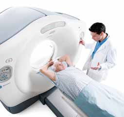
Breast cancer under control
          Thanks to improved screening programmes breast cancer is increasingly detected at an early stage, before
          the lymph nodes are involved. Furthermore, understanding about the spread of cancer in lymph nodes and the
          combination of nuclear medicine techniques such as sentinel lymph node detection, are improving the quality
          of life for patients.

          Juliana’s doctors used sentinel lymph node detection and found that her cancer hadn’t spread to
          the lymph nodes. She didn’t need to have the underarm lymph nodes removed, and was spared the
          pain involved with this surgery.

          Nuclear medicine in therapy                                 Nuclear medicine to relieve pain
          Rapidly dividing cells are particularly sensitive to        Sometimes certain types of cancers may progress to
          damage by radiation. For this reason, some cancerous        a painful disorder of the bone, known as metastatic
          growths can be controlled or eliminated by irradiating      bone disease. This occurs when cancer cells from the
          the area containing the growth.                             original tumour area travel through the body and move
                                                                      into the bone. Bone pain can make normal activities
          External radiation can be carried out using a beam from     challenging, sleep difficult and food unappetizing.
          a radioactive source. The important point however,          Most patients need some form of treatment to deal
          is to deliver the maximum amount of radiation to the        with this pain.
          cancer cells.Therapeutic radiopharmaceuticals do just
          that, killing the cancer cells, from the inside out.        Fortunately, important advances have been made
                                                                      using radiopharmaceuticals to ease the severe pain
          A biological substance tagged with a radioisotope will      related to metastatic bone disease when many bones
          follow its usual biological path to bring the radioactive   are affected.
          element to the malfunctioning cells. Treating these
          cells with a therapeutic dose of radiation, customized      Radiopharmaceuticals are injected into the body and
          for those cells, may lead to the regression — or cure       seek out areas of bone that are undergoing cell division,
          — of some diseases.                                         a central part of the bone’s response to new cancer cell
                                                                      growth. Radiopharmaceuticals accumulate in these
          Cancer therapies involving radioisotopes or radio-          affected areas and work to kill cancer cells, thereby
          immunotherapy may well lead the way into a more             lessening existing bone-related pain and possibly even
          promising future for millions around the world.             delaying the development of new areas of pain.

                                                                      The whole procedure takes only a few minutes and,
                                                                      in some cases, a single injection can relieve pain
                                                                      for an average of three to six months — without the
                                                                      disorientation, drowsiness and uncomfortable side
                                                                      effects of other types of pain treatment.

                                                                                                                           9

Beyond the visible.indd 9                                                                                                      19/9/06 11:12:27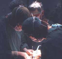
The Role of the IAEA
                        THE UNQUESTIONABLE BENEFITS OFFERED BY NUCLEAR MEDICINE ARE AVAILABLE TO
                        ALL MEMBER STATES.

              The IAEA has a unique mandate — to “accelerate and enlarge the contribution of atomic energy to peace,
              health, and prosperity throughout the world.” The Agency is committed to enhancing the capabilities of
              developing Member States in addressing major health problems like heart disease and cancer using nuclear
              medicine techniques.

              To achieve this goal, the IAEA establishes new practices, transfers updated technology, implements
              appropriate diagnostic and therapeutic applications, and provides targeted education and training for Member
              States.

              Advancing the field of nuclear medicine
              The IAEA’s Coordinated Research Projects (CRPs) support international research efforts to develop new
              scientific approaches in nuclear medicine. In these projects, the IAEA brings scientists together from
              developed and developing Member States to exchange knowledge and research capabilities while working
              together on areas of common interest.

              Recent CRPs in nuclear medicine have identified the best therapeutic strategy to use beta-emitting
              radiopharmaceuticals to treat inoperable liver cancer, and to support indigenous production of new
              radiopharmaceuticals for the early diagnosis of cancer.

              Distributing technology resources
              Nuclear medicine relies on advanced technologies such as SPECT and PET. However, the global reality today
              is one where technology and facilities are not evenly distributed between countries.

              Through Technical Cooperation projects, the IAEA provides equipment necessary to establish and maintain
              technical facilities in Member States. Recent projects have provided SPECT cameras to Ecuador, Malta,
              Nicaragua, Tajikistan, Uruguay and Uzbekistan, among others, as well as provided advice to implement PET
              programmes for cancer management in Argentina, Chile, Malaysia, Thailand and Vietnam.

              Implementing diagnostic and therapeutic applications
              The IAEA also provides Member States with technical expertise and infrastructure development in national and
              regional projects. Recently the IAEA helped establish completely new nuclear medicine programmes in Kenya,
              Mali, Senegal and Sudan, and helped re-establish facilities that had been shut down for more than ten years
              in the Former Yugoslav Republic of Macedonia, Montenegro and Serbia. The IAEA’s work to implement
              diagnostic and therapeutic applications has also raised the level of clinical practice in facilities in Malaysia,
              Thailand and Vietnam.

              Providing comprehensive education
              The IAEA provides on-site local training and professional advice from IAEA staff and external experts. It
              organizes several of these training courses and consultants meetings around the world each year to ensure the
              good practice of nuclear medicine.

                   10

Beyond the visible.indd 10                                                                                                 19/9/06 11:12:29
Distance Assisted Training (DAT) is a fellowship programme which provides support for doctors from
countries without formal university programmes in nuclear medicine. Under supervision from trained doctors,
participants follow learning modules to receive comprehensive training for two to three years. Recent pilot
programmes with participants from Bolivia, Colombia, Thailand and Vietnam have resulted in a significant
improvement in the level of local practice of nuclear medicine.
Sharing critical information
The IAEA has developed the Nuclear Medicine Database (NUMDAB) to gather and maintain information on
the nuclear medicine practices around the world. The database provides information on the current status of
nuclear medicine infrastructures and educational systems. It also assists in planning approaches to emerging
needs, as well as in prioritizing the educational and operational needs related to strengthening nuclear
medicine practices in Member States.
Working together
The IAEA brings together consultants from developed and developing countries to learn more about the needs of
Member States and how best to provide for them. For more information, please visit www-naweb.iaea.org/nahu/nm.
Cover :
Photo Disk
Photo Disk
SPECT scans light up in bright colour where blood flows to the heart; uneven rings here indicate reduced blood flow to the muscle tissue.
Credit: M. Dondi/IAEA
Inside Cover:
Early and accurate diagnosis is vital for effective treatment of both heart disease and cancer. Credit: GE Healthcare
Page 5:
Heart disease is on the rise in China, caused by lifestyle changes, poor diet and rapid urbanization. Credit: Getty Images
Doctors can use nuclear medicine techniques to determine if a patient has heart disease and decide on the best treatment.
Credit: Getty Images
A SPECT gamma camera takes images of a patient’s heart at rest and under stress to show blood flow to heart muscle tissue.
Credit: A. Soricelli/IAEA
Page 7:
A PET/CT scanner records the signals emitted by radioisotopes to show organ function and the development of the disease.
Credit: Getty Images
According to the American Cancer Society India Initiative, tobacco-related cancers account for almost one-third of all cancers in India.
Credit: Getty Images
Developing countries like India are experiencing a rapid rise in the incidence of cancer. Credit: Getty Images
Page 9:
Surgeons use a gamma probe to detect lymph nodes that are free of cancer. Credit: IAEA
Breast cancer cases are increasing in Brazil and throughout Latin America, where the disease is the second leading cause of cancer
deaths. Credit: Getty Images
Radiopharmaceuticals injected near the breast tumour site will lead doctors to identify the ‘sentinel’ lymph node. Credit: Getty Images
Page 11:
PET scans are painless procedures, and all traces of the radioisotopes leave the body naturally after a few hours. Credit: GE Healthcare
The IAEA helps to provide both the equipment and essential professional expertise to better diagnose and treat heart disease and cancer
in the developing world. Credit: Getty Images
IAEA headquarters at the Vienna International Centre in Austria. Credit: D. Calma/IAEA
11
Beyond the visible.indd 11 19/9/06 11:12:29Contributors from the Nuclear Medicine Section:
Maurizio Dondi, Section Head
Baldip Khan
Kishor Solanki
John Zaknun
Naoyuki Watanabe
Dagmar Umgeher
K. Shepherd (Writer)
For more information about the Nuclear Medicine Section of the IAEA,
please visit www-naweb.iaea.org/nahu/nm.
Division of Public Information:
D. Kinley III (Editor), A. Diesner-Kuepfer (Design)
Wagramer Strasse 5, P.O. Box 100, A-1400 Vienna, Austria
Tel.: (+43 1) 2600 21270/21275
Fax: (+43 1) 2600 29610
E-Mail: info@iaea.org / www.iaea.org
Printed by the IAEA in Austria, September 2006
IAEA/PI/A.91 / 06-29901
12
Beyond the visible.indd 12 19/9/06 11:12:30You can also read