Levels of DNA cytosine methylation in the Drosophila genome - PeerJ
←
→
Page content transcription
If your browser does not render page correctly, please read the page content below
Levels of DNA cytosine methylation in
                                        the Drosophila genome
                                        Saniya Deshmukh1 , VK Chaithanya Ponnaluri2 , Nan Dai2 , Sriharsa Pradhan2 and
                                        Deepti Deobagkar1
                                    1
                                      Molecular Biology Research Laboratory; Department of Zoology (Centre for Advanced Studies), Savitribai
                                      Phule Pune University (formerly University of Pune), Pune, Maharashtra, India
                                    2
                                      New England Biolabs, Ipswich, MA, United States of America

                                            ABSTRACT
                                            Insects provide an accessible system to study the contribution of DNA methylation
                                            to complex epigenetic phenotypes created to regulate gene expression, chromatin
                                            states, imprinting and dosage compensation. The members of genus Drosophila have
                                            been used as a model system to study aspects of biology like development, behaviour
                                            and genetics. Despite the popularity of Drosophila melanogaster as a genetic and
                                            epigenetic model organism, DNA methylation studies are limited due to low levels of
                                            genomic 5-methylcytosine. Our study employs a sensitive liquid chromatography-mass
                                            spectrometry (LCMS) based method to quantify the levels of 5-methylcytosine from
                                            the genomic DNA in different members of the genus Drosophila. Our results reveal
                                            that, despite being phylogenetically related, there is a marked variation in the levels
                                            of 5-methylcytosine between the genomes of the members of genus Drosophila. Also,
                                            there is a change in the genomic levels of 5-methylcytosine through each life cycle stage
                                            of holometabolous development in D. melanogaster.

                                        Subjects Biochemistry, Molecular Biology, Zoology
                                        Keywords DNA methylation, 5-methylcytosine, Drosophila , UHPLC-QQQ

Submitted 1 March 2018
Accepted 7 June 2018
                                        INTRODUCTION
Published 2 July 2018                   The epigenome of an organism constitutes histone modifications (Zentner & Henikoff,
Corresponding authors                   2013), non-coding RNA molecules (Busto et al., 2015; Stuwe, Tóth & Aravin, 2014) and
Sriharsa Pradhan, Pradhan@neb.com
Deepti Deobagkar,                       nucleotide modifications (Achwal, Ganguly & Chandra, 1984; Achwal, Iyer & Chandra,
deepti.deobagkar@gmail.com              1983; Gowher, Leismann & Jeltsch, 2000; Zhang et al., 2015). These modifications can
Academic editor                         together or independently influence the regulation of gene expression and conserve
Juan Riesgo-Escovar                     the energy resources by managing functional conformation of the genome (Zee et al.,
Additional Information and              2016). The epigenetic changes are important in the insect taxon which commonly exhibits
Declarations can be found on
page 10
                                        polyphenisms (Simpson, Sword & Lo, 2011). Unlike mammals and plants, the sparse
                                        methylation of gene bodies and transposons is characteristic of insect DNA methylation
DOI 10.7717/peerj.5119
                                        (Li-Byarlay, 2016). In social insects with the canonical DNMT1 and DNMT3A/3B
    Copyright                           methyltransferases, DNA methylation can be attributed to differential splicing, regulation
2018 Deshmukh et al.
                                        of expression and histone occupancy, whereas very little is known in solitary insects which
Distributed under                       mostly possess only DNMT2 methyltransferase (Glastad, Hunt & Goodisman, 2014).
Creative Commons CC-BY 4.0
                                           The members of genus Drosophila undergo holometabolous development beginning
OPEN ACCESS                             with the embryonic stage which eventually develops into an adult fly after passing through

                                        How to cite this article Deshmukh et al. (2018), Levels of DNA cytosine methylation in the Drosophila genome. PeerJ 6:e5119; DOI
                                        10.7717/peerj.5119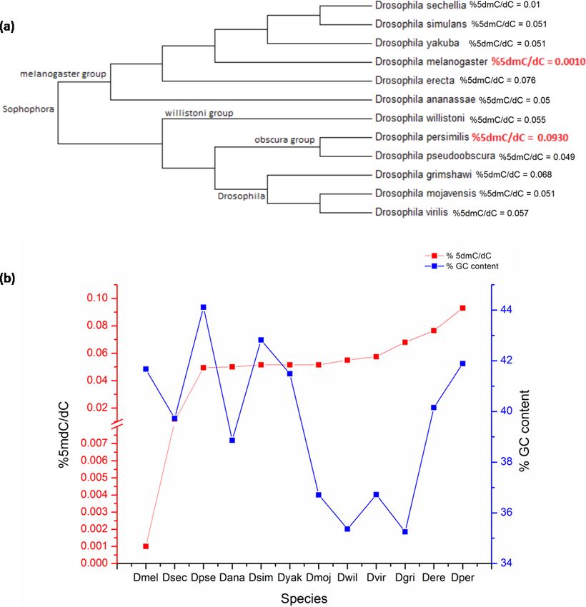
larval and pupal stages (Hartenstein, 1993). There are various changes observed in the DNA
                                  methylation patterns of model systems like the house mouse during their development
                                  from embryo to adulthood (Smith et al., 2012; Smith & Meissner, 2013). Some previous
                                  studies report the presence of low levels of 5-methylcytosine (5mC) and an active DNA
                                  methyltransferase in Drosophila melanogaster (Achwal, Ganguly & Chandra, 1984; Achwal,
                                  Iyer & Chandra, 1983; Gowher, Leismann & Jeltsch, 2000; Panikar et al., 2015). Recently, the
                                  presence of 5mC (less than 1%) in D. melanogaster genome was confirmed by 5mC-specific
                                  immunoprecipitation followed by bisulfite sequencing in stage 5 embryos (Takayama et
                                  al., 2014) and liquid chromatography/tandem mass spectrometry in adult stage (Capuano
                                  et al., 2014; Rasmussen et al., 2016). Bisulphite sequencing is a commonly used method for
                                  detection of genome-wide DNA methylation as it provides sequence context information
                                  of methylated cytosine residues. However, this method has limitations due to the lower
                                  concentration range, incomplete bisulphite conversion of unmethylated cytosines and
                                  misalignment of sequenced reads due to genomic repeats, telomeres and GC-rich regions
                                  (Warnecke et al., 2002). Using an alternative LCMS-based protocol, Capuano et al., 2014
                                  have reported DNA methylation over a large range from 0.034% of cytosines methylated
                                  in a sample with females and males in equal proportion of wt/w118 D. melanogaster adults
                                  to 7.6% in liver tissue from Mus musculus and 14% from leaf tissue of Arabidopsis thaliana.
                                  The level of DNA methylation in Drosophila is 10–100 folds below the detection limit of
                                  bisulphite sequencing. This study established the advantage of an LCMS-based method to
                                  assess the levels of DNA methylation in systems like Drosophila with low levels of 5mC.
                                      It is known that DNA methylation patterns change due to the life cycle stage, the tissue or
                                  cell type and the age of the specimen under consideration (Lokk et al., 2014). Independent
                                  studies mentioned above have demonstrated the presence of 5mC by different techniques
                                  in the embryo and adult stages of D. melanogaster. We have estimated the amount of 5mC
                                  in the genome of D. melanogaster (across all the life cycle stages) and other 11 species from
                                  genus Drosophila using ultra-high performance liquid chromatography/triple quadrupole
                                  mass spectrometry. Our analysis determines the change in the levels of 5mC during
                                  holometabolous development within a species and also between member species of genus
                                  Drosophila.

                                  MATERIALS & METHODS
                                  Sample preparation and DNA isolation
                                  The dechorionated 8–14 h old embryos from a synchronised batch of flies, larvae from third
                                  instar stage, male and female pupae, adult male and female flies (one day old) were collected
                                  from the laboratory culture of Oregon-R strain maintained on corn-meal medium under
                                  standard conditions. These samples were washed in 70% ethanol and 1X PBS (to surface
                                  sterilise the larva and adult), then processed for DNA isolation. Briefly, the samples were
                                  homogenised after snap-chilling in liquid nitrogen and incubated at 65 ◦ C with RNase A
                                  (12091–021) for 4 h; followed by standard phenol-chloroform extraction. Treatment with
                                  1:2.5 parts of 5M potassium acetate and 6M lithium chloride to remove any traces of RNA
                                  was given followed by precipitation by iso-propanol. The DNA samples were dissolved

Deshmukh et al. (2018), PeerJ, DOI 10.7717/peerj.5119                                                                       2/13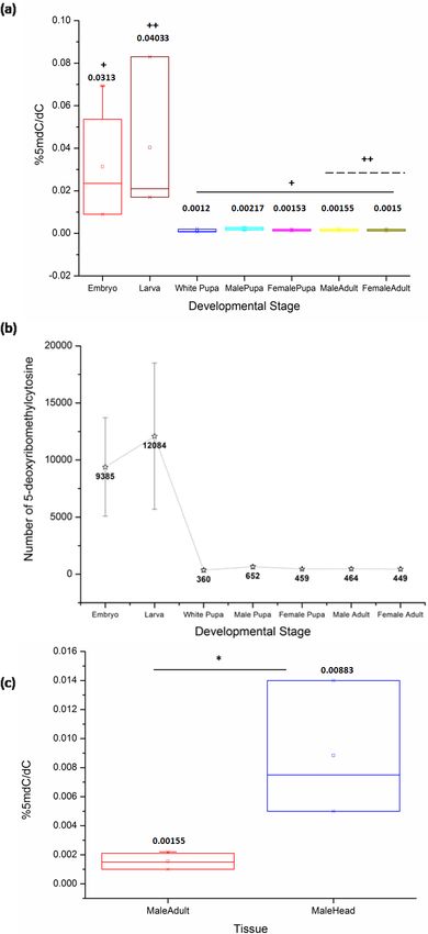
in TE buffer. The samples for all the 12 species of genus Drosophila analysed were also
                                  obtained in parallel from UCSD Drosophila Species Stock Center, USA. The DNA samples
                                  were checked for integrity using an agarose gel and NanoDrop full-spectrum, UV-Vis
                                  spectrophotometers which ensure quality output.

                                  Ultra-high performance liquid chromatography/triple quadrupole
                                  mass spectrometry analysis (UHPLC/QQQ)
                                  DNA (500 ng) was digested in 20 µL total volume with a NEB nuclease enzymes mix
                                  for 2 hr to obtain individual nucleosides. dU was spiked in as an internal control for all
                                  samples. The percentage of 5mdC/dC present in the sample was obtained by determining
                                  d5mC/dU and dU/dC values. The samples were resolved on a waters column (XSelect HSS
                                  T3 XP column (2.1 × 100 mm, 2.5 µm)) using ammonium formate and methanol buffer
                                  system on an Agilent 1290 UHPLC with 6490 QQQ Mass detector in MRM mode which
                                  is an established method for nucleoside detection (Estève et al., 2014). Similarly, the adult
                                  samples of 12 species obtained from UCSD Drosophila Species Stock Center, USA were
                                  analysed.
                                     The samples were run in minimum biological triplicates and technical duplicates
                                  to ensure reproducible values; the multiple and pair-wise sample comparisons were
                                  made by Kruskal-Wallis with post hoc tests (Mann–Whitney U and Dunn’s test) and
                                  Mann–Whitney U tests respectively using PAST (Hammer, Harper & Ryan, 2001) and
                                  GraphPad Prism version 5 for Windows (GraphPad Software, La Jolla, CA, USA;
                                  http://www.graphpad.com/). The statistical tests ensured within and between group
                                  variance and effect of outliers (if any) were accounted for in the comparisons.

                                  Nucleotide counting and estimation of GC content for twelve genus
                                  Drosophila
                                  The FlyBase is an extensive online sequence resource; encompassing fully sequenced
                                  genomes for twelve species of genus Drosophila which can be used for comparative
                                  genomic analyses (Crosby et al., 2006; Gramates et al., 2017). These are the twelve most
                                  commonly used, annotated Drosophila genomes. The latest whole genome sequences
                                  releases were downloaded from the FlyBase database. A Perl code was constructed and
                                  used for counting the number of individual nucleotides from each sequence fasta file and
                                  the percent GC was calculated. The written code was cross-checked with resource material
                                  available on Biostars (https://www.biostars.org/) and the veracity of the output count for
                                  the code was performed by comparing the nucleotide counts and genome length with the
                                  current release details of D. melanogaster genome. The UHPLC output counts methylated
                                  cytosines per 100 cytosines counted; this data was used to extrapolate the number of 5mC
                                  per haplogenome.

                                  Phylogenetic analysis for the for twelve genus Drosophila
                                  18S rRNA gene sequences for all the twelve species of genus Drosophila were extracted
                                  from the NCBI repository. The phylogenetic tree was constructed using MEGA version
                                  6 software (Tamura et al., 2013). The sequences were aligned using the Muscle multiple

Deshmukh et al. (2018), PeerJ, DOI 10.7717/peerj.5119                                                                     3/13
Table 1 The percentage of 5mC nucleotides per cytosine nucleotides counted in different developmen-
tal stages of D. melanogaster.
Developmental stage % 5mdC/dC Std. error
Late embryos 0.026 0.0137
3rd instar larva 0.025 0.0165
White pupa 0.001 0.0004
Male pupa 0.002 0.0003
Female pupa 0.001 0.0002
1d Male adult 0.001 0.0003
1d Female adult 0.001 0.0002
alignment option using the default parameters. The phylogenetic tree was constructed
using the neighbour-joining (NJ) method with 1,000 bootstrapping.
Identification of methyltransferases homologs in genus Drosophila
In order to identify the presence of a common methyltransferases protein present in the
twelve members of genus Drosophila. The protein sequence of the only identified DNA
methyltransferase, DNMT2 from D. melanogaster, was used as a query for the BLAST
algorithm. The proteins from the other eleven members with 70–100% identity were
selected. The selected twelve sequences were uploaded to the PROMALS3D server to
generate a multiple sequence alignment and identification of secondary protein structures
(Pei, Kim & Grishin, 2008).
RESULTS
In order to analyse the presence of DNA methylation in the members of genus Drosophila
we first standardised the protocol for detection of 5mC detection in D. melanogaster using
the UHPLC/QQQ.
The comparison of samples from the life cycle stages of D. melanogaster by Kruskal-
Wallis (KW) test shows a significant difference between the levels of 5mC (KW, p = 0.001).
The late stages of the embryo in our study represent the stages of development after
cellularization and the germ band elongation has been completed. The embryonic stages
from 12 to 16 used for representing the late embryos maintain the same general body plan
across the later stages in the life cycle. These embryonic stages exhibit significantly high
levels of 5mC amongst all the life cycle stages of D. melanogaster.
Also, there is a significant difference between DNA methylation of the third instar larva
and both sexes of the adult stage (MW, p = 0.04). The ratio of %5mdC/dC does not show
statistically significant difference amongst other stages in the life cycle (Fig. 1A) (Table 1,
Supplementary File). There is a decrease in the levels of 5mC after metamorphic transition
following the larval stage which is maintained throughout development. The early pupae
or white pupae, the sexually distinct pupal and adult stages show similar levels of global
DNA methylation. This indicates that there is no sex-specific difference in the levels of
5mC in age-matched samples of the same stage.
Deshmukh et al. (2018), PeerJ, DOI 10.7717/peerj.5119 4/13Figure 1 The percentage of 5mC per cytosine nucleotides across the developmental stages. (A) + in-
dicates a significant difference between the embryo and other stages, whereas ++ indicates a significant
difference between the larval and adult stages in both sexes (Using a combination of the current D. melanogaster genome release by FlyBase and our
results with a Perl script to count the number of deoxyribonucleotides in the genome, we
have extrapolated the number of modified cytosines in each developmental stage (details
in Supplementary File, sheet 2). The number of methylated cytosines per copy of genome
in each developmental stage and nucleotide composition of the fly genome are represented
in (Fig. 1B).
Although the overall levels remain comparable, the changes in the local patterns of
DNA cytosine methylation cannot be ruled out. The head tissue distinctly has a higher
level of 5mC when compared to the whole body of the adult (MW, p = 0.035) (Fig. 1C).
This indicates that in Drosophila methylation patterns can be rearranged and redistributed
between different tissues. There is no significant difference between the heads of male and
female adult D. melanogaster (Fig. S1). The presence of higher level of 5mC in the head also
eliminates the possibility of methylation being detected due to the possible contamination
from gut microflora or endosymbionts as a false positive in Drosophila genomic DNA.
D. melanogaster is a member of the melanogaster group of the subgenus Sophophora
and is one of 12 fruit fly species with completely sequenced genome for comparative
genomics. There is no date available on the presence of DNA methylation in the other
11 species. In order to assess the distribution of 5mC across genus Drosophila; DNA
from the adult stage of twelve prominent member species from subgenera Drosophila and
Sophophora was analysed. The bootstrap consensus tree for all the species is shown to
provide information on the most frequently appearing branch groupings using the 18S
rRNA sequences (original NJ tree and divergence time in Figs. S2 and S3) (Fig. 2A). There is
a significant difference between the values of the 5mC amongst the twelve-species analysed
(p = 0.0217, KW). The members of subgenus Drosophila carry more 5mC in their genome
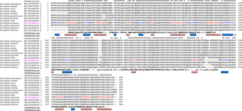
when compared to the Sophophora (Stage & Eickbush, 2007). Drosophila melanogaster has
the lowest while Drosophila persimilis has the highest levels of 5mC amongst the analysed
genomes. Our results show that even closely related species show a difference in the levels
of DNA methylation; D. simulans and D. sechellia are closely related species from the
melanogaster group but have a substantial difference in levels of 5mC in their genome.
The levels of 5mC observed across these species do not seem to show any correlation with
their phylogenetic relatedness. The members of the melanogaster group posses a diverse
profile of global DNA methylation. The lowest level DNA methylation value of 0.001%
5dmC/dC is observed in the genome of D. melanogaster and Drosophila erecta genome has
the 0.0765% 5dmC/dC between the members of the melanogaster group. However, the
subgenus Drosophila possesses a comparable distribution of 5mC among its members.
From the available genome releases and our data, we have compared the GC content,
the values of 5mC and C present and %5mdC/dC for the twelve species (Fig. 2B, Table
2, Figs. S1 and S2). Drosophila grimshawi and Drosophila willistoni possess the lowest
number of cytosines in their genomes while Drosophila pseudoobscura has the highest level
of cytosines. Despite the lowest GC content, D. grimshawi and D. willistoni have 0.068
and 0.055% 5dmC/dC which is comparable to 0.0495% 5dmC/dC in D. pseudoobscura.
Likewise, with a vast difference in the number of 5mC per genome copy, D. melanogaster
and D. persimilis have similar genomic GC content (Supplementary File, sheet 3).
Deshmukh et al. (2018), PeerJ, DOI 10.7717/peerj.5119 6/13Figure 2 The percentage of 5mC nucleotides per cytosine nucleotides and the percentage of GC
content in the genomes of members of Drosophila species. (A) Phyogenetic relationship between the
members of genus Drosophila. The two species with the highest and the lowest levels of 5mC are indicated
in red. (B) The left Y-axis represents percentage of 5mC nucleotides per cytosine nucleotides counted
and the right Y -axis represents the percentage of GC content of the genome; the X -axis represents D.
melanogaster (Dmel), D. sechellia (Dsec), D. pseudoobscura (Dpse), D. ananassae (Dana), D. simulans
(Dsim), D. yakuba (Dyak), D. mojavensis (Dmoj), D. willistoni (Dwil), D. virilis (Dvir), D. grimshawi
(D.gri), D. erecta (Dere), D. persimilis (Dper). (< 0.05, p = 0.0217, KW test for comparison of means).
Full-size DOI: 10.7717/peerj.5119/fig-2
D. melanogaster has been identified as one of the DNMT2 only organism, as there is an
absence of any other de novo or maintenance methyltransferases (Lyko & Maleszka, 2011).
DNMT2 protein from D. melanogaster as the query protein for the BLAST algorithm,
similar protein was identified from the other analysed Drosophila species. The sequences
with a high identity score between 70–100% and strong E value and at least 95% query
coverage were filtered from the obtained results. The multiple sequence alignment of the
extracted sequences similar to the DNMT2 of D. melanogaster show strongly conserved
Deshmukh et al. (2018), PeerJ, DOI 10.7717/peerj.5119 7/13Figure 3 The multiple alignments of protein sequences retrieved from the NCBI repository with conserved domain architecture with DNMT2
methyltransferase in D. melanogaster. The ‘e’ and ‘h’ regions represent beta strand and alpha helix consensus secondary structures from the
residues in the twelve proteins.
Full-size DOI: 10.7717/peerj.5119/fig-3
Table 2 The 5mC count per haplogenome, percentage of 5mC nucleotides per cytosine nucleotides
counted in genome and percent GC content for member species of genus Drosophila.
Species Total number of 5mC/haplogenome %5dmC/dC %GC content
D. melanogaster 300 0.001 42
D. sechellia 3,472 0.010 40
D. pseudoobscura 16,675 0.049 44
D. ananassae 22,468 0.050 39
D. simulans 13,774 0.051 43
D. yakuba 17,696 0.051 41
D. mojavensis 18,327 0.051 37
D. willistoni 22,910 0.055 35
D. virilis 21,769 0.057 37
D. grimshawi 24,046 0.068 35
D. erecta 23,451 0.076 40
D. persimilis 36,691 0.093 42
regions; they also bear identical secondary structures from the consensus residues aligned
using PROMALS3D server (Fig. 3) (Pei, Kim & Grishin, 2008). The possibility of the
existence of an unknown methyltransferase or a protein complex can be considered as
studies in D. melanogaster embryos with DNMT2 knockout has been shown to have
persistence of DNA methylation (Takayama et al., 2014).
Deshmukh et al. (2018), PeerJ, DOI 10.7717/peerj.5119 8/13DISCUSSION
HPLC-based analysis has shown the presence of 0.034% methylation in a mixed population
of w1118 adult flies and 0.002% 5-methylcytosine/cytosine in adult females of ore-R lab
strain in the genome of D. melanogaster. Also, there is a gene-specific distribution of
5mC methylation during D. melanogaster development (Panikar et al., 2017). Our current
analysis reports a change in the levels of global DNA methylation using UHPLC/QQQ
which allows detection of 5mC with greater accuracy as compared to previously used
antibody-based techniques. The method used overcomes limitations of bisulfite-sequencing
due to the low concentration of 5mC in Drosophila but does not provide sequence context
information on DNA methylation. Hence, we are unable to extrapolate the location of the
methylation with respect to annotated genomic features like the transposable elements,
promoters, repeats etc.
DNA methylation is present in many insects like hymenopterans, lepidopteran, etc., but
is much lower than in mammals and plants (Glastad, Hunt & Goodisman, 2014; Simola et
al., 2013; Xiang et al., 2010). The functional importance of DNA methylation in insects is
better understood in the eusocial systems like honey bees and wasps (Hunt et al., 2013).
The existing evidence suggests that DNA methylation in the eusocial systems delegates the
social order by means of alternative splicing or ploidy variation (Glastad et al., 2014; Lyko
et al., 2010).
Most Dipterans are known to lack DNMT1, 3A and 3B and appear to possess DNMT2
as the only known DNA methyltransferase (Glastad, Hunt & Goodisman, 2014). Despite
reports on the presence of DNA methylation in solitary or non-social insects like stick insects
and moths; not much is known about its role and importance. The exact involvement of
genomic methylation in molecular processes of Drosophila, if any, remains unexplored.
CONCLUSION
Our analysis of the complete life cycle of D. melanogaster and eleven other species
conclusively establishes the presence of DNA methylation in the members of genus
Drosophila. This modification undergoes change during holometabolous development.
Also, it suggests that there is no effect from factors like sex, phylogenetic relatedness and
genome GC content on the levels of 5mC in Drosophila. Despite the absence of typical
(DNMT1 and DNMT 3A/3B) DNA methyltransferases, there is DNA methylation in all
twelve species of genus Drosophila. These findings pose an interesting problem that demand
further investigation which can lead to the identification of a methyltransferase enzyme
with novel functions.
ACKNOWLEDGEMENTS
We thank Drs. Donald Comb, Rich Roberts, William Jack and Clotilde Carlow at NEB for
research support and encouragement.
Deshmukh et al. (2018), PeerJ, DOI 10.7717/peerj.5119 9/13ADDITIONAL INFORMATION AND DECLARATIONS
Funding
Saniya Deshmukh and Deepti Deobagkar are supported by UGC-UPE Phase II
Biotechnology [UGC-262(A)(1)] and SPPU-DRDP grants, respectively. The funders had
no role in study design, data collection and analysis, decision to publish, or preparation of
the manuscript.
Grant Disclosures
The following grant information was disclosed by the authors:
UGC-UPE Phase II Biotechnology: UGC-262(A)(1).
SPPU-DRDP.
Competing Interests
Chaithanya Ponnaluri, Nan Dai and Sriharsa Pradhan are employed by New England
Biolabs.
Author Contributions
• Saniya Deshmukh conceived and designed the experiments, performed the experiments,
analyzed the data, contributed reagents/materials/analysis tools, prepared figures and/or
tables, authored or reviewed drafts of the paper, approved the final draft.
• V K. Chaithanya Ponnaluri conceived and designed the experiments, performed the
experiments, analyzed the data, contributed reagents/materials/analysis tools, authored
or reviewed drafts of the paper, approved the final draft.
• Nan Dai conceived and designed the experiments, performed the experiments, analyzed
the data, contributed reagents/materials/analysis tools.
• Sriharsa Pradhan and Deepti Deobagkar conceived and designed the experiments,
contributed reagents/materials/analysis tools, authored or reviewed drafts of the paper,
approved the final draft.
Data Availability
The following information was supplied regarding data availability:
The raw data are provided in the Supplemental Files.
Supplemental Information
Supplemental information for this article can be found online at http://dx.doi.org/10.7717/
peerj.5119#supplemental-information.
REFERENCES
Achwal C, Ganguly P, Chandra HS. 1984. Estimation of the amount of 5-methylcytosine
in Drosophila melanogaster DNA by amplified ELISA and photoacoustic spec-
troscopy. The EMBO Journal 3:263–266.
Deshmukh et al. (2018), PeerJ, DOI 10.7717/peerj.5119 10/13Achwal CW, Iyer CA, Chandra HS. 1983. Immunochemical evidence for the presence
of 5mC, 6mA and 7mG in human, Drosophila and mealybug DNA. FEBS Letters
158:353–358 DOI 10.1016/0014-5793(83)80612-7.
Busto GU, Guven-Ozkan T, Fulga TA, Van Vactor D, Davis RL. 2015. microRNAs
that promote or inhibit memory formation in Drosophila melanogaster. Genetics
200:569–580 DOI 10.1534/genetics.114.169623.
Capuano F, Mülleder M, Kok R, Blom HJ, Ralser M. 2014. Cytosine DNA methylation is
found in Drosophila melanogaster but absent in Saccharomyces cerevisiae, Schizosac-
charomyces pombe, and other yeast species. Analytical Chemistry 86:3697–3702
DOI 10.1021/ac500447w.
Crosby MA, Goodman JL, Strelets VB, Zhang P, Gelbart WM, The FlyBase Consor-
tium. 2006. FlyBase: genomes by the dozen. Nucleic Acids Research 35:D486–D491
DOI 10.1093/nar/gkl827.
Estève P-O, Terragni J, Deepti K, Chin HG, Dai N, Espejo A, Corrêa IR, Bedford
MT, Pradhan S. 2014. Methyllysine reader plant homeodomain (PHD) finger
protein 20-like 1 (PHF20L1) antagonizes DNA (cytosine-5) methyltransferase 1
(DNMT1) proteasomal degradation. Journal of Biological Chemistry 289:8277–8287
DOI 10.1074/jbc.M113.525279.
Glastad KM, Hunt BG, Goodisman MA. 2014. Evolutionary insights into DNA methyla-
tion in insects. Current Opinion in Insect Science 1:25–30
DOI 10.1016/j.cois.2014.04.001.
Glastad KM, Hunt BG, Soojin VY, Goodisman MA. 2014. Epigenetic inheritance and
genome regulation: is DNA methylation linked to ploidy in haplodiploid insects?
Proceedings of the Royal Society of London B: Biological Sciences 281(1785):Article
20140411 DOI 10.1098/rspb.2014.0411.
Gowher H, Leismann O, Jeltsch A. 2000. DNA of Drosophila melanogaster contains 5-
methylcytosine. The EMBO Journal 19:6918–6923 DOI 10.1093/emboj/19.24.6918.
Gramates LS, Marygold SJ, Santos GD, Urbano J-M, Antonazzo G, Matthews BB, Rey
AJ, Tabone CJ, Crosby MA, Emmert DB. 2017. FlyBase at 25: looking to the future.
Nucleic Acids Research 45:D663–D671 DOI 10.1093/nar/gkw1016.
Hammer Ø, Harper D, Ryan P. 2001. PAST: paleontological statistics software package
for education and data analysis. Palaeontologia Electronica 4(1):1–9.
Hartenstein V. 1993. Atlas of Drosophila development. Cold Spring Harbor: Cold Spring
Harbor Laboratory Press.
Hunt BG, Glastad KM, Soojin VY, Goodisman MA. 2013. Patterning and regulatory
associations of DNA methylation are mirrored by histone modifications in insects.
Genome Biology and Evolution 5:591–598 DOI 10.1093/gbe/evt030.
Li-Byarlay H. 2016. The function of DNA methylation marks in social insects. Frontiers
in Ecology and Evolution 4:Article 57 DOI 10.3389/fevo.2016.00057 .
Lokk K, Modhukur V, Rajashekar B, Märtens K, Mägi R, Kolde R, Koltšina M, Nilsson
TK, Vilo J, Salumets A. 2014. DNA methylome profiling of human tissues identifies
global and tissue-specific methylation patterns. Genome Biology 15:Article 3248
DOI 10.1186/gb-2014-15-4-r54.
Deshmukh et al. (2018), PeerJ, DOI 10.7717/peerj.5119 11/13Lyko F, Foret S, Kucharski R, Wolf S, Falckenhayn C, Maleszka R. 2010. The honey bee
epigenomes: differential methylation of brain DNA in queens and workers. PLOS
Biology 8:e1000506 DOI 10.1371/journal.pbio.1000506.
Lyko F, Maleszka R. 2011. Insects as innovative models for functional studies of DNA
methylation. Trends in Genetics 27:127–131 DOI 10.1016/j.tig.2011.01.003.
Panikar CS, Paingankar MS, Deshmukh S, Abhyankar V, Deobagkar DD. 2017. DNA
methylation changes in a gene-specific manner in different developmental stages of
Drosophila melanogaster. Current Science 112(6):1165–1175
DOI 10.18520/cs/v112/i06/1165-1175.
Panikar CS, Rajpathak SN, Abhyankar V, Deshmukh S, Deobagkar DD. 2015. Presence
of DNA methyltransferase activity and CpC methylation in Drosophila melanogaster.
Molecular Biology Reports 42:1615–1621
DOI 10.1007/s11033-015-3931-5.
Pei J, Kim B-H, Grishin NV. 2008. PROMALS3D: a tool for multiple protein
sequence and structure alignments. Nucleic Acids Research 36:2295–2300
DOI 10.1093/nar/gkn072.
Rasmussen EM, Vågbø CB, Münch D, Krokan HE, Klungland A, Amdam GV, Dahl
JA. 2016. DNA base modifications in honey bee and fruit fly genomes suggest an
active demethylation machinery with species-and tissue-specific turnover rates.
Biochemistry and Biophysics Reports 6:9–15 DOI 10.1016/j.bbrep.2016.02.011.
Simola DF, Wissler L, Donahue G, Waterhouse RM, Helmkampf M, Roux J, Nygaard S,
Glastad KM, Hagen DE, Viljakainen L. 2013. Social insect genomes exhibit dramatic
evolution in gene composition and regulation while preserving regulatory features
linked to sociality. Genome Research 23:1235–1247 DOI 10.1101/gr.155408.113.
Simpson SJ, Sword GA, Lo N. 2011. Polyphenism in insects. Current Biology
21:R738–R749 DOI 10.1016/j.cub.2011.06.006.
Smith ZD, Chan MM, Mikkelsen TS, Gu H, Gnirke A, Regev A, Meissner A. 2012. A
unique regulatory phase of DNA methylation in the early mammalian embryo.
Nature 484:339–344 DOI 10.1038/nature10960.
Smith ZD, Meissner A. 2013. DNA methylation: roles in mammalian development.
Nature Reviews Genetics 14:204–220 DOI 10.1038/nrg3354.
Stage DE, Eickbush TH. 2007. Sequence variation within the rRNA gene loci of 12
Drosophila species. Genome Research 17:1888–1897 DOI 10.1101/gr.6376807.
Stuwe E, Tóth KF, Aravin AA. 2014. Small but sturdy: small RNAs in cellular memory
and epigenetics. Genes & Development 28:423–431 DOI 10.1101/gad.236414.113.
Takayama S, Dhahbi J, Roberts A, Mao G, Heo S-J, Pachter L, Martin DI, Boffelli
D. 2014. Genome methylation in D. melanogaster is found at specific short
motifs and is independent of DNMT2 activity. Genome Research 24:821–830
DOI 10.1101/gr.162412.113.
Tamura K, Stecher G, Peterson D, Filipski A, Kumar S. 2013. MEGA6: molecular evolu-
tionary genetics analysis version 6.0. Molecular Biology and Evolution 30:2725–2729
DOI 10.1093/molbev/mst197.
Deshmukh et al. (2018), PeerJ, DOI 10.7717/peerj.5119 12/13Warnecke PM, Stirzaker C, Song J, Grunau C, Melki JR, Clark SJ. 2002. Identifi-
cation and resolution of artifacts in bisulfite sequencing. Methods 27:101–107
DOI 10.1016/S1046-2023(02)00060-9.
Xiang H, Zhu J, Chen Q, Dai F, Li X, Li M, Zhang H, Zhang G, Li D, Dong Y. 2010.
Single base-resolution methylome of the silkworm reveals a sparse epigenomic map.
Nature Biotechnology 28:516–520 DOI 10.1038/nbt.1626.
Zee BM, Alekseyenko AA, McElroy KA, Kuroda MI. 2016. Streamlined discovery of
cross-linked chromatin complexes and associated histone modifications by mass
spectrometry. Proceedings of the National Academy of Sciences of the United States of
America 113:1784–1789 DOI 10.1073/pnas.1522750113.
Zentner GE, Henikoff S. 2013. Regulation of nucleosome dynamics by histone modifica-
tions. Nature Structural & Molecular Biology 20:259–266 DOI 10.1038/nsmb.2470.
Zhang G, Huang H, Liu D, Cheng Y, Liu X, Zhang W, Yin R, Zhang D, Zhang P, Liu
J. 2015. N6-methyladenine DNA modification in Drosophila. Cell 161:893–906
DOI 10.1016/j.cell.2015.04.018.
Deshmukh et al. (2018), PeerJ, DOI 10.7717/peerj.5119 13/13You can also read