Effect of self-assembling peptide p11-4 on orthodontic treatment-induced carious lesions - Nature
←
→
Page content transcription
If your browser does not render page correctly, please read the page content below
www.nature.com/scientificreports

            OPEN           Effect of self-assembling peptide
                           P11-4 on orthodontic treatment-
                           induced carious lesions
                           A. Welk1 ✉, A. Ratzmann2, M. Reich1, K. F. Krey2 & Ch. Schwahn3
                           This study aimed to evaluate the effect of self-assembling peptide P11-4 (SAP) in the therapy of initial
                           smooth surface caries (white spot lesions, WSL) following orthodontic multibracket treatment. Twenty-
                           three patients (13f/10m; average age 15.4 years) with at least two teeth with WSL were recruited for
                           the randomised controlled clinical trial with split-mouth design. In opposite to the control teeth, the
                           test teeth were treated with SAP on Day 0. The primary endpoint was the impedance measurement
                           of WSL using customised tray to ensure reproducibility of the measurement location. The secondary
                           endpoint was the morphometric measurement of WSL using a semi-automated approach to determine
                           the WSL size in mm2. Treatment effects were adjusted for site-specific baseline values using mixed
                           models adapted from the cross-over design. Test WSL showed a mean baseline impedance value of
                           46.7, which decreased to 21.1, 18.4, and 19.7 after 45, 90, and 180 days, respectively. Control WSL
                           showed a mean baseline value of 42.0, which decreased to 35.0, 29.5, and 33.7, respectively. The overall
                           treatment contrast was −13.7 (95% CI: −19.6 – −7.7; p < 0.001). For the secondary endpoint, the test
                           WSL size decreased from 8.8 at baseline to 6.5 after 180 days. The control WSL decreased from 6.8 to
                           5.7, respectively. The related treatment contrast was −1.0 in favour of test WSL (95% CI: −1.6 – −0.5;
                           p = 0.004). The treatment of initial carious lesions with self-assembling peptide P11−4 leads to superior
                           remineralisation of the subsurface lesions compared with the control teeth.

                           Orthodontic treatments with fixed multibracket appliances hindering oral hygiene, support plaque accumula-
                           tion, and caries progression1,2. These orthodontic treatment-induced carious lesions are typically visible first as
                           so-called white spot lesions (WSL) on the buccal surface of the tooth outlining the brackets3–6.
                               Modern treatment concepts for caries emphasise tooth preservation and remineralisation concepts especially
                           for initial non-cavitated carious lesions, in order to hinder or to delay the first invasive intervention, meaning
                           destruction of the natural tooth structure7.
                               Unique for buccal WSL is the addition of an aesthetic component to the cariological issue3,8. Fluorides pre-
                           vent the formation of so-called white spot lesions (WSL) but have shown little effect on the reduction of existing
                           WSL9–11. As their effect is restricted to the outer surface layer of the enamel (i.e. top 50 µm) and does not promote
                           the remineralisation throughout the demineralised lesion body. The WSL persist visually almost unchanged12–14.
                           Other remineralisation agents, often based on calcium phosphate, have been investigated, but could not show
                           clinically significant advantage over fluoride10,11,15–17.
                               As a consequence, new treatment approaches have been called for and biomimetic mineralisation seems
                           to be one promising possibility18–22. However, the only clinically available products at present are based on the
                           self-assembling peptide P11-4 (SAP P11-4)21.
                               The mode of action for the treatment of initial caries with SAP P11-4 is as follows23,24. Monomeric P11-4 dif-
                           fuses into the subsurface lesion, self-assembles into fibres to form a 3D-matrix and attracts calcium-ions from
                           saliva and templates the formation of hydroxyapatite crystals, thus supporting the natural remineralisation mech-
                           anism driven by saliva.
                               Clinically, SAP P11-4 has previously been investigated on buccal lesions. A first-in-man trial, mostly on
                           inactive lesions, demonstrated regression of the WSL size and a trend toward remineralisation25. Randomised
                           clinical trials could show superior regression of the lesion as judged by morphometric analysis compared with

                           1
                            Dental school of the University Medicine Greifswald, Department of Restorative Dentistry, Periodontology,
                           Endodontology, Preventive and Pediatric Dentistry, Greifswald, Germany. 2Dental school of the University
                           Medicine Greifswald, Department of Orthodontics, Greifswald, Germany. 3Dental school of the University Medicine
                           Greifswald, Department of Prosthodontics, Greifswald, Germany. ✉e-mail: welk@uni-greifswald.de

Scientific Reports |   (2020) 10:6819 | https://doi.org/10.1038/s41598-020-63633-0                                                            1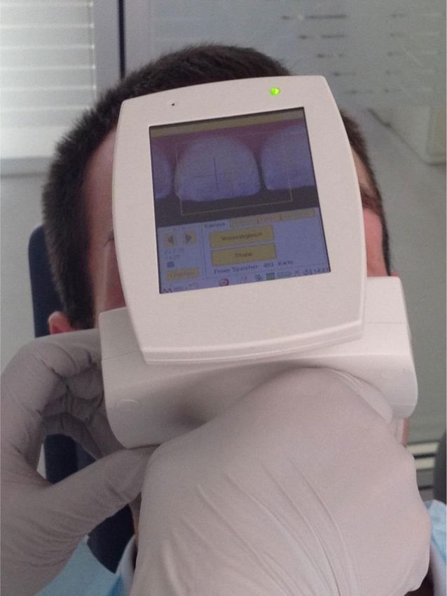
www.nature.com/scientificreports/                                                                     www.nature.com/scientificreports

                                            Observed values             Estimated values (adjusted for baseline values)
                                                                                   Control
                                                                        Test Group Group           Treatment effect
                                                          Control
                                            Test Group    Group         Mean ± SE Mean ± SE        Difference (95% CI)    p value
                            Baseline
                             Mean ± SD      46.7 ± 16.9   42.0 ± 15.7
                             Median (IQR)   50 (44–52)    47 (35–50)
                            Day 45
                             Mean ± SD      21.1 ± 23.2   35.0 ± 23.4   20.7 ± 5.1   35.4 ± 5.1    −14.7 (−22.8 – −6.6)   0.001
                             Median (IQR)   14 (7–23)     32 (17–51)
                            Day 90
                             Mean ± SD      18.4 ± 22.3   29.5 ± 25.0   18.2 ± 5.0   29.8 ± 5.0    −11.7 (−19.4 – −3.9)   0.005
                             Median (IQR)   10 (7–21)     19 (9–47)
                            Day 180
                             Mean ± SD      19.7 ± 24.2   33.7 ± 25.5   19.4 ± 5.4   34.0 ± 5.4    −14.6 (−24.5 – −4.8)   0.007
                             Median (IQR)   10 (5–27)     35 (10–49)
                            Baseline vs
                            Day 45
                             Mean ± SD      25.5 ± 23.3   7.0 ± 21.7
                             Median (IQR)   30 (18–43)    15 (−1–21)
                            Day 90
                             Mean ± SD      28.1 ± 25.1   12.2 ± 22.9
                             Median (IQR)   36 (16–43)    14 (−4–32)
                            Day 180
                             Mean ± SD      26.8 ± 28.0   8.0 ± 27.4
                             Median (IQR)   36 (17–45)    10 (1–28)

                           Table 1. Primary endpoint: Impedance measurement in test and control teeth (N = 21, split-mouth design, 122
                           observations in 2 sites or teeth at 3 time points). According to the manufacturer impedance values correspond to
                           the following: Sound: 0 = sound; 1–20 = Sound enamel, caries at the very outer enamel; Enamel caries 21–30 =
                           caries in the outer 1/3 of the enamel; 31–50 = caries in the middle 1/3 of the enamel; 51–90 = caries in the inner
                           1/3 of the enamel; 91–99 = caries at the dentine enamel junction; 100 = established dentine caries. Abbreviation:
                           CI, confidence interval; IQR, interquartile range; SD, standard deviation; SE, standard error of the mean.

                           both placebo26 and/or fluoride varnish26,27. As neither of the previous clinical trials investigated the effect of SAP
                           P11-4 following orthodontic treatment, it can be assumed that the lesions investigated in those clinical trials were
                           mostly inactive25–27.
                               Recently, the caries activity status of a lesion has become a new focus in cariology28.
                               Orthodontic WSL are of particular interest for clinical investigations of caries in this respect, as they are
                           assumed to be active until debonding of the brackets, whereupon they become inactive4,8 and good accessible for
                           several measurement methods16,29.
                               Therefore, the present split-mouth study investigated the effect of SAP P11-4 in addition to the conventional
                           treatment of early carious lesions after debonding of orthodontic brackets. The primary endpoint was the imped-
                           ance measurement29–31. Moreover, a semi-automated approach to measure the WSL size was used as done in other
                           clinical studies on SAP P11-425–27.

                           Results
                           Baseline characteristics.        The mean duration of the orthodontic treatment with fixed appliances was 27
                           months (min/max 13/39). Of the 23 recruited patients (13f 10 m; mean age 15.4 years), 21 could be analysed in
                           mixed models (12f/9 m; mean age 15.3 years). One patient showed established dentin caries (primary endpoint
                           = 100) on both teeth after 30 days and cavitated lesions after 90 days. The related four values of the primary end-
                           point after 90 and 180 days were set to 100. No further cavitated lesions were observed. The QHI at t0 was 2.2/2.2
                           (test/control teeth).

                           Impedance measurement of white spot lesion (primary endpoint).                         Both test and control teeth
                           exhibited a substantial decrease of impedance readings throughout the study period (Table 1). Control teeth
                           showed a mean baseline value of 42.0 at day 0, which decreased to 35.0 at day 45, 29.5 at day 90, and 33.7 at day
                           180. Test teeth showed a mean baseline value of 46.7 and exhibited a markedly larger decrease to 21.1 (day 45),
                           18.4 (day 90), and 19.7 (day 180).
                              The treatment effect was statistically significant (overall treatment contrast, which is the mean of the three
                           single contrasts in Table 1: -13.7, 95% CI: −19.6 – −7.7; p < 0.001). More importantly, the global F test of no treat-
                           ment effect was rejected (p = 0.003 for the test including one term for treatment and two terms for the interaction
                           between treatment and time). After treatment, the difference between test and control tooth changed only slightly

Scientific Reports |   (2020) 10:6819 | https://doi.org/10.1038/s41598-020-63633-0                                                              2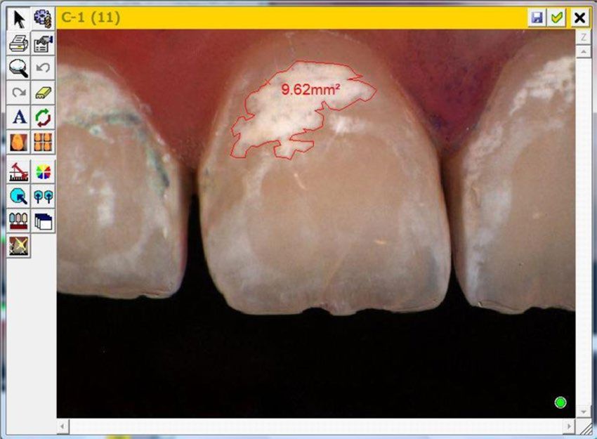
www.nature.com/scientificreports/                                                              www.nature.com/scientificreports

                           Figure 1. Impedance measurement of White Spot Lesion at different time points (black: test tooth/grey:
                           control tooth). As “temporally and logically, a baseline cannot be a response to treatment, so baseline and
                           response cannot be modeled in an integrated framework”43, baseline and response were graphed differently.
                           Consequently, the response and the 95% CI are adjusted for baseline values43. p = 0.001, p = 0.005, and p = 0.007
                           for treatment differences after 45, 90, and 180 days, respectively.

                           Figure 2. Morphometric measurement of White Spot Lesion Size in mm2 at different time points (black: test
                           tooth/grey: control tooth). As “temporally and logically, a baseline cannot be a response to treatment, so baseline
                           and response cannot be modeled in an integrated framework”43, baseline and response were graphed differently.
                           Consequently, the response and the 95% CI are adjusted for baseline values43. p = 0.969, p = 0.137, and p = 0.004
                           for treatment differences after 45, 90, and 180 days, respectively.

                           over time (p = 0.623; Fig. 1), yielding a large treatment effect of 43% after 180 days (14.6/34.0 in Table 1), which
                           is usually considered clinically relevant. Robust analyses using the means of the three time points after treatment
                           confirmed results of the mixed model in observed and imputed data with and without excluding dropouts (20, 21,
                           and 23 subjects, respectively; treatment differences −13.1 (95% CI: −18.8 – −7.4), −13.3 (95% CI: −20.2 – −6.3),
                           and −12.8 (95% CI: −22.4 – −3.2), respectively; for the “missing not at random” scenario by adding 50 to the
                           treatment site of the three dropouts: −9.7 (95% CI: −18.9 – −0.5)).

                           Morphometric measurement of white spot lesion (secondary endpoint).                            After treatment, the
                           decrease in size according to semi-automated morphometric measurements was more pronounced in test teeth
                           (p = 0.030 for the interaction between treatment and time; Fig. 2). After 180 days, the difference between test and
                           control group was statistically significant in mixed model analysis although the 95% CI was also consistent with a
                           weak to moderate effect (Table 2). The global test of no treatment effect was rejected (p = 0.013).
                               Missing values were also dealt with by multiple imputation, including sensitivity analysis for missing data
                           when true values are systematically lower or higher (nonignorable non-response). Both WSL of one patient were
                           too small to be measured semi-automatically. The two cavitated lesions were not measurable after 90 and 180
                           days. These paired nonignorable non-responses are levelled off by the split-mouth design. Missing values, for
                           which ignorable non-responses were assumed, occurred for a total of four subjects. One of the two patients who
                           were excluded in mixed models for missing baseline values could be re-included in robust analysis, resulting
                           in 15 patients available for complete case analysis. Robust analysis of contrasts between the first and third time
                           points (-t1 + t3 to model the interaction) confirmed the result of the mixed model by using complete and imputed

Scientific Reports |   (2020) 10:6819 | https://doi.org/10.1038/s41598-020-63633-0                                                           3
www.nature.com/scientificreports/ www.nature.com/scientificreports
Observed values Estimated values (adjusted for baseline values)
Control
Test Group Group Treatment effect
Control
Test Group Group Mean ± SE Mean ± SE Difference (95% CI) p value
Baseline
Mean ± SD 8.8 ± 7.8 6.8 ± 5.1
Median (IQR) 8.6 (2.0–11) 6.7 (2.8–9.6)
Day 45
Mean ± SD 8.1 ± 7.3 6.2 ± 4.7 7.2 ± 0.24 7.2 ± 0.24 0.0 (−0.6 – 0.6) 0.969
Median (IQR) 6.0 (1.9–11) 5.9 (2.4–9.9)
Day 90
Mean ± SD 7.5 ± 7.0 6.1 ± 4.6 6.5 ± 0.26 7.0 ± 0.26 −0.5 (−1.1 – 0.2) 0.137
Median (IQR) 5.4 (2.4–10) 5.4 (2.5-9.1)
Day 180
Mean ± SD 6.5 ± 6.5 5.7 ± 4.4 5.6 ± 0.24 6.6 ± 0.24 −1.0 (−1.6 – −0.4) 0.004
Median (IQR) 5.0 (0.5–11) 4.9 (2.4–9.4)
Baseline vs
Day 45
Mean ± SD 1.2 ± 1.0 0.9 ± 1.1
Median (IQR) 0.9 (0.4–1.9) 0.4 (0.1–1.3)
Day 90
Mean ± SD 1.8 ± 1.4 1.1 ± 0.9
Median (IQR) 1.7 (0.6–2.8) 0.9 (0.3–1.7)
Day 180
Mean ± SD 2.7 ± 1.7 1.5 ± 1.3
Median (IQR) 2.5 (1.3–3.8) 1.2 (0.4–2.5)
Table 2. Secondary endpoint: Morphometric measurement (mm2) in test and control teeth (N = 17, split-
mouth design, 94 observations in 2 sites or teeth at 3 time points). Missing values explain discrepancies between
differences of observed values and calculated changes from baseline (see results). Abbreviation: CI, confidence
interval; IQR, interquartile range; SD, standard deviation; SE, standard error of the mean.
data without dropouts (15 and 21 subjects, respectively). The treatment effect became uncertain if “ignorable
non-response” did not hold in sensitivity analyses after multiple imputation and by including dropouts (23
subjects).
Discussion
Our trial raised comparable parameters as the previously reported trials25–27,32 but differs from the other trials
on buccal caries, as active carious lesions (clinically visible as rough, chalky and matte surface) were treated,
and not predominantly inactive ones (clinically visible as a white spot under a hard, shiny surface)25–27. Both the
impedance values and morphometric measurements showed favourable WSL reduction results for the test group.
The greater difference between test and control lesions, however, occurred in the impedance values compared
with that in the morphometric measurements, thereby confirming that SAP P11-4 acts predominantly by regen-
erating the mineral structure of the enamel of the lesion body and not just the surface23,24. Moreover, at the last
study visit at day 180, the impedance values for test lesions indicated regression of caries into very outer enamel,
whereas the impedance values for the control lesions indicated still caries throughout the whole enamel layer31,33,
which agrees with previous data that fluoride (in the present study as fluoride-based professional prophylaxis
paste and home care toothpaste) only acts in the top 30-50 µm of the enamel surface13,14,34.
In recent years, caries activity has become a central topic within cariology, as activity defines the clinical need
and opportunity to intervene in the decay process. Active carious lesions, as treated in the present study, have
open pores allowing communication between the subsurface carious lesion body and the oral cavity. From the
lesion body, calcium and phosphate ions can diffuse out (demineralisation) or in (remineralisation). In a similar
fashion, the open pores allow SAP P11-4 to diffuse into the lesion body to support remineralisation and thus
regression of the tooth decay.
The active carious lesions in the conventional treatment control exhibited spontaneous regression of the meas-
ured WSL size over the study period until day 1803,8, which is comparable to literature data27,29. However, SAP
P11-4 enhanced the remineralisation to a higher decrease of the measured WSL size.
The present clinical trial is not without shortcomings. The possibility of random error because of the relatively
small sample size. Second, the trial was run within a university setting and the patients were Caucasians, thereby
limiting the generalizability of the findings. Third, information bias could have influenced our results. The morpho-
metric assessment represents only a part of the clinical situation because it is related only to the visible tooth surface.
Critical clinical assessment factors such as the hardness and gloss of the lesion could not be taken into account as
they could not be measured reliably within an in vivo study. It would be interesting to measure the enamel hardness
induced by SAP P11-4, and compare those to the previously reported in vitro microhardness results34,35.
Scientific Reports | (2020) 10:6819 | https://doi.org/10.1038/s41598-020-63633-0 4www.nature.com/scientificreports/ www.nature.com/scientificreports
• Anamnesis and patient information regarding study Recruiting
• Examination of inclusion and exclusion criteria (T0-30d)
N = 23
• Removal of the multibracket appliance (R-MBA)
• Written consent
R-MBA
• Professional dental hygiene incl. motivation & instruction (T0-2d)
• Impression of upper/lower jaw
(for retention appliance & measurement splint)
N = 23
• Placing of the retention appliance
• Checking of the dental hygiene procedure
• Professional dental hygiene incl. motivation & instruction
Baseline
• Measurements of the WSL (T0)
--------------------------------------------------------------------------
• Allocation
• Treatment of the test tooth with Curodont
N = 23
• Control of the retention appliance
• Checking of the dental hygiene procedure T1
• Professional dental hygiene incl. motivation & instruction (T0 + 45d)
• Measurements of the WSL on test- and control tooth
N = 21 2 dropouts
• Control of the retention appliance
• Checking of the dental hygiene procedure T2
• Professional dental hygiene incl. motivation & instruction (T0 + 90d)
• Measurements of the WSL on test- and control tooth
N = 21 for mixed models 1 both WSL filled (impedance = 100 )
N = 20 for complete case analysis 1 dropout
• Control of the retention appliance
• Checking of the dental hygiene procedure T3
• Professional dental hygiene incl. motivation & instruction (T0 + 180d)
• Measurements of the WSL on test- and control tooth
N = 21 for mixed models
N = 20 for robust analysis of complete cases
N = 23 for robust analysis after multiple imputation
Figure 3. Patient Flow Chart.
However, there are also several strengths. First, the source population and the chosen inclusion criteria
ensured that active carious lesions, not inactive ones, were investigated. Second, the split-mouth design is appeal-
ing since “each participant acts as their own control”36. By this favourable control mechanism, the split-mouth
design is related to counterfactual queries (would a subject have no caries had the subject been treated with
SAP given that the subject has in fact caries and is not treated with SAP), whereas the usual randomised clini-
cal trial in parallel-groups is merely related to intervention queries (would a subject have no caries if we make
sure that the subject is treated with SAP)37. Therefore, the split-mouth design is clearly superior to a parallel
group design (unless in identical twins) if the study design assumptions hold38. As SAP P11-4 acts solely on tooth
level23,24, it is biologically well justified to assume the absence of any cross-contamination or carry-across effect
Scientific Reports | (2020) 10:6819 | https://doi.org/10.1038/s41598-020-63633-0 5www.nature.com/scientificreports/ www.nature.com/scientificreports
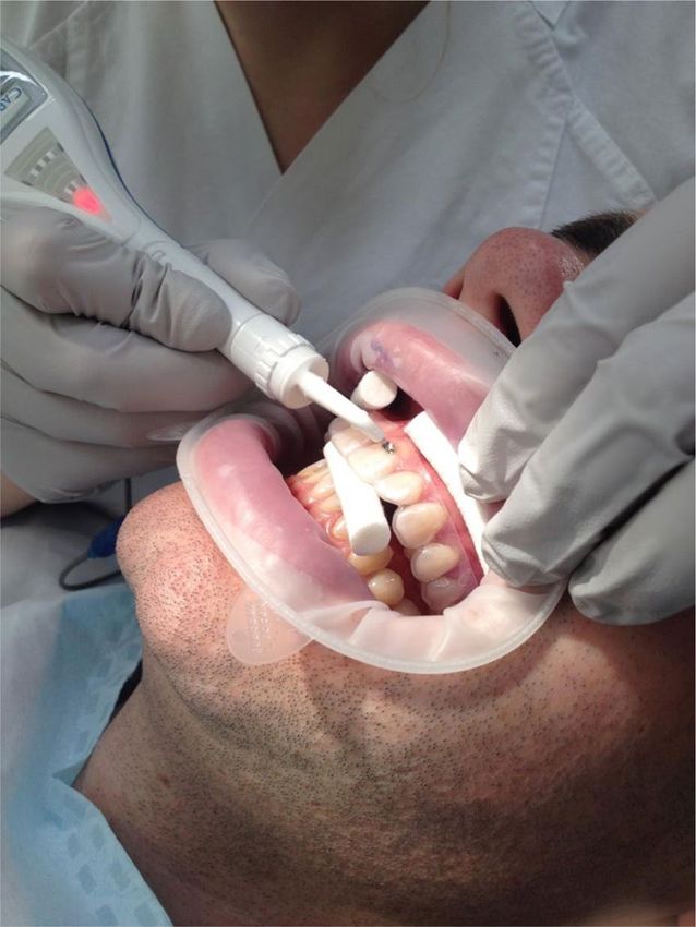
Figure 4. Image of Impedance measurement of WSL with CarieScan Pro (Orangedental/Biberach/Germany).
Figure 5. Overview image of Morphometric measurement of WSL with Shadepilot (DeguDent/Hanau/
Germany).
from one site to another39. Note that the theory of causality37 has been rapidly developed since the split-mouth
design was criticized for statistical reasons in 199040. Third, the split-mouth design was very efficient because
the within-patient correlation was clearly different from zero (p = 0.61 for the primary endpoint, p = 0.72 for
the secondary endpoint). For the primary and secondary endpoint, the sample sizes of n = 21 and n = 17 in our
Scientific Reports | (2020) 10:6819 | https://doi.org/10.1038/s41598-020-63633-0 6www.nature.com/scientificreports/ www.nature.com/scientificreports
Figure 6. Computer Screen image of Morphometric measurement of WSL with Shadepilot (DeguDent/Hanau/
Germany).
split-mouth design correspond to total sample sizes of 108 and 118 patients in a parallel group design, respec-
tively39. Fourth, three measurements of the primary endpoint were taken to increase reliability. Fifth, the duration
of the trial worked for both endpoints. It was long enough to gain important insights into the mechanism of the
treatment, especially for the secondary endpoint. Finally, the statistical analysis is state-of-the-art39,41–43, follows
rigorously the intention-to-treat principle by accounting for the uncertainty due to dropouts, and supports con-
fidence intervals44.
Overall, our results not only agree with other clinical trials on the effectiveness of SAP P11-420,25–27 but also
show that SAP P11-4 is effective in the treatment of active WSL26,27 supporting as already mentioned the proposed
mechanism of action within the subsurface lesion body23,24. Thus, SAP P11-4 treatment gets one step closer to a
regeneration ad integrum, that would be the ultimate goal in healing carious enamel.
One of the reasons that the treated enamel does not return to full translucency is seen in the fact that the
SAP P11-4-induced fibres support the formation of de novo hydroxyapatite crystals in a fan-type non-prismatic
arrangement around the fibres24,45, which have another refractive index than the prismatic hydroxyapatite crystals
produced by the Tomes processes from ameloblasts in the final stage of enamel deposition.
Although buccal carious lesions chosen in order to quantitatively assess the effect of the treatment are rare
outside the orthodontics, the clinical significance of the results relates to the treatment of caries in general. Thus,
the aesthetic shortcomings of the SAP P11-4 treatment46 are neglectable for almost all of the initial carious lesions
as most clinically relevant caries develops in proximal or occlusal sites and if buccal lesions occur, they are mostly
positioned on premolars and molars.
Professionals are trained to identify initial caries in any location, either visually, with caries diagnostics or
on x-rays. Based on those assessments the clinician will decide on whether invasive restorative treatments are
needed. Regenerative procedures should be considered whenever possible, in order to conserve natural tooth
structure and function and to avoid or at least to delay as far as possible the entry into the conventional filling
approach leading to ever larger fillings47. Bröseler et al. have shown that the SAP P11-4 treatment can be trans-
ferred into a practice setting27.
Conclusion
The treatment of initial carious lesions with self-assembling peptide P11-4 leads to superior remineralisation of the
subsurface lesions compared with the control teeth.
Material and Methods
Study design. The clinical trial was designed and performed as a split-mouth conventional treatment-con-
trolled trial investigating the effect of the treatment of buccal carious lesions following orthodontic treatment by
using SAP P11-4 as add-on with the conventional treatment control. Clinical study procedures were performed
parallel to regular appointments at the Orthodontic Department of the University Medicine Greifswald (Fig. 3)
according to the Declaration of Helsinki and in compliance with ISO 14155:2012. Approval for all clinical proce-
dures and the trial was obtained by the ethical committee of the University of Greifswald (Code BB 99/12, date of
approval: 28th August 2012).
The clinical trial was registered in the German Database for clinical trials (DRKS00016501, date of registry:
30th January 2019).
Patient and study tooth selection. Twenty-three patients (13f/10 m; average age 15.4 years) with at least
two WSL after removal of fixed orthodontic appliances could be recruited into the trials from the Orthodontic
Department of the University Medicine Greifswald and treated by the Restorative Department. The written
informed consent was obtained from every participance or from a parent of every participance under 18 years
prior to any study-related procedures two days before baseline (Fig. 3).
Patients had to fulfil all the following selection criteria:
Inclusion
Scientific Reports | (2020) 10:6819 | https://doi.org/10.1038/s41598-020-63633-0 7www.nature.com/scientificreports/ www.nature.com/scientificreports
• At least two active carious lesions around the bracket area with a rough, chalky and matte surface;
• Size and form of the active carious lesion: The carious lesion must be fully visible and assessable and accessible;
• Able and willing to observe good oral hygiene throughout the study;
• ≥20 teeth and a BP score ofwww.nature.com/scientificreports/ www.nature.com/scientificreports
to correct for small-sample inference. Mixed models can deal with imbalanced or missing data if the “missing
at random” assumption holds43. The complex mixed models were analysed using Stata software (release 14.2,
Stata Corporation, College Station, TX, USA) and checked using SAS software (release 9.4, Cary, NC, USA). In
robust analyses of the direct treatment effect (not including baseline values), the two-sample t-test was used for
differences in linear combinations of repeated measurements between groups defined by the site of treatment52.
To cover the uncertainty due to dropouts, multiple imputations were generated by fully conditional specification
using R software43. Scenarios other than “missing at random” were examined in sensitivity analysis after multiple
imputation.
Data availability
The datasets of the current study are available from the corresponding author on request.
Received: 17 July 2019; Accepted: 31 March 2020;
Published: xx xx xxxx
References
1. Staudt, C. B., Lussi, A., Jacquet, J. & Kiliaridis, S. White spot lesions around brackets: in vitro detection by laser fluorescence. Eur J
Oral Sci 112, 237–243, https://doi.org/10.1111/j.1600-0722.2004.00133.x (2004).
2. Mattousch, T. J., van der Veen, M. H. & Zentner, A. Caries lesions after orthodontic treatment followed by quantitative light-induced
fluorescence: a 2-year follow-up. Eur J Orthod 29, 294–298, https://doi.org/10.1093/ejo/cjm008 (2007).
3. Höchli, D., Hersberger-Zurfluh, M., Papageorgiou, S. N. & Eliades, T. Interventions for orthodontically induced white spot lesions:
a systematic review and meta-analysis. Eur J Orthod 39, 122–133, https://doi.org/10.1093/ejo/cjw065 (2017).
4. Ren, Y., Jongsma, M. A., Mei, L., van der Mei, H. C. & Busscher, H. J. Orthodontic treatment with fixed appliances and biofilm
formation–a potential public health threat? Clin Oral Investig 18, 1711–1718, https://doi.org/10.1007/s00784-014-1240-3 (2014).
5. Gorelick, L., Geiger, A. M. & Gwinnett, A. J. Incidence of white spot formation after bonding and banding. Am J Orthod 81, 93–98
(1982).
6. Richter, A. E., Arruda, A. O., Peters, M. C. & Sohn, W. Incidence of caries lesions among patients treated with comprehensive
orthodontics. Am J Orthod Dentofacial Orthop 139, 657–664, https://doi.org/10.1016/j.ajodo.2009.06.037 (2011).
7. Featherstone, J. D. B. & Chaffee, B. W. The Evidence for Caries Management by Risk Assessment (CAMBRA(R)). Adv Dent Res 29,
9–14, https://doi.org/10.1177/0022034517736500 (2018).
8. Gao, S. S., Zhang, S., Mei, M. L., Lo, E. C. & Chu, C. H. Caries remineralisation and arresting effect in children by professionally
applied fluoride treatment - a systematic review. BMC Oral Health 16, 12, https://doi.org/10.1186/s12903-016-0171-6 (2016).
9. Petersson, L. G., Twetman, S. & Pakhomov, G. N. The efficiency of semiannual silane fluoride varnish applications: a two-year
clinical study in preschool children. J Public Health Dent 58, 57–60 (1998).
10. de Oliveira, B. H. & Dos Santos, A. P. Semiannual Fluoride Applications in Low-Risk Toddlers May Not Be More Effective Than
Toothbrushing Instruction and Dietary Counseling in Controlling Dental Caries. J Evid Based Dent Pract 16, 246–248, https://doi.
org/10.1016/j.jebdp.2016.11.006 (2016).
11. de Oliveira, P. R., Fonseca, A. B., Silva, E. M., Coutinho, T. C. & Tostes, M. A. Remineralizing potential of CPP-ACP cremes with and
without fluoride in artificial enamel lesions. Aust Dent J, https://doi.org/10.1111/adj.12305 (2015).
12. Muller, F. et al. Elemental depth profiling of fluoridated hydroxyapatite: saving your dentition by the skin of your teeth? Langmuir
26, 18750–18759, https://doi.org/10.1021/la102325e (2010).
13. Bergman, G. & Lind, P. O. A quantitative microradiographic study of incipient enamel caries. J Dent Res 45, 1477–1484 (1966).
14. Pandya, M. & Diekwisch, T. G. H. Enamel biomimetics-fiction or future of dentistry. Int J Oral Sci 11, 8, https://doi.org/10.1038/
s41368-018-0038-6 (2019).
15. Rechmann, P., Chaffee, B. W., Rechmann, B. M. T. & Featherstone, J. D. B. Changes in Caries Risk in a Practice-Based Randomized
Controlled Trial. Adv Dent Res 29, 15–23, https://doi.org/10.1177/0022034517737022 (2018).
16. Urquhart, O. et al. Nonrestorative Treatments for Caries: Systematic Review and Network Meta-analysis. J Dent Res,
22034518800014, https://doi.org/10.1177/0022034518800014 (2018).
17. Fernandez-Ferrer, L. et al. Enamel remineralization therapies for treating postorthodontic white-spot lesions: A systematic review.
J Am Dent Assoc 149, 778–786 e772, https://doi.org/10.1016/j.adaj.2018.05.010 (2018).
18. Jablonski-Momeni, A. & Heinzel-Gutenbrunner, M. Efficacy of the self-assembling peptide P11-4 in constructing a remineralization
scaffold on artificially-induced enamel lesions on smooth surfaces. J Orofac Orthop 75, 175–190, https://doi.org/10.1007/s00056-
014-0211-2 (2014).
19. Amaechi, B. T. Remineralisation - the buzzword for early MI caries management. Br Dent J 223, 173–182, https://doi.org/10.1038/
sj.bdj.2017.663 (2017).
20. Alkilzy, M., Santamaria, R. M., Schmoeckel, J. & Splieth, C. H. Treatment of Carious Lesions Using Self-Assembling Peptides. Adv
Dent Res 29, 42–47, https://doi.org/10.1177/0022034517737025 (2018).
21. Philip, N. State of the Art Enamel Remineralization Systems: The Next Frontier in Caries Management. Caries Res 53, 284–295,
https://doi.org/10.1159/000493031 (2018).
22. Jablonski-Momeni, A. et al. Randomised in situ clinical trial investigating self-assembling peptide matrix P11-4 in the prevention of
artificial caries lesions. Sci Rep 9, 269, https://doi.org/10.1038/s41598-018-36536-4 (2019).
23. Kind, L. et al. Biomimetic Remineralization of Carious Lesions by Self-Assembling Peptide. J Dent Res 96, 790–797, https://doi.
org/10.1177/0022034517698419 (2017).
24. Kirkham, J. et al. Self-assembling peptide scaffolds promote enamel remineralization. J Dent Res 86, 426–430, 86/5/426 (2007).
25. Brunton, P. A. et al. Treatment of early caries lesions using biomimetic self-assembling peptides - a clinical safety trial. Br Dent J 215,
E6, https://doi.org/10.1038/sj.bdj.2013.741 (2013).
26. Mannaa, A., Sedlakova, P., Bommer, C., di Bella, E. & Krejci, I. In IADR Vol. 96th General Session (London, GB, 2018).
27. Bröseler, F. et al. Randomised clinical trial investigating self-assembling peptide P11-4 in the treatment of early caries. Clin Oral
Investig, https://doi.org/10.1007/s00784-019-02901-4 (2019).
28. Nyvad, B. & Baelum, V. Nyvad Criteria for Caries Lesion Activity and Severity Assessment: A Validated Approach for Clinical
Management and Research. Caries Res 52, 397–405, https://doi.org/10.1159/000480522 (2018).
29. Mortensen, D., Gizani, S., Salamara, O., Sifakakis, I. & Twetman, S. Monitoring regression of post-orthodontic lesions with
impedance spectroscopy: a pilot study. Eur J Orthod, https://doi.org/10.1093/ejo/cjy075 (2018).
30. Longbottom, C., Huysmans, M. C., Pitts, N. B., Los, P. & Bruce, P. G. Detection of dental decay and its extent using a.c. impedance
spectroscopy. Nat Med 2, 235–237 (1996).
31. Cohen, J. E. The association between CarieScan Pro readings and histologic depth of caries in non cavitated occlusal lesion in vitro. MS
(Master of Science) thesis, University of Iowa, (2013).
Scientific Reports | (2020) 10:6819 | https://doi.org/10.1038/s41598-020-63633-0 9www.nature.com/scientificreports/ www.nature.com/scientificreports
32. Alkilzy, M., Tarabaih, A., Santamaria, R. M. & Splieth, C. H. Self-assembling Peptide P11-4 and Fluoride for Regenerating Enamel.
J Dent Res 97, 148–154, https://doi.org/10.1177/0022034517730531 (2018).
33. Tassery, H. et al. Use of new minimum intervention dentistry technologies in caries management. Aust Dent J 58(Suppl 1), 40–59,
https://doi.org/10.1111/adj.12049 (2013).
34. Schmidlin, P., Zobrist, K., Attin, T. & Wegehaupt, F. In vitro re-hardening of artificial enamel caries lesions using enamel matrix
proteins or self-assembling peptides. J Appl Oral Sci 24, 31–36, https://doi.org/10.1590/1678-775720150352 (2016).
35. Kamal, D., Hassanein, H., Elkassas, D. & Hamza, H. Comparative evaluation of remineralizing efficacy of biomimetic self-
assembling peptide on artificially induced enamel lesions: An in vitro study. J Conserv Dent 21, 536–541, https://doi.org/10.4103/
JCD.JCD_123_18 (2018).
36. Pandis, N., Chung, B., Scherer, R. W., Elbourne, D. & Altman, D. G. CONSORT 2010 statement: extension checklist for reporting
within person randomised trials. BMJ 357, j2835, https://doi.org/10.1136/bmj.j2835 (2017).
37. Pearl, J. Causality. Models, Reasoning, and Inference. 2nd edn, (Cambridge University Press, 2009).
38. Harrell, F. E., Jr. & Slaughter, J. C. In Biostatistics for Biomedical Research (2001–2019).
39. Lesaffre, E., Philstrom, B., Needleman, I. & Worthington, H. The design and analysis of split-mouth studies: what statisticians and
clinicians should know. Stat Med 28, 3470–3482, https://doi.org/10.1002/sim.3634 (2009).
40. Hujoel, P. P. & Loesche, W. J. Efficiency of split-mouth designs. J Clin Periodontol 17, 722–728, https://doi.org/10.1111/j.1600-
051x.1990.tb01060.x (1990).
41. Kenward, M. G. & Roger, J. H. Small sample inference for fixed effects from restricted maximum likelihood. Biometrics 53, 983–997
(1997).
42. Committee for Proprietary Medicinal Products (CPMP). Points to consider on adjustment for baseline covariates. Statistics in
Medicine 23, 701–709, https://doi.org/10.1002/sim.1647 (2004).
43. Harrell, F. E., Jr. Regression modeling strategies. With applications to linear models, logistic and ordinal regression, and survival analysis.
2nd edn, ix, 5, 19, 104, (Springer, 2015).
44. Wasserstein, L. R. & Lazar, N. A. The ASA’s Statement on p-Values: Context, Process, and Purpose. The American Statistician 70,
129–133, https://doi.org/10.1080/00031305.2016.1154108 (2016).
45. Takahashi, F. et al. Ultrasonic assessment of the effects of self-assembling peptide scaffolds on preventing enamel demineralization.
Acta Odontol Scand 74, 142–147, https://doi.org/10.3109/00016357.2015.1066850 (2016).
46. Wierichs, R. J., Kogel, J., Lausch, J., Esteves-Oliveira, M. & Meyer-Lueckel, H. Effects of Self-Assembling Peptide P11-4, Fluorides,
and Caries Infiltration on Artificial Enamel Caries Lesions in vitro. Caries Res 51, 451–459, https://doi.org/10.1159/000477215
(2017).
47. Krejci, I., Lieber, C. M. & Lutz, F. Time required to remove totally bonded tooth-colored posterior restorations and related tooth
substance loss. Dent Mater 11, 34–40, https://doi.org/10.1016/0109-5641(95)80006-9 (1995).
48. Senn, S. Cross-over Trials in Clinical Research. 2nd edn, (John Wiley & Sons, Ltd, 2002).
49. Boersma, J. G., van der Veen, M. H., Lagerweij, M. D., Bokhout, B. & Prahl-Andersen, B. Caries prevalence measured with QLF after
treatment with fixed orthodontic appliances: influencing factors. Caries Res 39, 41–47, https://doi.org/10.1159/000081655 (2005).
50. van der Veen, M. H., Attin, R., Schwestka-Polly, R. & Wiechmann, D. Caries outcomes after orthodontic treatment with fixed
appliances: do lingual brackets make a difference? Eur J Oral Sci 118, 298–303, https://doi.org/10.1111/j.1600-0722.2010.00733.
xEOS733 (2010).
51. Greenland, S. et al. Statistical tests, P values, confidence intervals, and power: a guide to misinterpretations. Eur J Epidemiol 31,
337–350, https://doi.org/10.1007/s10654-016-0149-3 (2016).
52. Jones, B. & Kenward, M. G. Design and Analysis of Cross-Over Trials. 3rd edn, (CRC Press, 2015).
Acknowledgements
Our gratitude goes to our statisticians of the University Medicine Greifswald, Dr. P. Kolyschkow, Dr. C. Holtfreter
for their help. Further, we would like to thank Dr. M. Müller for his clinical support and credentis ag, Procter
& Gamble Germany GmbH & Co Operations oHG, DeguDent GmbH, orangedental GmbH & Co. KG for its
material support of the study.
Author contributions
Conceived, designed and managed the clinical study: A.W.; treated the patients: A.R.; conducted measurements:
M.R.; conducted statistical analysis: Ch.S.; wrote the manuscript: A.W.; reviewing the manuscript: Ch.S., A.R.,
M.R. and K.F.K.
Competing interests
The authors declare no competing interests.
Additional information
Correspondence and requests for materials should be addressed to A.W.
Reprints and permissions information is available at www.nature.com/reprints.
Publisher’s note Springer Nature remains neutral with regard to jurisdictional claims in published maps and
institutional affiliations.
Open Access This article is licensed under a Creative Commons Attribution 4.0 International
License, which permits use, sharing, adaptation, distribution and reproduction in any medium or
format, as long as you give appropriate credit to the original author(s) and the source, provide a link to the Cre-
ative Commons license, and indicate if changes were made. The images or other third party material in this
article are included in the article’s Creative Commons license, unless indicated otherwise in a credit line to the
material. If material is not included in the article’s Creative Commons license and your intended use is not per-
mitted by statutory regulation or exceeds the permitted use, you will need to obtain permission directly from the
copyright holder. To view a copy of this license, visit http://creativecommons.org/licenses/by/4.0/.
© The Author(s) 2020
Scientific Reports | (2020) 10:6819 | https://doi.org/10.1038/s41598-020-63633-0 10You can also read