Dynamic cell transition and immune response landscapes of axolotl limb regeneration revealed by single-cell analysis
←
→
Page content transcription
If your browser does not render page correctly, please read the page content below
Protein Cell 2021, 12(1):57–66
https://doi.org/10.1007/s13238-020-00763-1                                                               Protein & Cell

LETTER
Dynamic cell transition and immune response
landscapes of axolotl limb regeneration
revealed by single-cell analysis

Dear Editor,                                                       (Table S3), reinforcing the previous study that this cell type
                                                                   may originate from other types of cells by dedifferentiation,

                                                                                                                                       Protein & Cell
The axolotl, Ambystoma mexicanum, has extraordinary                such as CT cells, and may serve as progenitor cells for re-
capability to fully recover multiple tissues after lost, whereas   differentiation as previously suggested (Gerber et al., 2018).
such capability has disappeared in mammals. Thus, deci-                More interestingly, the time-course clustering results
phering detailed mechanisms underlying axolotl regenera-           revealed a clear inverse correlation of cell populations
tion could provide valuable lessons for regenerative               between epithelial, Prdx2+ blastema and CT cells at two
medicine. However, many questions, such as the origin of           phases (first: 1 d to 7 d; second: 7 d to 22 d) (Fig. 1C and
essential progenitor cells and key responses of individual         1D). The number of basal epidermis (BE) and intermediate
types of cells for regeneration remain elusive (Haas and           epidermis (IE) cells, dropped rapidly from 1 d to 7 d and then
Whited, 2017). Newly developed single-cell RNA sequenc-            gradually returned from 7 d to 33 d (Fig. 1C and 1D). In
ing (scRNA-seq) method enables researchers to observe              contrast, the number of mesenchymal and Prdx2+ blastema
cellular and molecular dynamics in axolotl regeneration at         cells increased in the first phase, and then gradually returned
the single-cell resolution (Gerber et al., 2018; Leigh et al.,     to normal level in the second phase (Fig. 1C and 1D). Such
2018), but the reported transcriptome landscapes are only          results suggest a potential cell state transformation from
for certain cell types or in certain regenerative stages. A        epidermal cells into mesenchymal-state cells, and the sec-
complete overview of the regeneration process for all cell         ond phase indicates a reversed process, while it is also
types is still lacking.                                            possible the rates of cell proliferation/death are distinct
    To this end, we performed axolotl limb amputation fol-         between different cell types.
lowed by scRNA-seq (10× Genomics) on upper forearm                     To further evaluate such two possibilities, we pooled dif-
tissues at the homeostatic stage (uninjured control, 0 h), and     ferent cell populations from various regeneration time points
7 regeneration stages (3 h–33 d) (Fig. 1A, Supplementary           for pseudotime trajectory analysis (Figs. 1E and S3A). While
Material). In total, we obtained high-quality sequencing data      the majority of epidermal (IE and BE) and mesenchymal cell
for over 41,000 cells (Table S1). After clustering all cells and   populations were located at two ends of the pseudotime
reflecting them in uniform manifold approximation and pro-          trajectory axis, an evident proportion of both the IE and BE
jection (UMAP), we identified 12 putative cell types                was positioned along the conversion route, and their num-
expressing specific markers (Figs. 1B and S1A; Table S2),           bers increased toward the mesenchymal-state end from 1 d
which were further confirmed by differentially expressed            to 14 d (Fig. 1F). Prdx2+ blastema cells were located
genes (DEG) analysis (Fig. S1B). By performing integrative         between the epidermal-state and mesenchymal-state ends
comparison of our dataset with ones from Gerber et al. and         with some overlaps (Fig. 1F), suggesting they may possess
Leigh et al. (Gerber et al., 2018; Leigh et al., 2018), we found   cellular characteristics of both CT cells and epidermal cells.
a remarkable similarity of cell-type distribution between our          To further investigate the cellular correlation, we clustered
and Leigh et al.’s data (Fig. S2A and S2B), thus further           Prdx2+ blastema, CT, BE and IE cells into 8 states along the
supporting the reproducibility of our experimental procedure.      pseudotime axis (Figs. 1G and S3B), and projected them
    Of note, cells expressing the embryonic limb marker            back onto UMAP (Fig. S3C). CT cells were mainly in State 1
Peroxiredoxin 2 (Prdx2) (Gerber et al., 2018) were identified       (S1) and S2, which are the mesenchymal-like states, while
in all three datasets (Fig. S2C). Located at the center of the     IE and BE mostly located at S7 and S8, which are the epi-
UMAP plot, Prdx2+ blastema cells were associated with              dermal-like states. The intermediate states, S3, S4, S5 and
connective tissue (CT) cells from 1 to 7 d (Fig. 1B and 1C)        S6, were mixed with CT, epidermal and Prdx2+ blastema
and expressed genes related to stem cell maintenance, cell         cells (Figs. 1G and S3B). At individual sampling time point,
morphogenesis and multi-tissue development pathways                the cell number in the epidermal-like states continuously

© The Author(s) 2020
LETTER        Hanbo Li et al.
Protein & Cell

                 58       © The Author(s) 2020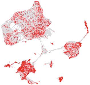
Single-cell atlas analysis of axolotl limb regeneration                                                                    LETTER

b    Figure 1. Integrated transcriptional cell state Atlas of the          for the changes in cell number of epidermal and mes-
     axolotl limb regeneration revealed a putative EMT-MET                 enchymal tissues, in addition to variant cell proliferation/
     process. (A) Schematic of the scRNA-seq workflow used for              death rate, suggesting that there is a sequential epithelial to
     the upper arm tissues collected from the homeostatic (uninjured       mesenchymal transition (EMT), followed by a mesenchymal
     control, 0 h), trauma (3 h), wound healing (1 d), early-bud           to epithelial transition (MET) during limb regeneration.
     blastema (3 d), mid-bud blastema (7 d), late-bud blastema (14             Therefore, key EMT genes (Kalluri and Weinberg, 2009;
     d), palette stage (22 d) and re-differentiated stage (33 d).          Zhang et al., 2013) were then analyzed to further confirm this
     (B) UMAP visualization of the scRNA-seq data from 41,376              process. The expression patterns of EMT genes showed a
     single cells collected from 8 sampling stages. Each dot               similar tendency with the cell population distribution, as the
     represents a single cell; the colors distinguish clusters. The        expression levels of genes positively related to EMT con-
     cell-type annotation is determined by published cell-lineage          tinued to increase after limb amputation and peaked at 7 d,
     specific markers. (C) UMAP distribution of cells from each             and then reduced after 14 d (Fig. 1I). In contrast, genes
     sampling stage. Red dots represent the cells from the corre-          negatively related to EMT, including cadherin 1 (E-cad) and
     sponding time point, and gray dots represent all the cells from 8
                                                                           epidermal growth factor receptor (Egfr), exhibited the exact
     stages. (D) Cell number proportion of each cell type at the
                                                                           opposite pattern (Fig. 1I). These facts, complied with our
     corresponding time point. The x-axis represents the time points.
                                                                           cellular dynamics data, provide evidence of a potential
     The y-axis represents the cell ratio. (E) Pseudotime analysis of
                                                                           dynamic EMT-MET process in axolotl limb regeneration with
     epidermal and mesenchymal populations during the regenera-

                                                                                                                                              Protein & Cell
                                                                           turning point between 7 d and 14 d. However, since epithelial
     tion process. Each dot corresponds to a single cell. The
                                                                           and CT cells are both linked to the Prdx2+ blastema cells, it is
     gradient bar reflects the pseudotime. The cell-type components
                                                                           difficult to distinguish between two possibilities that epider-
     are indicated by different colors. (F) Pseudotime analysis
                                                                           mal cells directly transformed into CT lineage cells, and that
     reveals the distribution of Prdx2+ blastema, BE, IE and CT
                                                                           the Prdx2+ blastema cells functioned as a transient stage
     cells along the pseudotime trajectory. The cell-type components
                                                                           linking these two groups of cells.
     are indicated by different colors. (G) Prdx2+ blastema, BE, IE
     and CT cells are divided into 8 separate pseudotime states in
                                                                               It has been established that EMT plays a critical role in
     the pseudotime analysis. Colored dots represent different             physiologic tissue repair, yet sustained EMT promotes
     states. (H) Cell type shift patterns according to real-time points.   fibrosis of multiple organs under a variety of pathological
     Red dots represent the cells from the corresponding time point,       conditions (Stone et al., 2016; Forte et al., 2017). By
     and gray dots represent all involved cells. (I) Expression of EMT     examining the co-expression of marker genes, our data
     and MET related genes on a single-cell trajectory plot. Colored       revealed that cells characterized as fibroblasts were present
     dots represent single cells from individual sampling time points.     during axolotl limb regeneration, most of which were in CT
     Black curves reflect the fitted smooth spline curves.                   cluster (Fig. 2A). Importantly, the number of this group of
                                                                           cells also first increased, peaked at 7 d and 14 d, and then
                                                                           decreased till absence. In contrast, during digit wound
                                                                           healing process of mouse, the number of fibroblasts at the
    dropped from 1 d to 7 d, while the cell number in the inter-           wound area appeared to be consistent and relatively high at
    mediate and mesenchymal-like states gradually increased                all stages according to our scRNA-seq data (Figs. 2A, S4A
    (Fig. 1G and 1H) and reached their peaks at 7 d (Fig. 1C, 1D           and S4B). Altogether, axolotl limb regeneration seems to
    and 1H). From 14 d to 33 d, as the number of mesenchymal               experience a temporal EMT, which may prevent uncontrolled
    cells decreased, epidermal cells were repopulated (Fig. 1C,            fibrosis.
    1D and 1H). Interestingly, a significant portion of IE and BE               Consistently at the molecular level, the expression levels
    with the epidermal-like state transformed into the intermedi-          of extracellular matrix (ECM) deposition related genes,
    ate-like state from 1 d to 7 d (Fig. S3B), and while most of           including spalt like transcription factor 4 (Sall4) (Erickson
    those cells remained in the intermediate state, a minor part           et al., 2016) and collagen type XII alpha 1 chain (Col12a1)
    further transformed into mesenchymal-like state at 7 d and             and ECM remodel related genes, including matrix metal-
    14 d (Fig. S3B). At last, those cells shifted back into an             lopeptidase 2 (Mmp2) and laminin subunit alpha 1 (Lama1)
    epidermal-like state from 14 d to 33 d (Fig. S3B). Altogether,         (Rayagiri et al., 2018) were low at the beginning of axolotl
    these digital cell state transitions provide an extra possibility      regeneration, but increased and peaked at 7 d and 14 d
                                                                           (Fig. 2B). In contrast, the same genes in the healing mouse

    © The Author(s) 2020                                                                                                                59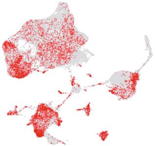
LETTER                               Hanbo Li et al.
Protein & Cell

                          Figure 1. continued.

                 60                              © The Author(s) 2020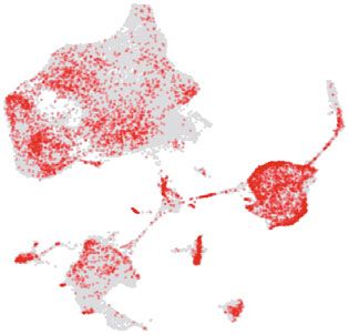
Single-cell atlas analysis of axolotl limb regeneration                                                              LETTER

digits were low or undetectable throughout the healing pro-       gene expression pattern (Fig. 2E and 2F; Table S2), and its
cess (Fig. 2B). This result suggests that different from mouse    expression of both anti-infection and anti-inflammatory
wound healing, axolotl simultaneously undergoes a rapid           genes (Table S4). In addition, the majority of our Macro-
ECM redeposition and remodeling, leading to a complete            phage 3 cells were found overlapped with neutrophils iden-
ECM recovery.                                                     tified by Leigh et al. (2018) (Fig. S5C), possibly due to their
   To further explore the mechanism of gene regulation for        shared expression patterns in genes related to anti-inflam-
fibrosis, we examined the expression of key genes in fibro-         mation function (Marwick et al., 2018). To our interest, DEG
genic transforming growth factor beta (TGF-β) pathway             analysis showed that the expression of Nuclear Factor
(Meng et al., 2016). We observed the expression level of          Kappa B (Nf-κb) was decreased in Macrophage 3 from 1 d to
Tgf-β1 was relatively low and only increased at 7d and 14d        7 d (Table S4). Once activated, NF-κB acts as a key tran-
during axolotl regeneration, while the expression of TGF-β1       scription factor that induces expression of inflammatory
receptor, transforming growth factor beta receptor 2 (Tgfβr2)     mediators to enrich the pro-inflammatory macrophage phe-
and downstream factors RUNX family transcription factor 2         notype (Doyle and O'Neill, 2006; Sharif et al., 2007). Inacti-
(Runx2) and SMAD family member 2 (Smad2) remained                 vation of NF-κB suppresses the release of inflammatory
consistently low at all stages (Fig. 2C). In sharp contrast, in   cytokines, thus generating anti-inflammatory/pro-resolving
the mouse model, cells that expressed these genes were            macrophage phenotype (Marwick et al., 2018).
greater in number and showed a wider distribution over time           Macrophages are required for wound healing or epimor-

                                                                                                                                    Protein & Cell
than those in axolotl (Fig. 2C). Also, as another downstream      phic regeneration in mammals, in which inflammatory mac-
consequence of TGF-β pathway, the expression levels of            rophages play an important role in stimuli response in the
cell senescence markers cyclin dependent kinase inhibitor         early stages before weakened and replaced by pro-resolving
1A (P21) and nuclear factor of kappa light polypeptide gene       macrophages at the wound site (Wynn and Vannella, 2016).
enhancer in B-cells 3 (P65) were constantly low, whereas in       Such a time-dependent process greatly promotes fibrosis
mouse cells the expression of these genes remained high           and scarring at the wounding site, ensuring wound closure
throughout the wound healing process. Altogether, our             and healing in mouse. Our mouse digit wound healing model
results strongly suggest that fibrosis and cellular senescence     confirmed that only Macrophage 1 and Macrophage 2 were
are restrained at minimal level in regenerating axolotl cells.    observed at the early stages in injured mouse digit (Figs. 2G,
The low level of TGF-β pathway activity, together with unique     S4A and S4B), implying a need for inflammatory and
ECM redeposition and remodel pattern, may contribute to           immune wound healing responses. In contrast, axolotl limb
the scarless regeneration of axolotl limbs.                       regeneration seemed to utilize an additional mechanism to
   Macrophages play critical roles during fibrosis and scar        balance the inflammatory response by recruiting a third type
formation, and thus are key players in regulating wound           of pro-resolving-like macrophage to the site of limb structure
healing and tissue repair (Stone et al., 2016). Godwin et al.     recovery at early stages (Fig. 2G). Only a few Macrophage 2
reported that macrophages are required for proper limb            at the wound site were observed in the initial and early
regeneration in axolotl and showed that systemic macro-           stages of axolotl regeneration until 7 d (Figs. 1D and 2G).
phage depletion resulted in scarred wound closure and the         Instead, Macrophage 1 and Macrophage 3 appeared to
permanent failure of limb regeneration (Godwin et al., 2013).     respond to injury quickly and accumulated as early as 1 d,
In our data, three clusters of macrophages were distin-           until their cell number decreased after 14 d (Figs. 1D and
guished by the differential expression of marker genes dur-       2G). Both Macrophage 2 and Macrophage 3 expressed high
ing regeneration (Figs. 1B, 2E and 2F). The expression            levels of cell migration factors as well as proliferation-regu-
pattern of marker genes of Macrophage 1 was similar to that       lating factors (Fig. S6A and S6B), implying that accumulation
in the reported macrophage types with known tissue repair         of these macrophages at regeneration site might be due to
and anti-bacterial functions (Fig. 2E and 2F; Table S2)           both recruitment and proliferation. Whether the accumulation
(Wynn and Vannella, 2016). The distribution of Macrophage         of Macrophage 3 would be at least partially responsible for
1 was also found overlapped with Macrophage and Recrui-           the suppression of the inflammation and fibrosis that leads to
ted Macrophage identified by Leigh et al. (2018) in our            scar formation, warrants further functional investigation.
integrated analysis (Fig. S5A and S5B). Macrophage 2                  In sum, our scRNA-seq covers the regenerative process
expressed markers similar to the typical pro-inflammatory          from the immediate response stage until the complete
macrophage (Fig. 2E and 2F; Table S2) (Wynn and Vannella,         recovery stage, providing data for a thorough landscape of a
2016). Macrophage 3 appeared to be a type of pro-resolving        highly dynamic cell reprogramming and microenvironment.
and anti-inflammatory macrophage according to its marker           In comparison to the mouse wound healing process, we

© The Author(s) 2020                                                                                                          61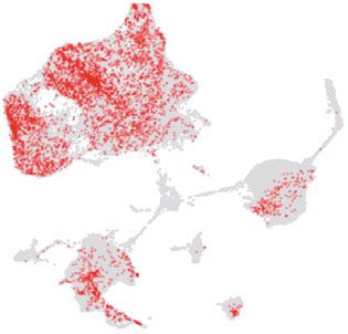
LETTER        Hanbo Li et al.
Protein & Cell

                 62       © The Author(s) 2020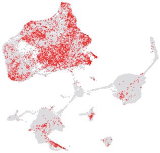
Single-cell atlas analysis of axolotl limb regeneration                                                                        LETTER

b    Figure 2. The cellular and molecular dynamics of ECM and              transition process is also linked to unique immune responses
     immune microenvironment during axolotl limb regenera-                 mediated by three types of macrophages, mechanistically
     tion. (A) UMAPs reflect the cellular distribution of fibroblasts        different from that in mouse. Our data provide valuable
     during different stages of axolotl limb regeneration and mouse        mechanistic and biological insights into how multi-tissue
     digit wound healing. Each dot represents a cell. Violin plots         structures regenerate in a spatial–temporal manner, which
     reflect the expression of the cell type identify markers. (B) Violin   may guide future efforts in regenerative medicine.
     plots reflect the gene expression of ECM deposition and
     modeling related genes during axolotl limb regeneration and           FOOTNOTES
     mouse digit wound healing. (C) Violin plots reflect the gene
     expression of fibrosis pathway related genes during axolotl limb       We would like to acknowledge the contributions and support of all
     regeneration and mouse digit wound healing. (D) Violin plots          BGI-Shenzhen, BGI-Qingdao and China National Gene Bank
     reflect the gene expression of cellular senescence related             employees, in particular the many highly skilled individuals who
                                                                           make it possible to generate high-quality data. This work was sup-
     genes during axolotl limb regeneration and mouse digit wound
                                                                           ported by the Strategic Priority Research Program of the Chinese
     healing. (E) Expression and distribution of marker genes and
                                                                           Academy of Sciences (Grant No. XDA16010000), the National Key
     key genes of macrophages. Each dot represents a cell, and the
                                                                           Research and Development Program of China (2018YFC2000100,
     color corresponds to the expression level (log2(CPM+1)).
                                                                           2017YFA0103304, 2017YFA0102802), the National Natural Science
     (F) Expression of macrophage marker genes in each macro-
                                                                           Foundation of China (81921006, 81625009), Youth Innovation Pro-
     phage cluster at different time points. Error bars indicate the

                                                                                                                                                   Protein & Cell
                                                                           motion Association of CAS (2016093) and the Guangdong Provin-
     standard deviation to the expression level (log2(CPM+1)).
                                                                           cial Key Laboratory of Genome Read and Write (No.
     (G) Cell number distribution of the macrophage subtypes at
                                                                           2017B0303011). The data that support this study have been
     each time point during axolotl limb regeneration (left) and
                                                                           deposited in the CNSA (https://db.cngb.org/cnsa/) and SRA (https://
     mouse digit wound healing (right).
                                                                           www.ncbi.nlm.nih.gov/sra/), under accession code CNP0000706
                                                                           (CNSA of the CNGBdb), and PRJNA589484 (SRA of the NCBI).
                                                                               Hanbo Li, Xiaoyu Wei, Li Zhou, Weiqi Zhang, Chen Wang, Yang
                                                                           Guo, Denghui Li, Jianyang Chen, Tianbin Liu, Yingying Zhang,
    report here a unique molecular feature in axolotl that may             Shuai Ma, Congyan Wang, Fujian Tan, Jiangshan Xu, Yang Liu, Yue
    contribute to the complete regeneration, containing a                  Yuan, Liang Chen, Qiaoran Wang, Jing Qu, Yue Shen, Shanshan
                                                                           Liu, Guangyi Fan, Longqi Liu, Xin Liu, Yong Hou, Guang-Hui Liu,
    sequential EMT and MET process followed by a unique
                                                                           Ying Gu, Xun Xu declare that they have no conflict of interest. All
    temporal ECM reconstruction and suppressed activation of
                                                                           institutional and national guidelines for the care and use of labora-
    fibrosis and cellular senescence pathways. This cell fate
                                                                           tory animals were followed.

    © The Author(s) 2020                                                                                                                     63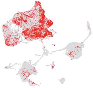
LETTER                               Hanbo Li et al.
Protein & Cell

                          Figure 2. continued.

                 64                              © The Author(s) 2020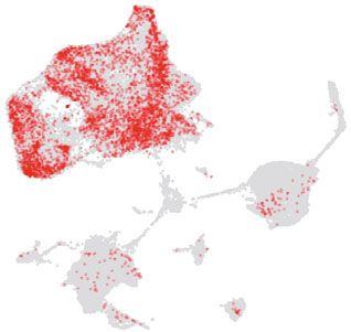
Single-cell atlas analysis of axolotl limb regeneration                                                                            LETTER

Hanbo Li1,2, Xiaoyu Wei1,3,4, Li Zhou1,2, Weiqi Zhang5,6,7,              indicated otherwise in a credit line to the material. If material is not
Chen Wang1,2, Yang Guo2, Denghui Li2, Jianyang Chen2,                    included in the article's Creative Commons licence and your
Tianbin Liu1,4,8, Yingying Zhang2, Shuai Ma6,7,9,                        intended use is not permitted by statutory regulation or exceeds
Congyan Wang2, Fujian Tan2, Jiangshan Xu1,3,4,                           the permitted use, you will need to obtain permission directly from
Yang Liu1,3,4, Yue Yuan1,3,4, Liang Chen10, Qiaoran Wang5,6,             the copyright holder. To view a copy of this licence, visit http://
Jing Qu6,7,11, Yue Shen1,4,8, Shanshan Liu1,2,                           creativecommons.org/licenses/by/4.0/.
Guangyi Fan1,2, Longqi Liu1,4, Xin Liu1,2, Yong Hou1,4,
                     &              &                &
Guang-Hui Liu6,7,12 , Ying Gu1,4,8 , Xun Xu1,4,7,8,13

1
   BGI-Shenzhen, Shenzhen 518083, China
2
   BGI-Qingdao, Qingdao 266555, China                                    REFERENCES
3
   BGI Education Center, University of Chinese Academy of                Doyle SL, O'Neill LA (2006) Toll-like receptors: from the discovery of
   Sciences, Shenzhen 518083, China                                         NFkappaB to new insights into transcriptional regulations in
4
   China National Gene Bank, BGI-Shenzhen, Shenzhen 518120,                 innate immunity. Biochem Pharmacol 72:1102–1113
   China                                                                 Erickson JR, Gearhart MD, Honson DD, Reid TA, Gardner MK,
5
   CAS Key Laboratory of Genomic and Precision Medicine, Beijing            Moriarity BS, Echeverri K (2016) A novel role for SALL4 during
   Institute of Genomics, China National Center for Bioinformation,         scar-free wound healing in axolotl. NPJ Regener Med 1:1–11
   Chinese Academy of Sciences, Beijing 100101, China

                                                                                                                                                    Protein & Cell
                                                                         Forte E, Chimenti I, Rosa P, Angelini F, Pagano F, Calogero A,
6
   University of Chinese Academy of Sciences, Beijing 100049,               Giacomello A, Messina E (2017) EMT/MET at the crossroad of
   China                                                                    stemness, regeneration and oncogenesis: he Ying-Yang equilib-
7
   Institute for Stem Cell and Regeneration, Chinese Academy of             rium recapitulated in cell spheroids. Cancers 9:98
   Sciences, Beijing 100101, China                                       Gerber T, Murawala P, Knapp D, Masselink W, Schuez M, Hermann S,
8
   Guangdong Provincial Key Laboratory of Genome Read and                   Gac-Santel M, Nowoshilow S, Kageyama J, Khattak et al (2018).
   Write, BGI-Shenzhen, Shenzhen 518083, China                              Single-cell analysis uncovers convergence of cell identities during
9
   National Laboratory of Biomacromolecules, CAS Center for                 axolotl limb regeneration. Science (New York, NY) 362
   Excellence in Biomacromolecules, Institute of Biophysics, Chinese     Godwin JW, Pinto AR, Rosenthal NA (2013) Macrophages are
   Academy of Sciences, Beijing 100101, China                               required for adult salamander limb regeneration. Proc Natl Acad
10
    Hubei Key Laboratory of Cell Homeostasis, College of Life               Sci USA 110:9415–9420
    Sciences, Wuhan University, Wuhan 40072, China                       Haas BJ, Whited JL (2017) Advances in decoding axolotl limb
11
    State Key Laboratory of Stem Cell and Reproductive Biology,             regeneration. Trends Genet 33:553–565
    Institute of Zoology, Chinese Academy of Sciences, Beijing           Kalluri R, Weinberg RA (2009) The basics of epithelial-mesenchy-
    100101, China                                                           mal transition. J Clin Investig 119:1420–1428
12
    State Key Laboratory of Membrane Biology, Institute of Zoology,      Leigh ND, Dunlap GS, Johnson K, Mariano R, Oshiro R, Wong AY,
    Chinese Academy of Sciences, Beijing 100101, China                      Bryant DM, Miller BM, Ratner A, Chen A et al (2018) Transcrip-
13
    Synthetic Biology Technology Innovation Center of Shandong              tomic landscape of the blastema niche in regenerating adult
    Province, Qingdao 266003, China                                         axolotl limbs at single-cell resolution. Nat Commun 9:5153
& Correspondence: ghliu@ioz.ac.cn (G.-H. Liu), guying@ge-                Marwick JA, Mills R, Kay O, Michail K, Stephen J, Rossi AG,
     nomics.cn (Y. Gu), xuxun@genomics.cn (X. Xu)                           Dransfield I, Hirani N (2018) Neutrophils induce macrophage anti-
                                                                            inflammatory reprogramming by suppressing NF-κB activation.
                                                                            Cell Death Dis 9:665
OPEN ACCESS                                                              Meng XM, Nikolic-Paterson DJ, Lan HY (2016) TGF-beta: the
                                                                            master regulator of fibrosis. Nat Rev Nephrol 12:325–338
This article is licensed under a Creative Commons Attribution 4.0        Rayagiri SS, Ranaldi D, Raven A, Mohamad Azhar NIF, Lefebvre O,
International License, which permits use, sharing, adaptation,              Zammit PS, Borycki AG (2018) Basal lamina remodeling at the
distribution and reproduction in any medium or format, as long as           skeletal muscle stem cell niche mediates stem cell self-renewal.
you give appropriate credit to the original author(s) and the source,       Nat Commun 9:1075
provide a link to the Creative Commons licence, and indicate if          Sharif O, Bolshakov VN, Raines S, Newham P, Perkins ND (2007)
changes were made. The images or other third party material in this         Transcriptional profiling of the LPS induced NF-kappaB response
article are included in the article's Creative Commons licence, unless      in macrophages. BMC Immunol 8:1

© The Author(s) 2020                                                                                                                          65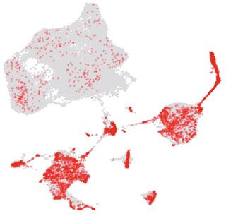
LETTER                                                                                                                    Hanbo Li et al.

                 Stone RC, Pastar I, Ojeh N, Chen V, Liu S, Garzon KI, Tomic-Canic    Zhang L, Huang G, Li X, Zhang Y, Jiang Y, Shen J, Liu J, Wang Q,
                    M (2016) Epithelial-mesenchymal transition in tissue repair and     Zhu J, Feng X et al (2013) Hypoxia induces epithelial-mesenchy-
                    fibrosis. Cell Tissue Res 365:495–506                                mal transition via activation of SNAI1 by hypoxia-inducible factor -
                 Wynn TA, Vannella KM (2016) Macrophages in tissue repair,              1alpha in hepatocellular carcinoma. BMC Cancer 13:108
                    regeneration, and fibrosis. Immunity 44:450–462
Protein & Cell

                 Hanbo Li, Xiaoyu Wei, Li Zhou and Weiqi Zhang have contributed
                 equally to this work.

                 Electronic supplementary material The online version of this
                 article (https://doi.org/10.1007/s13238-020-00763-1) contains sup-
                 plementary material, which is available to authorized users.

                 66                                                                                                                  © The Author(s) 2020
You can also read