Skin endometriosis: A case report and review of the literature - Spandidos Publications
←
→
Page content transcription
If your browser does not render page correctly, please read the page content below
EXPERIMENTAL AND THERAPEUTIC MEDICINE 21: 532, 2021

        Skin endometriosis: A case report and review of the literature
              ANDREEA‑MARIANA MATEI1,2*, ANA-MARIA DRAGHICI‑IONESCU1*, MIRELA CIOPLEA3,4,
            SABINA ANDRADA ZURAC3,4, DANIEL BODA5,6, IOANA SERBAN6, CONSTANTIN CARUNTU2,5,
                          MIHAELA ADRIANA ILIE6 and LÁSZLÓ FEKETE GYULA7

                           1
                            Department of Dermatology, Colentina Clinical Hospital, 020125 Bucharest;
          Departments of 2Physiology, 3Pathology, ‘Carol Davila’ University of Medicine and Pharmacy, 050474 Bucharest;
              4
                Department of Pathology, Colentina Clinical Hospital, 020125 Bucharest; 5Department of Dermatology,
             ‘Prof. N.C. Paulescu’ National Institute of Diabetes, Nutrition and Metabolic Diseases, 011233 Bucharest;
           6
             Dermatology Research Laboratory, ‘Carol Davila’ University of Medicine and Pharmacy, 050474 Bucharest;
                  7
                   Department of Dermatology, Dermatology Clinic, ‘George Emil Palade’ University of Medicine,
                                Pharmacy, Science and Technology, 540139 Târgu Mureș, Romania

                                    Received October 19, 2020; Accepted November 18, 2020

                                                   DOI: 10.3892/etm.2021.9964

Abstract. Skin endometriosis is a rare disease with variable        Rokitansky in 1860 (2), endometriosis is characterized by the
clinical and histopathological characteristics that depend on       presence of endometrial stroma outside the uterus, with the
hormonal stimuli. The skin is not a common location, as most        same reactivity to hormonal stimuli as the normal stroma (3,5).
cases of endometriosis involve pelvic sites, such as the ovaries,   The disease can be primary, where the endometrial tissue is
peritoneum and bowel. However, the most common extrapelvic          found outside the uterus without any external intervention and
site affected is the abdominal wall and this location of the        secondary or iatrogenic, following obstetric and gynecological
disease is frequently associated with obstetric and gynecologic     procedures (uterine wall opening), of which Caesarean‑section
surgery. Here we report a case of skin endometriosis emerged        (C‑section) is the most frequent (4).
as a painful subcutaneous nodule located near to the left side of       In patients with C‑sections, the incidence can be as high
an obstetrical surgery procedure scar. The patient affected was     as 1% (5). Usually, the diagnosis is made several months or
a woman in her reproductive age, with a history of right ovary      up to several years following the procedure (5). The clinical
endometriotic cyst laparoscopically removed and histologi‑          appearance varies with the depth and localization of the
cally confirmed as a primary endometriosis. Dermatologists          tumor (6). The symptomatology is not always present, and the
should be aware of this condition in any woman with a painful       diagnosis is difficult. However, the most frequent symptom is
lump located in the proximity of a pelvic surgery‑induced scar.     cyclic pain and a positive history for surgery may be a clue for
Its non‑specific clinical appearance may confuse the clinician      the correct diagnosis (5,7).
and may delay the diagnosis and management.                             Herein, we present the case of skin endometriosis
                                                                    presenting as a subcutaneous nodule in the proximity of a
Introduction                                                        C‑section surgical scar and review existing literature in order
                                                                    to increase the index of suspicion in the case of painful lesions
Endometriosis is a gynecological disease affecting fertile          appearing close to surgical scars following gynecological or
women, with a prevalence of 2‑10% in the general female             obstetrical procedures.
population (1), which can reach up to 50% in patients with pain,
infertility or abdomino‑pelvic surgery (2‑4). First described by    Case report

                                                                    A 29‑year‑old female presented to the Dermatology
                                                                    Department of ‘Prof. Dr. Nicolae C. Paulescu’ National Institute
                                                                    of Diabetes, Nutrition and Metabolic Diseases, Bucharest,
Correspondence to: Dr Mihaela Adriana Ilie, Dermatology
Research Laboratory, ‘Carol Davila’ University of Medicine and      Romania for the investigation of a painful nodule located in
Pharmacy, 8 Eroii Sanitari Street, 050474 Bucharest, Romania        the inferior abdominal wall. Written informed consent was
E‑mail: mihaelaadriana2005@yahoo.com                                provided and the patient agreed to undergo diagnostic and
                                                                    therapeutic procedures included in the study protocol that was
*
    Contributed equally                                             conducted in accordance with the Declaration of Helsinki with
                                                                    approval of the Ethics Committee of the Colentina Clinical
Key words: endometriosis, skin, immunohistochemistry nodule,        Hospital (approval no. 25/27.11.2017).
diagnosis                                                               The patient medical history was significant for a right
                                                                    ovarian endometrial cyst, laparoscopically removed 4 years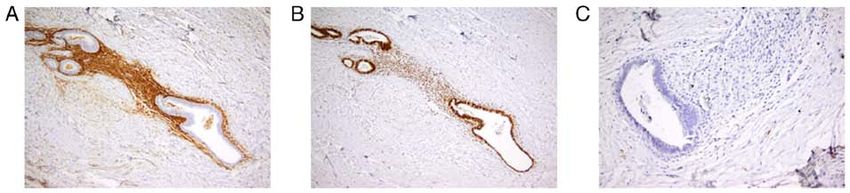
2 MATEI et al: SKIN ENDOMETRIOSIS: CASE REPORT AND REVIEW
prior to this presentation, and pathologically confirmed as with obstetric and gynecologic surgery (9). Abdominal wall
primary endometriosis. The patient presented menstrual endometriosis usually presents at surgery departments,
cycle‑dependent lower abdominal pain after the first surgery. being misdiagnosed as incisional hernia or granuloma (10).
A second laparoscopy was performed, showing multiple sites The clinical differential diagnoses of skin endometriosis
of endometriosis. The lesions were thermocoagulated followed are represented by incisional hernia, lipoma, dermoid cyst,
by treatment with triptorelin 3.75 mg, 1 monthly injection for abscess, suture granuloma, keloid, melanoma, hematoma and
5 months. After hormonal treatment withdrawal, the patient others (11,12).
became pregnant. During pregnancy, the patient experienced In our case, based on characteristic history and examination
diffuse low abdominal pain, but without any complications findings, behind the most probable diagnosis of endometriosis,
and she delivered by term C‑section. Two years after giving other diagnoses including lipoma, granuloma and desmoid
birth, she presented at our department for a painful nodule tumor, were also deliberated.
(the pain was worsening prior menses), located in the lower Multiple diagnostic tools have been used in the diagnosis
abdominal wall. The patient also described an intermittent, of skin endometriosis. Examination techniques usually used for
rather than cyclic, C‑section scar pain which started one year diagnosis include ultrasonography, computed tomography (CT),
after she gave birth. magnetic resonance imaging (MRI), and Doppler sonography.
The physical examination showed a palpable subcutaneous Ultrasonography seems to be the first choice for evaluation
nodule of approximately 1.5 cm in diameter, round, well of any abdominal lesion, but CT and MRI may exclude other
defined and mobile, tender on palpation, located 2 cm superior possible diagnoses such as a lipoma, hernia or a tumor (13).
to the left side of the C‑section scar (Pfannenstiel incision). Tumor markers such as CA19‑9 and CA125 may or may not be
The suprajacent skin was normal. The C‑section scar was elevated; however histologic examination after excision of the
well‑healed, supple and whitish, without any pathological lesion can confirm the diagnosis (14). Kinkel et al reported the
finding upon palpation. A diagnosis of cutaneous endometri‑ sensitivity and specificity of MRI in diagnosing endometriomas
osis was suspected given the patient's history of laparoscopic to be 90‑92 and 91‑98%, respectively (15).
intervention to the ovary and flares of pain with menstrual In regards to skin imaging techniques, standardized
periods. dermoscopic features of skin endometriosis have not yet been
The lesion was surgically excised. The macroscopic aspect established. Multiple factors such as the site of occurrence, histo‑
of the nodule excluded the diagnosis of a cystic lesion or lipoma logical subtype, depth of the lesion or patient phototype may
and the specimen was referred to the pathology department for influence the dermoscopic aspect of skin endometriosis (16).
microscopic examination. However, this examination technique may provide additional
The specimen was routinely processed for paraffin- information useful for clinical diagnosis. A study published by
embedding; then 3‑µm‑thick sections were cut and routinely de Giorgi et al revealed dominant dermoscopic features in skin
stained with hematoxylin and eosin (H&E), and immunohisto‑ endometriosis such as homogeneous reddish pigmentation,
chemistry analysis for CD10, estrogen receptor and Ki67 was containing small red globular structures, which they termed
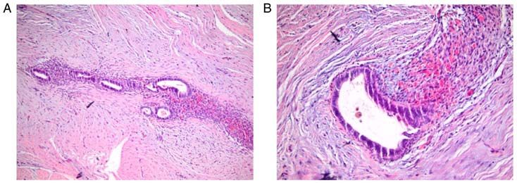
performed. The pathologic report showed adipose connective ‘red atolls’ (7). Moreover, Costa et al described the dermos‑
tissue, neuro‑vascular and fascial tissue including multiple copy pattern of cutaneous endometriosis in the follicular
glandular structures of variable dimensions with a simple phase as erythematous‑violaceous polypoid projections with
columnar focal ciliated epithelium, surrounded by an endome‑ light brown spots and areas of active bleeding; further in the
trial stroma (Fig. 1) with multiple hematic extravasations and luteal phase the dermoscopic feature of the lesion was as an
rare siderophages. The immunohistochemical testing showed erythematous‑bluish aspect (17).
positivity for the estrogen receptor in the nuclei of epithelial Skin endometriosis usually affects women in their
cells lining glandular structure endometrial‑like cells, CD10 reproductive age (mean age 30‑40 years) and its clinical
diffuse and intense positivity in the endometrial stroma and presentation starts with a pigmented or skin‑colored papule or
Ki67 positivity below 1% of epithelial cell nuclei (Fig. 2). nodule with an average diameter of 2 cm. The symptoms asso‑
These findings sustained the diagnosis of skin endometriosis. ciated with this disease are pain, tenderness or bleeding during
The post‑operatory evolution was good, with disappearance menstrual cycle and their persistence ranges from 2 months to
of the nodule‑associated symptoms. After 1 year, the patient 2 years (18). After surgery the average period to the onset of
remains without recurrence of the disease or appearance of symptoms is between 3 months to 18 years (19).
endometriosis. In our case report, there are many similarities to previously
reported cases. Our female patient was 29 years of age, thus she
Discussion was in her reproductive age; the site of the lesion was close to
the C‑section scar; the lesion appeared approximately 2 years
Skin endometriosis is a rare disorder with variable clinical after surgery as a subcutaneous nodule; and associated symp‑
and histopathological appearance that depends on hormonal tomatology included abdominal pain that started one year after
stimuli and it primarily affects women of reproductive age. C‑section procedure. In this case, characteristic symptoms of
More than 300 cases of skin endometriosis have been described endometriosis such as bleeding or monthly swelling were not
in the literature and in a review published by Stojanovic et al, present and considering previously described differentials
they found 210 cases of skin endometriosis located on surgical near a scar (20,21), this lesion was not easy to diagnose.
scars, whereof 119 cases followed a C‑section procedure (8). The surgical excision revealed a non‑cystic appearance,
In regards to skin endometriosis, the most frequent extra‑ and the specimen was referred to the anatomopathologist. The
pelvic site affected is the abdominal wall and it is associated characteristic features of ectopic endometrial tissue includeEXPERIMENTAL AND THERAPEUTIC MEDICINE 21: 532, 2021 3
Figure 1. Hematoxylin and eosin‑stained histopathological images [(A) (x10 magnification) and (B) (x20 magnification)] showing endometrial glandular
structures, bordered by variable amounts of endometrial stroma.
Figure 2. Immunohistochemical staining for (A) CD10 (x10 magnification), (B) estrogen receptor (x10 magnification) and (C) Ki67 (x20 magnification).
glands with cylindrical epithelium and endometrial stroma the abdominal wall with saline solution before closing the
which may be influenced by cyclic hormonal changes. This is abdominal layers (33). Other recommendations published in
one of the reasons why all stages of the menstrual cycle could the literature include isolation of the surgical scar, changing
be found in the ectopic tissue. Immunohistochemical testing needles during the closure of the superficial layer of the
in our case showed positive CD10, a similarity found with abdomen wall, and replacing the instruments used during
other reported cases (13,22). Studies have reported that CD10 C‑section to prevent the iatrogenic grafting of cells (37).
as a useful marker for diagnosis as it is strongly expressed in A retrospective study of 198 Caesarean scar‑related
endometrial stromal nodules (23). endometriosis cases by Zhang et al found more than 70% of
Furthermore, in our case, Ki67 positivity was found in less the endometrial cases in superficial regions of the abdominal
than 1% of the nuclei of the cells. According to the literature, wall; 5.7% were found in the adipose layer and 64.6% between
an increased Ki67 and CD10 positivity indicates that the the adipose layer and the fascia layer, and 83% were located in
stroma is decidualized (24). Another finding of our case was a corner of the Pfannenstiel incision scar (38). Ding and Zhu
the positivity for the estrogen receptor in endometroid‑like conducted another retrospective study with similar results;
cells. This was also previously reported (25‑28). The produc‑ 77.1% of the cases were located in the corners of the scars (39).
tion of estrogen can be stimulated in endometrial lesions by One argument for this particular location may be that endo‑
aromatase activity (29). metrial cells are less easily detached from the corners of the
Concerning the possible pathophysiological mechanisms incisions during C‑section (38).
involved, one accepted theory suggests that primitive pluripo‑ The most frequently used abdominal skin incisions
tent mesenchymal cells that have undergone differentiation are Pfannenstiel incision and vertical midline; therefore
metaplasia are one of the cause of abdominal wall endome‑ C‑section seems to be one of the most popular surgical proce‑
triosis (30). In addition, some authors propose that retrograde dures utilized on the female population (38,39). According
menstruation could be a cause for the implantation of endo‑ to Zhang et al, more blood loss in the Pfannenstiel incision
metrial cells in the peritoneal area (31), while others advocate would supply a relatively rich nutritional background for the
the theory of lymphatic or hematogenic dissemination (32), implantation and expansion of residual endometrial cells,
the role of genetics (33) and other probable pathophysiological facilitating the development of skin endometriosis. Therefore,
mechanisms (34,35). Another theory explains the possibility this type of incision would increase the risk for skin endome‑
that during a C‑section procedure endometrial tissue could triosis (38).
be iatrogenically grafted into ectopic sites such as the skin, In our patient, the cause for this ectopic tissue may have
muscles or other layers of the abdominal wall, this being been the iatrogenic transportation of endometrial cells during
the reason why the nodule appears frequently above the the C‑section incision. The site of the lesion was found in the
scar. In addition, these grafted endometrial cells are able to superficial abdominal wall, in the proximity of the left side of
proliferate due to hormonal stimulation (36). Therefore, one the scar, after Pfannenstiel incision, as similarly mentioned in
recommendation according to various authors is to irrigate the studies above.4 MATEI et al: SKIN ENDOMETRIOSIS: CASE REPORT AND REVIEW
Our treatment for this patient was surgical excision of the Ethics approval and consent to participate
lesion with clear margins to prevent recurrence. Generally,
surgical excision is the first‑line treatment as it is the best Ethics approval was obtained from the Ethics Committee of
way to establish a clear diagnosis and to ensure a low rate the Colentina Clinical Hospital (approval no. 25/27.11.2017).
of recurrence. The recurrence rate is generally low, but Written informed consent of the patient was obtained.
other publications have described recurrence in 6‑11% of
patients (40,41). The malignancy rate is also extremely low Patient consent for publication
with less than 1% of endometriosis cases reported in associa‑
tion with cancer; one of the most common types is clear‑cell Written informed consent of the patient was obtained for
carcinoma with a survival rate of 80% (39,42). publication of the information.
Hormonal therapy with gonadotropin releasing hormone
(GnRH) agonists, danazol, preoperative or postoperative Competing interests
progesterone is also advocated in the literature. Preoperative
therapy is effective in ameliorating symptoms such as pain The authors declare that they have no competing interests.
and minimizing the lesion size. The goal of postoperative
hormonal therapy is to prevent recurrence, but its use is still References
under debate as the overall result has been poor (43). Moreover,
considering the psychological and social impact of the disease, 1. Dunselman GAJ, Vermeulen N, Becker C, Calhaz‑Jorge C,
patient counseling should also be considered (44,45). D'Hooghe T, De Bie B, Heikinheimo O, Horne AW, Kiesel L,
Nap A, et al: ESHRE guideline: Management of women with
In conclusion, skin endometriosis is a rare and benign endometriosis. Hum Reprod 29: 400‑412, 2014.
condition, with an unknown mechanism and a very low rate of 2. Giudice LC and Kao LC: Endometriosis. Lancet 364: 1789‑1799,
malignancy. Its clinical appearance is very unspecific which 2004.
3. Blanco RG, Parithivel VS, Shah AK, Gumbs MA, Schein M
may hinder the dermatologist's diagnosis, delaying the right and Gerst PH: Abdominal wall endometriomas. Am J Surg 185:
management of the lesion. The rate of C‑section is increasing 596‑598, 2003.
in the female population and the associated incidence in 4. Scholefield HJ, Sajjad Y and Morgan PR: Cutaneous endometri‑
osis and its association with caesarean section and gynaecological
skin endometriosis (found on or in close proximity to a scar procedures. J Obstet Gynaecol 22: 553‑554, 2002.
associated with this procedure) may increase in the future. 5. Ozel L, Sagiroglu J, Unal A, Unal E, Gunes P, Baskent E, Aka N,
Dermatologists should be aware of this condition in any Titiz MI and Tufekci EC: Abdominal wall endometriosis in the
cesarean section surgical scar: A potential diagnostic pitfall.
women with pain and a lump close to an incisional scar after J Obstet Gynaecol Res 38: 526‑530, 2012.
pelvic surgery. 6. Tognetti L, Cinotti E, Tonini G, Habougit C, Cambazard F,
The first‑line treatment of skin endometriosis is surgical Rubegni P and Perrot JL: New findings in non‑invasive imaging
of cutaneous endometriosis: Dermoscopy, high‑frequency ultra‑
excision and the gold standard for its diagnosis is histopatho‑ sound and reflectance confocal microscopy. Ski Res Technol 24:
logic and, if necessary, immunohistochemical examination. 309‑312, 2018.
7. De Giorgi V, Massi D, Mannone F, Stante M and Carli P:
Cutaneous endometriosis: Non‑invasive analysis by epilumines‑
Acknowledgements cence microscopy. Clin Exp Dermatol 28: 315‑317, 2003.
8. Stojanovic M, Brasanac D and Stojicic M: Cutaneous inguinal
Not applicable. scar endosalpingiosis and endometriosis: Case report with review
of literature. Am J Dermatopathol 35: 254‑260, 2013.
9. Bektaş H, Bilsel Y, Sar YS, Ersöz F, Koç O, Deniz M, Boran B
Funding and Huq GE: Abdominal wall endometrioma; a 10‑year experi‑
ence and brief review of the literature. J Surg Res 164: e77‑e81,
2010.
This research and review was funded by a grant from 10. Loh SH, Lew BL and Sim WY: Primary cutaneous endometriosis
the Romanian Ministry of Research and Innovation, of umbilicus. Ann Dermatol 29: 621‑625, 2017.
CCCDI-UEFISCDI (project no. 61PCCDI ⁄2018 PN‑III‑P1‑1.2- 11. Curry TW: Subcutaneous endometriomas: Two case reports and
review of the literature. J Gynecol Surg 14: 31‑34, 1998.
PCCDI‑2017‑0341) within PNCDI‑III. 12. Tatu AL: Umbilicated blue‑black lesion on the lateral thorax.
J Cutan Med Surg 21: 252, 2017.
Availability of data and materials 13. Neri I, Tabanelli M, Dika E, Valeria G and Patrizi A: Diagnosis
and treatment of post‑Caesarean scar endometriosis. Acta Derm
Venereol 87: 428‑429, 2007.
All findings generated or analyzed during this study are 14. Mechsner S, Bartley J, Infanger M, Loddenkemper C, Herbel J
included in this published article and the literature findings and Ebert AD: Clinical management and immunohistochemical
analysis of umbilical endometriosis. Arch Gynecol Obstet 280:
are documented by relevant references. 235‑242, 2009.
15. Kinkel K, Frei KA, Balleyguier C and Chapron C: Diagnosis of
Authors' contributions endometriosis with imaging: A review. Eur Radiol 16: 285‑298,
2006.
16. Jaime TJ, Jaime TJ, Ormiga P, Leal F, Nogueira OM and
AMM and AMDI contributed equally to the conceptualiza‑ Rodrigues N: Endometriose umbilical: Relato de um caso e
tion, data analysis, and writing of the manuscript. MC, SAZ, seus achados dermatoscópicos. An Bras Dermatol 88: 121‑124,
2013.
DB, IS and LFG conducted the literature research and data 17. Costa IMC, Gomes CM, Morais OO, Costa MC, Abraham LS
analysis. CC and MAI conducted the data analysis, critical and Argenziano G: Cutaneous endometriosis: Dermoscopic
review, editing of the manuscript and supervision of the findings related to phases of the female hormonal cycle. Int J
Dermatol 53: e130‑e132, 2014.
project. All authors read and gave approval for publication of 18. Friedman PM and Rico MJ: Cutaneous endometriosis. Dermatol
the final manuscript. Online J 6: 8, 2000.EXPERIMENTAL AND THERAPEUTIC MEDICINE 21: 532, 2021 5
19. Kazakov DV, Ondic O, Zamecnik M, Shelekhova KV, 33. Witz CA. Pathogenesis of endometriosis. Gynecol Obstet
Mukensnabl P, Hes O, Dvorak V and Michal M: Morphological Invest 53 (Suppl 1): S52‑S62, 2002.
variations of scar‑related and spontaneous endometriosis of the 34. Nwabudike LC and Tatu AL: Reply to Happle R et al. Koebner's
skin and superficial soft tissue: A study of 71 cases with emphasis sheep in Wolf's clothing: Does the isotopic response exists as
on atypical features and types of müllerian differentiations. J Am a distinct phenomenon? J Eur Acad Dermatology Venereol 32:
Acad Dermatol 57: 134‑146, 2007. e336‑e337, 2018.
20. Tatu AL, Kluger N and Nwabudike LC: Pain and shingles on an 35. Aida Maranduca M, Liliana Hurjui L, Constantin Branisteanu D,
old scar. Int J Dermatol 59: 1158‑1159, 2020. Nicolae Serban D, Elena Branisteanu D, Dima N and Lacramioara
21. Ardeleanu V, Jecan CR, Tatu AL and Motoc AG: A recurrent soli‑ Serban I: Skin‑a vast organ with immunological function
tary glomus tumor of the forearm. Rom J Morphol Embryol 60: (Review). Exp Ther Med 20: 18‑23, 2020.
1019‑1023, 2019. 36. Seydel AS, Sickel JZ, Warner ED and Sax HC: Extrapelvic
22. Kerr OA, Mowbray M and Tidman MJ: An umbilical nodule due endometriosis: Diagnosis and treatment. Am J Surg 171: 239‑241,
to endometriosis. Acta Derm Venereol 86: 277‑278, 2006. 1996.
23. Van den Nouland D and Kaur M: Primary umbilical endome‑ 37. Agarwal A and Fong YF. Cutaneous endometriosis. Singapore
triosis: A case report. Facts Views Vis Obgyn 9: 115‑119, 2017. Med J 49: 704‑709, 2008.
24. Val‑Bernal JF, Val D, Gómez‑Aguado F, Corcuera MT and 38. Zhang P, Sun Y, Zhang C, Yang Y, Zhang L, Wang N and Xu H:
Garijo MF: Hypodermal decidualized endometrioma with aber‑ Cesarean scar endometriosis: Presentation of 198 cases and
rant cytokeratin expression. A lesion mimicking malignancy. Am literature review. BMC Womens Health 19: 14, 2019.
J Dermatopathol 33: e58‑e62, 2011. 39. Ding Y and Zhu J: A retrospective review of abdominal wall endo‑
25. Kholová I, Ryska A and Dedic K: Composite tumor consisting metriosis in Shanghai, China. Int J Gynecol Obstet 121: 41‑44,
of dermatofibrosarcoma protuberans and giant cell fibroblastoma 2013.
associated with intratumoral endometriosis. Report of a case. 40. Chatterjee SK: Scar endometriosis: A clinicopathologic study of
Pathol Res Pract 197: 263‑267, 269‑270, 2001. 17 cases. Obstet Gynecol 56: 81‑84, 1980.
26. Dragoumis K, Mikos T, Zafrakas M, Assimakopoulos E, 41. Steck WD and Helwig EB: Cutaneous endometriosis. JAMA 191:
Stamatopoulos P and Bontis J: Endometriotic uterocutaneous 167‑170, 1965.
fistula after cesarean section. A case report. Gynecol Obstet 42. Petca A, Radu N and Petca R: Insights into malignant potential
Invest 57: 90‑92, 2004. of ovarian endometriomas. In: The 17th National Congress of
27. Frischknecht F, Raio L, Fleischmann A, Dreher E, Lüscher KP the Romanian Society of Obstetrics and Gynecology and First
and Mueller MD: Umbilical endometriosis. Surg Endosc 18: 347, Advanced Colposcopy Course. pp614‑619, 2019.
2004. 43. Raffi L, Suresh R, McCalmont TH and Twigg AR: Cutaneous
28. Farooq U, Laureano AC, Miteva M and Elgart GW: Cutaneous endometriosis. Int J Womens Dermatol 5: 384‑386, 2019.
endometriosis: Diagnostic immunohistochemistry and clinico‑ 44. Rebegea L, Firescu D, Baciu G and Ciubara A: Psycho‑oncology
pathologic correlation. J Cutan Pathol 38: 525‑528, 2011. support. BRAIN Broad Res Artif Intell Neurosci 10: 77‑88, 2019.
29. Bulun SE, Imir G, Utsunomiya H, Thung S, Gurates B, Tamura M 45. Aerts L, Grangier L, Streuli I, Dällenbach P, Marci R, Wenger JM
and Lin Z: Aromatase in endometriosis and uterine leiomyomata. and Pluchino N: Psychosocial impact of endometriosis: From
J Steroid Biochem Mol Biol 95: 57‑62, 2005. co‑morbidity to intervention. Best Pract Res Clin Obstet
30. Dwivedi AJ, Agrawal SN and Silva YJ: Abdominal wall endome‑ Gynaecol 50: 2‑10, 2018.
triomas. Dig Dis Sci 47: 456‑461, 2002.
31. Vinatier D, Orazi G, Cosson M and Dufour P: Theories of endo‑
metriosis. Eur J Obstet Gynecol Reprod Biol 96: 21‑34, 2001.
32. Horton JD, DeZee KJ, Ahnfeldt EP and Wagner M: Abdominal
wall endometriosis: A surgeon's perspective and review of
445 cases. Am J Surg 196: 207‑212, 2008.You can also read