(Some) Medical Applications of Microwaves
←
→
Page content transcription
If your browser does not render page correctly, please read the page content below
Bellairs Workshop on Microwave Imaging and Detection
 for Biomedical Applications
 Barbados, March 2020

(Some) Medical Applications of Microwaves

 Prof. Zoya Popovic
 Department of Electrical, Computer and Energy Engineering
 University of Colorado, Boulder

 Zoya Popovic, University of Colorado, Boulder, 2020
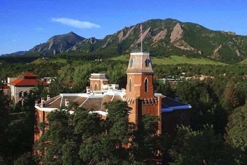
University of Colorado, Boulder
 • 32,000 students
 • 5 Nobel prizes
 • > $500M in funding
 • Largest NASA funding of
 any US university
 • Main campus: 600 acres in
 Boulder, Colorado (Forbes
 magazine named America's
 smartest city two years in a
 row)
 • ~100k people, 93k bikes

• Several
 national
 institutes:
• NIST (science
 metrology)
• NREL (energy)
• NOAA, NCAR,
 CIRES
 (weather)
• USGS
 Zoya Popovic, University of Colorado, Boulder, 2020 2
Microwaves at CU Boulder
Prof. Em. Ed Prof. Zoya Prof. Melinda Prof. Dejan Prof. Albin Prof. Taylor Prof. Dimitra Prof. Gregor
Kuester Popovic Piket-May Filipovic Gasiewski Barton Psychogiou Lasser

• 50+ graduate
 students Zoya’s research group
• >$10M current
 research funding
• 15 Best Paper
 Awards, 2
 Microwave Prizes
• Distinguished IEEE
 MTT Educator
• 3 IEEE Fellows, 2
 Endowed Chairs,
 one Distinguished
 Professor

 Zoya Popovic, University of Colorado, Boulder, 2020 3
Research Focus
 High-efficiency transmitters for
 communications and radar

 Wireless power transfer
from µW/cm2 far-field to kW near-field

Medical applications of microwaves

 Other projects, e.g.
 High-performance passives
 Multi-beam and broadband antennas
 Zoya Popovic, University of Colorado, Boulder, 2020 4
Overview

1. High-field MRI: excitation and bore design
 • 7T, 10.5T, 16.5T travelling wave
2. Tissue ablation and cauterization:
 transmitter design
 • High-efficiency with variable load
3. Measuring core body temperature
 • Microwave radiometry

 Zoya Popovic, University of Colorado, Boulder, 2020 5
High-field MRI • Precession (Larmor) frequency = B DC magnetic Larmor = Gyromagnetic ratio field frequency For protons: = 42.58 MHz/T 1.5T 64 MHz • B1+ transverse right handed circular 3T 128 MHz polarized field excites atoms 7T 297 MHz 10.5T 447 MHz • High B0 offers increased spatial resolution, better parallel imaging performance, improved SNR κγ2 02 TW MRI Near = ≈ κγ 0 3/2 Field γ 02 + 3 0, 0 MRI • Problems: – quasi-static approximation (coils) – waveguide effects appear causing image quality degradation D. Brunner, N. D. Zanche, J. Frohlich, J. Paska, and K. P. Pruessmann, “Travelling-wave nuclear magnetic resonance,” Nature, vol. 457, no. 7232, pp. 994–998, Feb. 2009. Zoya Popovic, University of Colorado, Boulder, 2020 6
Existing 16.4-T MRI loop coil probe
 Loop Coil:
 Axial and saggital measurements
 of cylindrical water phantom

 Axial
 Saggital

 Coronal

 Work with Harvard and CMRR:
 Thanks to Andrew Kiruluta;
Larmor frequency: Pierre-Francois Van de
 Moortele and Gregor Adriany
698 MHz

 Zoya Popovic, University of Colorado, Boulder, 2020 7
16.4T MRI patch probe
• Uneven slots enable RHCP Circular Slot-Loaded Patch Probe
Free space:
• 0.3 dB axial ratio
• 8 dB return loss
In Bore:
• 5 dB axial ratio (simulated)
• 15 dB return loss with matching
 network (measured)

 |Js|[A/m]
 0 10 20 30 40 50

 • Slots on ground plane
 perform as quadrature
 dipoles
 Zoya Popovic, University of Colorado, Boulder, 2020 8
Coil vs. patch probe

 Loop Coil Patch Probe

 Zoya Popovic, University of Colorado, Boulder, 2020 9
16.4T MRI improvements
 • Approximation of an
 electrically hard surface
 • 3 cm copper tape spaced
 1 cm apart placed on
 Mylar sheet

 • Comparable to the simulation
 • Demonstrated 16.4 T MR
 images in a phantom excited by
 a travelling-wave field patch
 probe far from the phantom.
 • Anisotropic copper strip array is
 inserted into the bore,
 modifying the modal content.
 • New design shows a seven-fold
 increase in SNR as compared to
 loop probe.

 Zoya Popovic, University of Colorado, Boulder, 2020 10
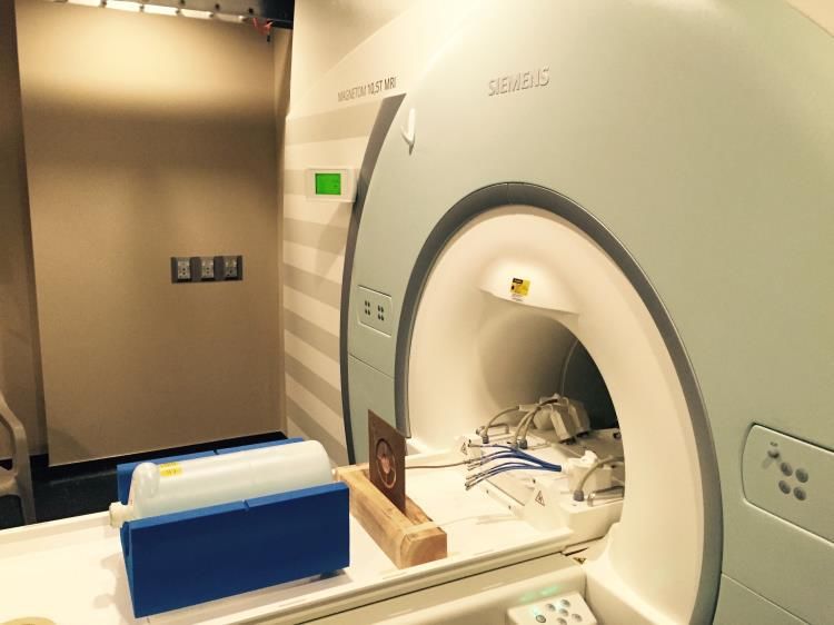
10.5T human-size bore MRI

 • Siemens MRI, 4.1 meter long
 • 447 MHz excitation
 • Wide-bore to accommodate human patient
 • Phantom contains DI water (εr=81 and σ=0.4 S/m)
 • ~40 cm encoding region under gradient coil

 Zoya Popovic, University of Colorado, Boulder, 2020 11
10.5T human-size bore MRI

 Zoya Popovic, University of Colorado, Boulder, 2020 12
Patch + C-array

Phase excitation, relative to patch array

 Zoya Popovic, University of Colorado, Boulder, 2020 13
Methods to improve uniformity

Helix

 Zoya Popovic, University of Colorado, Boulder, 2020 14
Nonuniform phantoms

 Zoya Popovic, University of Colorado, Boulder, 2020 15
10.5T human simulations

• Simulated performance with phantom
 compares well with measurements
• Measured inhomogeneous phantom (NIST)
 and pineapple
• Simulated B1+ for Duke and determined
 spatial SAR
• Improved uniformity with modified
 boundary conditions

 Zoya Popovic, University of Colorado, Boulder, 2020 16
References

1. P. Bluem, A. Tonyushkin, D. Deelchand, G. Adriany, P. F. V. de Moortele, A. J. M. Kiruluta, and Z.
 Popovic, “Travelling-wave excitation for 16.4T small-bore MRI," in Proc. IEEE MTT-S Int.
 Microwave Symp. (IMS), May 2015.
2. P. Bluem and Z. Popovic, "10.5-T MRI volume excitation using traveling-wave microstrip
 probes," 2017 IEEE MTT-S Intern. Microwave Symp. (IMS), Honololu, HI, 2017, pp. 1396-1399.
3. P. Bluem, A. Kiruluta, P. F. Van de Moortele, A. Duh, G. Adriany and Z. Popović, "Patch-Probe
 Excitation for Ultrahigh Magnetic Field Wide-Bore MRI," IEEE Transactions on Microwave Theory
 and Techniques, vol. 65, no. 7, pp. 2547-2557, July 2017.
4. P. Bluem, P. Van de Moortele, G. Adriany and Z. Popović, "Excitation and RF Field Control of a
 Human-Size 10.5-T MRI System," in IEEE Transactions on Microwave Theory and Techniques, vol.
 67, no. 3, pp. 1184-1196, March 2019.

 Zoya Popovic, University of Colorado, Boulder, 2020 17
Overview

1. High-field MRI: excitation and bore design
 • 7T, 10.5T, 16.5T travelling wave
2. Tissue ablation and cauterization:
 transmitter design
 • High-efficiency with variable load
3. Measuring core body temperature
 • Microwave radiometry

 Zoya Popovic, University of Colorado, Boulder, 2020 18
Motivation
 Targeted heating using RF

 Primary liver cancer
 - 6th leading cause
 - 2nd leading cause of death
 Secondary liver cancer
 - Liver is the most common site
 of metastases
Joseph Brannan, Medtronic, private comm.
 Zoya Popovic, University of Colorado, Boulder, 2020 19
Liver tumor ablation
 Raptor 2450 MHz Generation 3 Rev 2: 60 Watts, 5 minutes
 5
 Ablation 1
 4.5 Ablation 2
 Ablation 3
 4 Ablation 4
 Ablation 5
 3.5 Ablation 6

 3

 Power [W]
 2.5

 2

 1.5
 Measured reflected
 power from
 1 applicator (courtesy:
 J Brannan, Medtronic,
 0.5
 Boulder)
 0
 0 50 100 150 200 250 300
 Time [sec]

 • Microwave tumor ablation and cauterization performed
 with coaxial probe connected to 50-100W PA
 • As tissue heats and burns, VSWR changes (1.5-5),
 power lost to heat in circulator load

 Zoya Popovic, University of Colorado, Boulder, 2020 20
Outphasing for load mismatch
 C. Sanchez-Perez, D. Sardin, M. Roberg, J. de Mingo, Z. Popovic, “Tunable Outphasing for Power
 Amplifier Efficiency Improvement under Load Mismatch,” IEEE MTT International Microwave Symp.
 Digest, June 2012, Montreal.

 Zoya Popovic, University of Colorado, Boulder, 2020 21
Design and test procedure GaN HEMT Cree demo boards, Pout= 39dBm, ηd=55% at 2.14GHz, ZL=50Ω • 1,2 varied manually C=(0.1 pf – 1.5 pF) • L= (1 nH – 2.7 nH) 3.5 mm L (nH) • For each 1,2 combination, 39 mm adjusted for maximum efficiency at ZL = 50Ω • Load swept over 236 complex impedances within VSWR < 10 Non isolated combiner fabricated on C (pF) RO4350 (εr=3.48, tanδ=0.004) Zoya Popovic, University of Colorado, Boulder, 2020 22
Measured results +j1.0 +j1.0 +j0.5 +j2.0 +j0.5 +j2.0 45 40 36 35 37 50 3 40 55 38 387 39 30 40 39 +j0.2 45 +j5.0 +j0.2 +j5.0 36 45 36 55 59 50 30 40 50 59 35 3938 7 40 59 3 35 59 40 37 0.0 55 0.0 38 40 40 20 55 50 25 39 50 40 30 45 39 40 45 39 -j0.2 30 -j5.0 35 40 -j0.2 -j5.0 36 15 35 36 38 38 25 37 20 25 30 37 36 20 3534 35 15 33 -j0.5 -j2.0 -j0.5 -j2.0 -j1.0 -j1.0 • The measured output power is higher than 40 dBm for all swept impedances corresponding to an efficiency around 55% • Maximum efficiency obtained is 63% Zoya Popovic, University of Colorado, Boulder, 2020 23
Overview

1. High-field MRI: excitation and bore design
 • 7T, 10.5T, 16.5T travelling wave
2. Tissue ablation and cauterization:
 transmitter design
 • High-efficiency with variable load
3. Measuring core body temperature
 • Microwave radiometry

 Zoya Popovic, University of Colorado, Boulder, 2020 24
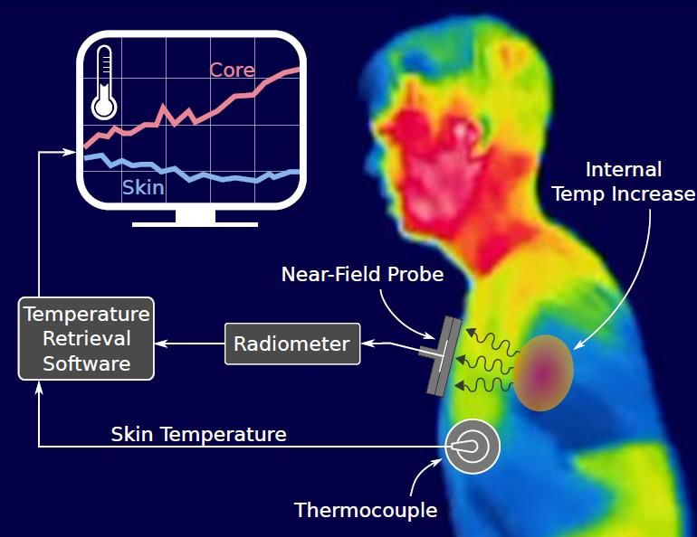
Motivation example

• Muscle temperature
 increases with increasing
 exercise intensity

• Skin temperature
 decreases (sweating cools
 down the skin)

• The internal temperature
 of the human body and
 the skin temperature can
 be quite different.

 Zoya Popovic, University of Colorado, Boulder, 2020 24
Other needs
 Motivation
 In vivo subsurface tissue Non-invasive and versatile, with applications to numerous medical diagnoses and
 treatments (cancers, arthritis, hypoxia ischemia; hypothermia and hyperthermia
 temperature treatment, ablation.
 Determine burn severity and monitor burn healing and infection status through
 In vivo skin layer temperature existing medical dressings. Skin grafts, artificial skin growth monitoring.

 In vivo athletic and high-stress Non-invasive and portable temperature tracking to improve physical
 performance.
 physical performance
 Track flow of fluids, such as urine, lymph, IV administered medications, or
In vivo internal fluid distribution antineoplastic drugs. Also detect plaque build-up in blood vessels (as with
 atherosclerosis).
 In vivo circadian rhythm Identify and alert patient of disruptions in circadian rhythms that have been
 correlated with conditions such as diabetes and heart failure, as well as sleep
determination; sleeping disorders disorders.
 Organ tissue transportation and Monitor tissue temperature during transport and during procedure for patients
 giving and receiving organs (especially brain-dead donors) to ensure safety of
 transplantation patient and success of transplant.
 Accurately determine core body temperature at scene of crime to determine
 Forensics postmortem interval.
 Further the study mechanisms of endothermic and ectothermic animals to
 Animal applications survive in extreme cold conditions.
 Monitoring temperature in industrial microwave heating cavities (textile drying,
 Industrial microwave heating ceramic manufacturing, pharmaceuticals, waste conversion to fuel, etc.)

 Obtain a temperature profile of food and sensitive chemical compounds during
 Manufacturing and Food transportation to ensure safety

 Zoya Popovic, University of Colorado, Boulder, 2020 26
Some Application Scenarios
 Medical treatment, for
 example in cancer
Diagnostics detection and
in patients with monitoring drug delivery
sleep disorders

 Tracking the
 difference
 between
 internal and
 skin temp.

 Athletes, soldiers, fire
 fighters and astronauts →
 track core temperature to
 prevent overheating and
 permanent organ damage.

 Zoya Popovic, University of Colorado, Boulder, 2020 27
Available Internal Thermometers
Oral, rectal and ear thermometers
• Invasive
• Non-wearable
• Not convenient for long-term
 temperature monitoring

Ingestible - pill radios
• Measure the temperature in the
 digestive track for a limited time
• Not reusable
• Unknown position
• After drinking liquid, accuracy Pill radios
 reduced HQInc.

Surgically inserted thermometers
• Invasive
• Cause irritation
 BioThermo
 Implant

 Zoya Popovic, University of Colorado, Boulder, 2020 27
Available External Thermometers
Magnetic Resonance Imaging
(MRI)
• High spatial resolution
• Very expensive
• Not portable MRI

Zero heat flux S. Sheehan, Accurate temperature imaging based on intermolecular coherence in

• 2 thermometers, insulator,
 magnetic resonance, Science 17 October 2008, Vol. 322. no. 5900.

 heater: keep the 2 temps
 identical, an isothermal 3M Health Care Inc.
 tunnel develops below the
 skin surface, skin temp
 becomes the same as
 subcutaneous temperature
• Less accurate for patients
 Y. Eshraghi, et. al, “An evaluation of a zero-heat-flux cutaneous thermometer in cardiac
 with thick isothermal fat surgical patients,” Anesth. Analg. , vol. 119, no. 3, pp. 543–549, Sep 2014.

 layer

 Zoya Popovic, University of Colorado, Boulder, 2020 28
Microwave Thermometry
• Black body radiation: all materials at
 non-zero temperature emit
 electromagnetic energy across the entire Infrared
 spectrum.

• For a human, the black-body curve (red)
 peaks in the infrared (penetration into
 tissues: ~1mm)
 • Microw
• At lower microwave frequencies, ave
 penetration is a few cm
• Thermal noise power on tail of curve is
 low (< -100dBm)

 IR image shows only
 “Quiet” radio astronomy band: skin temperature
 1.4-1.427GHz (2% BW)
 Compromise between sensing depth and low
 RF interference

 Zoya Popovic, University of Colorado, Boulder, 2020 29
Existing Microwave Thermometers
 5-band radiometer with 2
 WG probes for measuring
 infant’s brain temperature

 Radiometer

 Cylindrical
 Head model WG Probe
BW: 400MHz
• Probe Size: 4.4 cm × 6.7cm (Rectangular WG)
• Freq: 1.2GHz/1.65GHz/2.3GHz/3GHz/3.6GHz
• Method: 5-frequency radiometry, combined with
 thermal knowledge of the body gained from animal Method: 2-freq
 experiment • Freq: 1.15GHz/3.8GHz
• Integration time: 5sec • Device mass: 4kg
• Phantom: Skull/Bone/Brain • Power consumption: 5W
• Accuracy: ±1.5°C • Integration time: 6sec
 K. Maruyma et al., "Feasibility of noninvasive measurement of • Limited to hospital use RTM Diagnostics LTD
 deep brain temperature in newborn infants by multifrequency • For tumor detection
 microwave radiometry,“ T-MTT,, Nov 2000.

 Zoya Popovic, University of Colorado, Boulder, 2020 30
Wearable Microwave Thermometer
 Our goal: wearable device for
 monitoring temperature in
 congested RF environments

 Black-body radiation at
 microwave frequencies:

 =

 Radiometer resolution or
 sensitivity:

 +
 Δ = 3

 : Radiometer output power, : Antenna/probe radiometric temperature; k:
 Boltzmann constant; B: Radiometer bandwidth; : integration time

32
 Zoya Popovic, University of Colorado, Boulder, 2020 31
Challenges

Tissues Probe Radiometer Temperature retrieval
• Black-body noise power is • Maximize received • High sensitivity • Solving the near-field
 low (about -100dBm at noise power from • Stability because of inverse problem
 1.4GHz) deep tissue layers high G requirement • Knowledge of tissue
• Power loss due to • Reduce RFI, provide • Shielding from RFI layer thickness and
 attenuation in tissue shielding and • Narrowband electromagnetic
 layers filtering functions • Small size and low properties
• Detecting small power • Small size power consumption • RFI cancellation
 variation (
Temperature Estimation • The total power received from the tissue stack corresponds to a probe radiometric temperature = • Probe radiometric temperature = , , , , ( ) • is the power dissipated in each layer ( ) = σ • For 3 layers: Zoya Popovic, University of Colorado, Boulder, 2020 33
Emission and Absorption
 Reciprocity

Simulate probe in TX mode, find dissipated power in each layer
 → predict emitted power from that layer.
 Zoya Popovic, University of Colorado, Boulder, 2020 34
Tissue Phantoms
For probe design and experimental validation, various solid and liquid tissue phantoms are developed.
 Real Tissue Permittivity tan
 Skin 39.661 0.335
 Agar
 Fat 5.395 0.154
 Muscle 54.112 0.270
 Can be used as
 Tissue Phantom Permittivity tan a transparent
 skin or muscle
 Skin phantom [1] 42.92 0.404 tissue phantom
 Muscle phantom [1] 52.791 0.389 Developed based on the recipe [2]
 Agar [2] 76 0.39
 Chicken breast [3] 56 0.321
 Salmon [4] 52.5 0.37
 FR4 4.4 0.02
 Muscle Skin
 Rohacell 1.05 0.0003 Phantom Phantom
 Water 78 0.058
 Salt water (Salinity=9ppt) 78 0.26 Developed based on the recipe [1]

 [1] T. Yilmaz, et.al, "Broadband Tissue Mimicking Phantoms and a Patch Resonator for Noninvasive Monitoring of Blood Glucose Levels," IEEE Trans. on Ant. and Propag, June 2014.
 [2] A. T. Mobashsher, and A. M. Abbosh. “Artificial Human Phantoms” IEEE Microwave Magazine. July 2015.
 [3] H. Zhuang, et.al, “ Dielectric Properties of Uncooked Chicken Breast Muscles from Ten to One Thousand Eight Hundred Megahertz”, Poult Sci, Nov. 2007.
 [4] Y. Wang, et al, “Dielectric properties of salmon fillets as a function of temperature and composition,” Journal of Food Engineering, p. 236–246, December 2007.

 36
 Zoya Popovic, University of Colorado, Boulder, 2020 35
Probe design considerations
 Issues:
 • High dielectric constant contrast
 between skin, fat, muscle
 • High conductivity of skin and
 muscle → High power loss
 • A good design enhances the
 power reception from the core
 tissue layer (muscle)!

Weighting function defined as a metric _
to compare different probes: =
 _

 Zoya Popovic, University of Colorado, Boulder, 2020 36
Probe
Superstrate/Shorting-pin Circular Patch WF (%)
 S11
• Superstrate (dielectric coating) Architecture Skin Fat Muscle
 (dB)
• back-ground plane (Agar) (FR4) (Agar)
• Reduced sensitivity to the surrounding media Superstrate
• Protected against corrosion -15.5 62.1 3.3 34.6
 short-pin patch
• Shorting-pin
• Reduces the patch size Substrate Superstrate
 (Rogers 6010) (Rogers 6010)

 Dia= 1.55cm

 38
 Zoya Popovic, University of Colorado, Boulder, 2020 37
Validation
• HFSS and FDTD commercial code (Sim4Life)
• Return loss from both software tools and
 measurement show the same resonance
• Normalized volume power loss density is
 compared under the center and edge of the
 patch probe and shows good agreement
 between both software tools

 Zoya Popovic, University of Colorado, Boulder, 2020 38
Experimental Validation
 Probe is tested in transmitting mode when placed on a layered phantom gel.
 Sensitive liquid crystal sheet
 Measurement setup (25-30◦C, Edmund Scientific)
 placed inside a transparent
 muscle phantom.

A power amplifier is used for ~60s to
feed the probe with 5W.

 Simulation result Measurement result
 After 60s, the
 5W 5W field penetrates
 15 mm into the
 muscle layer
 following the
 profile from VPLD
 simulation.

 Zoya Popovic, University of Colorado, Boulder, 2020 39
Dicke Radiometer
Hot noise
Agilent 346A
noise source
 ℎ

 V∝

 Power
 Dicke SW Radiometer detector

Cold noise
Standard 50 Ω Load

 • The input signal is received by the probe, amplified by a chain of LNAs and filters
 • A diode detector produces a DC voltage proportional to the input temperature
 • In this system, any gain fluctuation will translate to T estimation error
 • To minimize the gain fluctuation, radiometer is periodically switched
 • The known calibration Ts and the corresponding output Vs give the linear relation between V and T
 • Calibrated radiometer T can found from the measured output V

 Zoya Popovic, University of Colorado, Boulder, 2020 40
Radiometer Characterization

 Zoya Popovic, University of Colorado, Boulder, 2020 41
Homogenous Phantom Thermometry
 Measurement in low-RFI environment

 ∆ = 0.4°

 Measurement in non-shielded
 environment
 ∆ = 0.25°

GOAL: Determine unknown water
 temperature with a radiometer.

 Zoya Popovic, University of Colorado, Boulder, 2020 42
RFI Mitigation
 Muscle phantom

 RFI probe

x[n] : the measured total signal
d[n] : desired noise signal
v1[n] : radio frequency interference (RFI)
v2[n] : reference signal that is correlated with v1[n]
e[n] : least square error

 Zoya Popovic, University of Colorado, Boulder, 2020 43
Buried Layer Temperature Tracking in
 a Two-Layer Tissue Phantom
• Muscle phantom

 • Skin phantom

 • Challenge: measure the temperature
 of a tissue layer that is not in direct
 contact with the probe.
 • 2-mm smoked salmon layer is used
 GOAL: Estimate unknown muscle phantom
 temperature with the radiometer
 as a skin phantom, and saline as
 muscle phantom.
Phantom Skin Muscle Probe • Two plastic bags filled with cold/hot
 WF 0.21 0.69 0.1 muscle phantoms are cycled and
 placed on the skin tissue phantom
 Zoya Popovic, University of Colorado, Boulder, 2020 44
Three-Layer Buried Tissue Temperature
 Measurements

 GOAL: Estimate unknown
 Phantom Skin Fat Muscle Probe
 muscle phantom temperature
 with radiometer WF 0.63 0.025 0.24 0.1

• Thermal conduction characterized by a much longer time constant (tens of seconds)
• The radiometric measurement is instantaneous (speed of light)

 Zoya Popovic, University of Colorado, Boulder, 2020 45
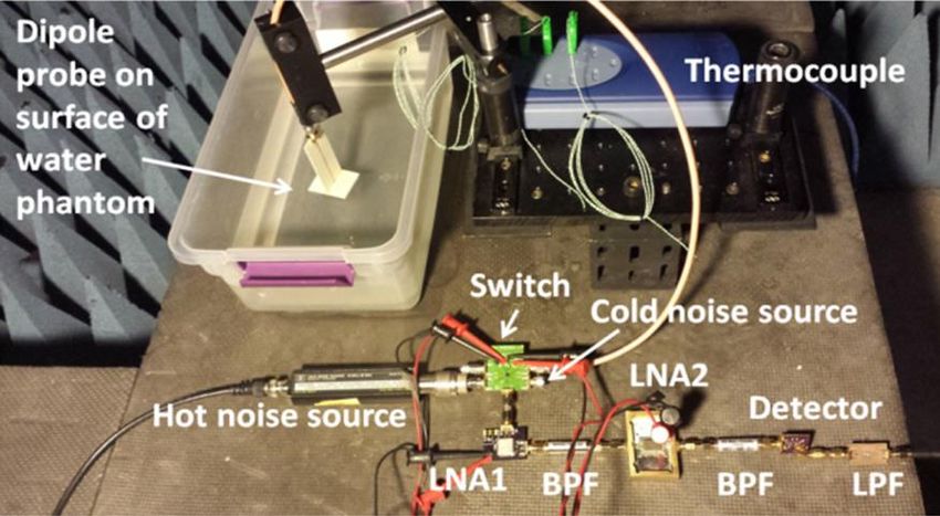
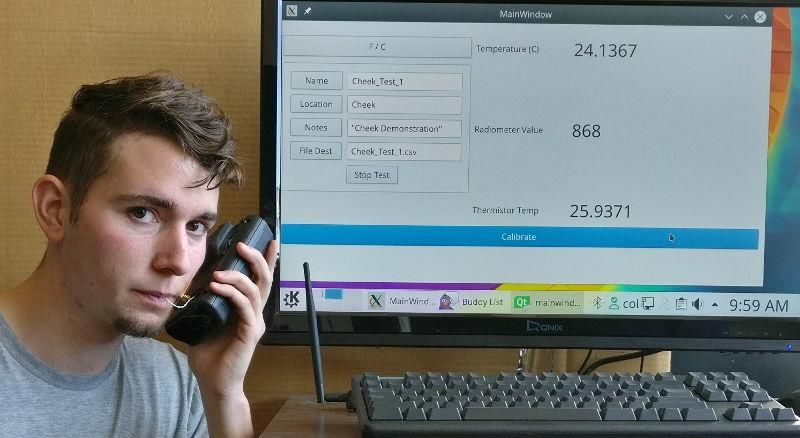
In-Vivo Measurements
 Goal: track temperature variations of the water inside the
 mouth from the skin
 V Radiometer
 Probe Radiometer

 Thermocouple
 in mouth
 Cold,
 Room temp,
 Warm Water

 Avg. Tissue
 thickness
Skin: 1.8mm,
 Fat: 4mm,
Muscle: 2mm

Klemetsen Ø, Jacobsen S and Birkelund Y “Radiometric temperature reading of a hot ellipsoidal object inside the oral
cavity by a shielded microwave antenna put flush to the cheek” Phys. Med. Biol. 57 2633–52, 2012.

 Zoya Popovic, University of Colorado, Boulder, 2020 46
Publications
1. R. Scheeler, E. Kuester, Z. Popovic, “Sensing depth of microwave radiation for internal body
 temperature measurements,” IEEE Trans. Antennas and Propagation, Vol. 62, pp.1-12, 2014
2. W. Haines, P. Momenroodaki, E. Berry, M. Fromandi and Z. Popovic, "Wireless system for
 continuous monitoring of core body temperature," 2017 IEEE MTT-S International Microwave
 Symposium (IMS), Honololu, HI, 2017, pp. 541-543.
3. P. Momenroodaki, W. Haines and Z. Popović, "Non-invasive microwave thermometry of multilayer
 human tissues," 2017 IEEE MTT-S International Microwave Symposium (IMS), Honololu, HI, 2017,
 pp. 1387-1390.
4. P. Momenroodaki, W. Haines, M. Fromandi and Z. Popovic, "Noninvasive Internal Body
 Temperature Tracking With Near-Field Microwave Radiometry," in IEEE Transactions on
 Microwave Theory and Techniques, vol. PP, no. 99, pp. 1-11, 2018.

 Zoya Popovic, University of Colorado, Boulder, 2020 48
Conclusions
A lot of opportunities for interesting and hard electromagnetics
and microwave research in areas related to medical applications!

• High-field MRI:
• Ablation and cauterization:
• Relative internal body temperature measurements

 Thank you!

 Zoya Popovic, University of Colorado, Boulder, 2020
Spare slides

Zoya Popovic, University of Colorado, Boulder, 2020 50
Reciprocity
To predict the amount of power radiated from the
tissue, we can use the Lorentz reciprocity theorem
and convert field to circuit quantities

 Power received by the probe equal to power dissipated in the tissue

 51
 Zoya Popovic, University of Colorado, Boulder, 2020
Temperature Estimation
 • The radiometer temperature first needs to be
 corrected for probe mismatch and loss in the
 cable:

 • The radiometer voltage when switch selects
 probe:

 • Where the total conversion from T(K) to V(V) is

 • Voltage of ref sources
 • Sequential measurements:

(Probe BW narrower than receiver BW)

 Zoya Popovic, University of Colorado, Boulder, 2020
Temperature Estimation (2)
 • Use of two references at two known temperatures
 allows for finding conversion factor and T’rec:

 • Solve for T’’:

 • Invert to get:

 • To extract temperature, layer 2 (fat) has mean
 temperature between layer 1 (skin) and layer 3
 (muscle):

Use measured surface T1 (thermocouple)
and calculated Wi’s to find radiometric
temperature of muscle:
 Zoya Popovic, University of Colorado, Boulder, 2020
Probe Architecture Investigation
Circular Patch WF (%)
 S11
 Architecture Skin Fat Muscle
• Simple, narrow-band (dB)
 (Agar) (FR4) (Agar)
• Ground plane for mitigating RFI
 Patch -13 65.5 1.1 33.4
• Integration with radiometer
• High dielectric constant substrate
 Rogers6010( =10) for size reduction

 1.00 × 10⁶
 3.98 × 10⁵
 1.58 × 10⁵
 6.30 × 10⁴ 1W
 2.50 × 10⁴
 1.00 × 10⁴
 3.98 × 10³
 1.58 × 10³
 6.30 × 10²
 2.50 × 10²
 1.00 × 10²
 3.98 × 10
 1.58 × 10
 6.30
 2.50
 Volume Loss Density (W/ )

 Zoya Popovic, University of Colorado, Boulder, 2020
Probe Architecture Investigation
Circular Patch with superstrate S11
 WF (%)
• Keeps the advantages of the Architecture
 (dB) Skin Fat Muscle
 (Agar) (FR4) (Agar)
 patch(NB/back-ground plane)
 Patch -13 65.5 1.1 33.4
• Reduced sensitivity to the
 Superstrate patch -6.17 38.1 1.2 60.7
 surrounding media
• Protected against corrosion
 Patch on
 Superstrate
 Substrate
 Rogers 6010 Rogers 6010

 Improved power absorption in the muscle!
 Zoya Popovic, University of Colorado, Boulder, 2020
Effect of Superstrate

 E normal = E transverse =

 = 40 = 5 = 55
 Skin Fat Fat Muscle
 Muscle Skin Fat Muscle

 w/o Superstrate w/ Superstrate

 • En attenuates at boundary of fat and muscle (due to
 high relative permittivity contrast)
 • Et is continuous
 • Add superstrate → confines non-propagating En in
 low DK superstrate → reduces skin WF

 Zoya Popovic, University of Colorado, Boulder, 2020
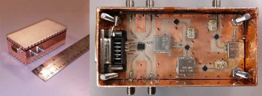
1.4-GHz Radiometers
 Radiometer
 ℎ

 #2
 Power
 detector
 #1
5 cm

 8.2 cm #1

 Radiometers (1&2) from ”A Microwave Radiometer for Internal Body Temperature Measurement”,
 PhD dissertation, R. Scheeler, CU Boulder.

 #3

 Zoya Popovic, University of Colorado, Boulder, 2020 38
1.4-GHz Radiometer
• A low-loss substrate (Rogers 6010)
 reduces loss of input TL from 0.5dB in
 • Att
 FR4 to 0.05dB
• Grounded pads on the substrate reduce
 cap coupling that can cause oscillations
• The minimum spacing between the gnd
 pad & signal line is found from EM
 simulation
• Filters are reflective out of band, LNAs
 are not well matched out of their band.
 • Input3 Input4 • Outpu
• A 1-3 dB pi-pad network is added t
 between the LNA and the filter for • Filt
 • A
 defining out-of-band imp. • •1 21 Filter1er3
 • 1 • A
• Adding shunt capacitor to the supplies • L
 • A 4 LNA3 5cm
 can improve the immunity from RFI. • Swit N
 2 Filter2
 Filtered ch A1 • LN • A
• At each of the IC supplies, small cap at sub-D • 3
 • s A2
 3
 the pin (C3) is added to mitigate the connector
 effect of high freq RFI. C4 which can be a
 Input2 Input1
 bigger cap, helps with lower freq RFI
 10cm
• Filtered D-Sub connector reduces the
 interference from the bias lines
• Grounded metal enclosure
 Zoya Popovic, University of Colorado, Boulder, 2020
Narrowband 1.4-GHz Radiometer
• Improve immunity to RFI: quiet frequency band of 1.4-1.427 GHz
• Commercial filters at 1.4 GHz are not small or high Q
• Possible solution : heterodyne architecture
• Easier to implement filter and amplifiers at a lower IF frequency
• A commercial SDR is cascaded (has mixer with an adjustable LO and adjustable BW that
 can reduce the BW of radiometer to 27MHz)
 Radiometer AD9364 (NI-USRP-2900)
 LNAs Mixer LNAs
 Att to PC
 ADC

 LO

 Zoya Popovic, University of Colorado, Boulder, 2020
Homogeneous Phantom Thermometry
 GOAL: Determine unknown water
 temperature with a radiometer.

 Measurement in anechoic chamber

 ∆ = 0.5°

Folded dipole from ”A Microwave Radiometer for Internal Body Temperature Measurement”, PhD dissertation, R. Scheeler, CU Boulder.

 Zoya Popovic, University of Colorado, Boulder, 2020
Fat-Layer Thickness Variations
What if an incorrect fat thickness is used to estimate the temperature?

• Simulations performed in HFSS for
 different fat-layer thicknesses
• Resonant frequency and WFs are recorded
 1. Resonant frequency increases with fat
 thickness (can change the freq. and find
 the maximum power for a rough
 estimation of fat thickness)
 2. Fat WF only slightly varies (low loss)
 3. Muscle WF reduces

 Zoya Popovic, University of Colorado, Boulder, 2020
Fat-Layer Thickness Variation
• Temperature estimated based on assumed WFs for
 various fat thicknesses
• Muscle temperature is calculated
• Temperature estimate does not deviate from the real
 muscle temp. more than 0.6°C for fat thickness 2-8 mm
• Core tissue temp at rest is assumed to be 37°C

 Zoya Popovic, University of Colorado, Boulder, 2020
Probe Position on Body: Sternum
• The human-body has a non-uniform composition
• Human heart is situated behind and slightly on the left side
 of the sternum
• Six-layer stack is considered in the analysis
• The VPLD in the heart layer is only 2% of total power
• This is due to thick cancellous bone which has a higher
 amount of loss compared to cortical bone and fat layers
 Simulation of probe in HFSS

 Avg.
 th.
 13.2
 mm
[1] G. Tortora, and B. Derrickson “Introduction to the human body”, 8th edition, WILEY Inc, 2010.
[2] Tissue Electromagnetic Properties. https://www.itis.ethz.ch
[3] http://www.ipms.fraunhofer.de/en/press-media/press/2013/2013-01-09.html

 Zoya Popovic, University of Colorado, Boulder, 2020
Probe Placement on Body: Forehead
• Next, the probe is placed on the forehead
• The brain is situated under the stack of scalp and skull
• Pattern of volume loss density shows that 28% of the
 total power is received from the brain layer.
• This is due to thin cancellous bone in forehead compared
 with the heart.

 Simulation of probe in HFSS

 Avg. Th. 9.4mm

 [1] G. Tortora, and B. Derrickson “Introduction to the human body”, 8th edition, WILEY Inc, 2010.
 [2] https://www.reference.com/science/thick-human-skull
 [3] http://www.ipms.fraunhofer.de/en/press-media/press/2013/2013-01-09.html

 Zoya Popovic, University of Colorado, Boulder, 2020
RFI Mitigation
To reduce RFI:
• Measurement in a shielded environment
• NB probe (to pick up thermal noise in a quiet
 band)
• Hybrid PCB radiometer
• Designed a stable, NB radiometer
• Random man-made RFI still presents in the quiet
 band of 1.4-1.427GHz!

 Buried muscle (water phantom) under skin
 (salmon) and fat (Rohacell) measurements in
 the presence of RFI shows larger errors in
 temperature estimation

 Zoya Popovic, University of Colorado, Boulder, 2020 41
You can also read