Insights into Activation Mechanisms of Store-Operated TRPC1 Channels in Vascular Smooth Muscle - MDPI
←
→
Page content transcription
If your browser does not render page correctly, please read the page content below
cells
Review
Insights into Activation Mechanisms of
Store-Operated TRPC1 Channels in
Vascular Smooth Muscle
Miguel A. S. Martín-Aragón Baudel 1 , Jian Shi 2 , William A. Large 3 and Anthony P. Albert 3, *
 1    Department of Pharmacology, University of California, Davis, CA 95615, USA; martinaragon@ucdavis.edu
 2    LIGHT Laboratories, Leeds Institute of Cardiovascular and Metabolic Medicine, University of Leeds,
      Leeds LS2 9JT, UK; J.Shi1@leeds.ac.uk
 3    Vascular Biology Research Centre, Molecular and Clinical Research Institute, St. George’s,
      University of London, London SW17 0RE, UK; largew@sgul.ac.uk
 *    Correspondence: aalbert@sgul.ac.uk

 Received: 25 November 2019; Accepted: 5 January 2020; Published: 10 January 2020

 Abstract: In vascular smooth muscle cells (VMSCs), the stimulation of store-operated channels (SOCs)
 mediate Ca2+ influx pathways which regulate important cellular functions including contraction,
 proliferation, migration, and growth that are associated with the development of vascular diseases.
 It is therefore important that we understand the biophysical, molecular composition, activation
 pathways, and physiological significance of SOCs in VSMCs as these maybe future therapeutic
 targets for conditions such as hypertension and atherosclerosis. Archetypal SOCs called calcium
 release-activated channels (CRACs) are composed of Orai1 proteins and are stimulated by the
 endo/sarcoplasmic reticulum Ca2+ sensor stromal interaction molecule 1 (STIM1) following store
 depletion. In contrast, this review focuses on proposals that canonical transient receptor potential
 (TRPC) channels composed of a heteromeric TRPC1/C5 molecular template, with TRPC1 conferring
 activation by store depletion, mediate SOCs in native contractile VSMCs. In particular, it summarizes
 our recent findings which describe a novel activation pathway of these TRPC1-based SOCs, in which
 protein kinase C (PKC)-dependent TRPC1 phosphorylation and phosphatidylinositol 4,5-bisphosphate
 (PIP2 ) are obligatory for channel opening. This PKC- and PIP2 -mediated gating mechanism is
 regulated by the PIP2 -binding protein myristoylated alanine-rich C kinase (MARCKS) and is coupled
 to store depletion by TRPC1-STIM1 interactions which induce Gq/PLCβ1 activity. Interestingly,
 the biophysical properties and activation mechanisms of TRPC1-based SOCs in native contractile
 VSMCs are unlikely to involve Orai1.

 Keywords: TRPC1; PKC; PIP2 ; Gq; PLC; MARCKS; STIM1; Orai1; store-operated channels; vascular
 smooth muscle

1. Introduction
      In vascular smooth muscle cells (VSMCs), neurotransmitters and hormones such as noradrenaline,
adrenaline, angiotensin II (Ang II), and endothelin 1 (ET-1) produce an increase in cytosolic Ca2+
concentration (Ca2+ i ) (see [1–7] for reviews on Ca2+ signaling mechanisms in smooth muscle). This
rise in Ca2+ i is important for initiating vasoconstriction, which determines vascular resistance and
blood pressure and ultimately regulates blood flow to our tissues and organs. In addition, an excessive
increase in Ca2+ i is linked to cell proliferation, migration and growth, phenotypes which are involved
in the development of vascular disease. Therefore, understanding cellular pathways which mediate
increases in Ca2+ i may help to identify future therapeutic targets for conditions such as hypertension
and atherosclerosis.

Cells 2020, 9, 179; doi:10.3390/cells9010179                                        www.mdpi.com/journal/cells
Cells 2020, 9, 179 2 of 13
Vasoconstrictors induce a rise in Ca2+ i by activating the classical phosphoinositol signaling
pathway involving: stimulation of Gq-protein-coupled receptors, phospholipase C (PLC) activity,
phosphatidylinositol 4,5-bisphosphate (PIP2 ) hydrolysis, and inositol 1,4,5-trisphosphate (IP3 ) and
diacylglycerol (DAG) generation [1–7]. IP3 mediates an increase in Ca2+ i by causing the release of Ca2+
from sarcoplasmic reticulum (SR) Ca2+ stores, and this subsequent rise in Ca2+ i and DAG-mediated
pathways induce Ca2+ influx from the extracellular medium. A component of this Ca2+ influx
occurs through activation of voltage-gated Ca2+ channels (VGCCs), but there is also a significant
contribution from voltage-independent Ca2+ -permeable non-selective cation channels. Stimulation
of Ca2+ -permeable cation channels are thought to mediate Ca2+ entry pathways through direct Ca2+
influx, and through Na+ influx which leads to depolarization and activation of voltage-gated Ca2+
channels and stimulation of the Na+ /Ca2+ exchanger in reverse mode.
There is substantial evidence that Ca2+ -permeable non-selective cation channels classified as
receptor-operated (ROCs) and store-operated (SOCs) are expressed in VSMCs [1,2]. ROCs are
defined as ion channels activated by receptor stimulation independently of IP3 -mediated depletion
of SR Ca2+ stores, whereas SOCs are activated by pathways coupled to depletion of SR Ca2+
stores but not by the subsequent increase in Ca2+ i . These definitions infer that stimulation of
Gq-protein-coupled receptors will activate both ROCs and SOCs. In addition, it is proposed
that SOCs may be activated by Gq-protein-coupled receptor stimulation independently of store
depletion, hence SOCs may also function as ROCs [8]. Together, these findings make it difficult
to study ROCs and SOCs independently of each other using macroscopic measurements such as
whole-cell patch clamp and Ca2+ signal recordings but possible using single channel recordings [9–11].
Moreover, SOCs can be selectively activated by depleting SR Ca2+ stores in the absence of receptor
stimulation using SR Ca2+ -ATPase inhibitors (e.g., cyclopiazonic acid (CPA) and thapsigargin),
and high (e.g., 1,2-bis(o-aminophyoxy)ethane-N,N,N’,N’-tetraacetic acid (BAPTA)) and low affinity
(e.g., N,N,N’,N’,-tetrakis(2-pyridmethyl)-1,2-ethnediamine (TPEN)) Ca2+ chelators to passively deplete
Ca2+ i and SR Ca2+ levels respectively.
This article reviews our current understanding of the biophysical properties, molecular
composition, and activation mechanisms of SOCs in VSMCs, in particular, it focuses on recent studies
from our laboratory which have described a novel activation pathway of SOCs composed of canonical
transient receptor potential 1 (TRPC1) proteins in native contractile VSMCs. In the majority of these
studies SOCs were activated by SR Ca2+ ATPase inhibitors or Ca2+ chelators to prevent complications
from activation of ROCs, although in the final section we discuss the potential physiological significance
of this store-operated activation pathway in vasoconstrictor-induced TRPC1-based SOCs.
2. Biophysical Properties and Molecular Composition of SOCs in VSMCs
It is well-established that archetypal SOCs, termed calcium release-activated channels (CRACs),
are characterized by high Ca2+ permeabilities (PCa2+ :PNa+ > 1000:1), pronounced inward rectification
with reversal potentials (Erev ) greater than +50 mV, unitary conductances in the fS range, and are
composed of Orai1 proteins (see [12] for review on properties and functions of Orai channels and their
associated activation mechanisms). The fundamental activation mechanism of Orai1-based CRACs is
also clearly outlined, with an essential role for stromal interaction molecule 1 (STIM1) which senses
Ca2+ levels within ER/SR Ca2+ stores and following store depletion undergoes oligmerisation and
translocation to cytosolic surface of the plasma membrane where it interacts with Orai1 to induce
channel assembly and gating.
However, it is apparent that many cell types express SOCs with very different characteristics to
Orai1-based CRACs such as much lower Ca2+ permeabilities, relatively linear or outward rectification,
and considerably larger unitary conductances (see [13] for review on role of TRPC proteins in mediated
SOCs). Given the properties of these SOCs, they are unlikely to be composed of Orai proteins. The
molecular composition of these SOCs is controversial but there is increasing evidence that they are
formed by the TRPC family of Ca2+ -permeable non-selective cation channels (TRPC1–C7) [13]. TheseCells 2020, 9, 179 3 of 13
TRPC-based SOCs are likely to form a diverse group of channels with differing expression, properties,
and functions since TRPC subunits form heterotetrameric structures [7,13]. There is further controversy
over whether TRPC proteins form SOCs due to limited understanding, unlike with Orai proteins, of how
store depletion couples to channel activation that is an essential requirement when defining SOCs.
These issues are beginning to be unraveled by findings which suggest that store depletion activates
TRPC-based SOCs through diverse STIM1-mediated processes including direct interactions between
TRPC and Orai1 proteins, activation of Orai1-based CRACs as a prerequisite for TRPC1 opening,
and direct interactions between TRPC and STIM1 [13,14]. For example, in overexpression studies it is
proposed that direct interactions between STIM1 and TRPC channels govern activation [13–18], whereas
elegant studies in salivary glands have shown that activation of TRPC1-based SOCs require prior
activation of Orai1-based CRACs to induce insertion of TRPC1 subunits into the plasma membrane,
with both activation of Orai1-based CRACs and TRPC1-based SOCs coupled to store depletion by
STIM1 (see [19] for review on activation mechanisms of TRPC1-based SOCs in salivary glands). The
studies highlighted in this review indicate that activation of STIM1-mediated TRPC1-based SOCs in
native contractile VSMCs involves a Gq/PLCβ1 pathway and interactions between protein kinase C
(PKC) activity and PIP2 which do not require Orai1.
The molecular composition and activation mechanisms of SOCs in VSMCs reflects the controversies
highlighted above as there is significant evidence to indicate that both Orai1-based CRACs and
TRPC-based SOCs are present in VSMCs, but that these channels are differentially expressed according
to cell phenotype. In freshly isolated VSMCs or primary cultured VSMCs maintained in low
serum, conditions in which VSMCs retain their native contractile phenotype, evidence suggests
that SOCs have biophysical properties which resemble TRPC-based SOCs and not Orai1-based
CRACs with relatively linear, S-shaped, or slightly outward rectification, low Ca2+ permeabilities
(PCa2+ :PNa+ of 1–50), and unitary conductances of 2–3 pS and 2–8 pS in 1.5 mM and 0 mM external
Ca2+ respectively [1,2,20–29]. Moreover, studies using an array of different techniques such as
blocking anti-TRPC antibodies (e.g., T1E3), TRPC1−/− mice, molecular knockdown (e.g., siRNA),
and pharmacology (e.g., the selective TRPC1/C4/C5 inhibitor Pico145) [30,31] indicate that these
SOCs are composed of a heteromeric TRPC1/C5 molecular template, which can also involve other
TRPC subunits depending on the vascular bed [7,20–29,32,33]. As such, these studies show that
TRPC1/C5-based SOCs are functionally expressed in pial arterioles and mesentery artery VSMCs,
whereas TRPC1/C5/C6- and TRPC1/C5/C7-based SOCs are reported in coronary artery and portal
vein VSMCs respectively. It is predicted that differences in the molecular compositions of these
TRPC-based SOCs are reflected in the different properties of TRPC-based SOCs such as rectification,
Ca2+ permeability and unitary conductance. For example, inclusion of TRPC6 within the TRPC1/C5
template produces biophysical properties which indicate a decreased Ca2+ permeability [22], whereas
the presence of TRPC7 produces a facilitatory action of IP3 on TRPC1-based SOCs [22,34]. Studies
using TRPC1−/− VSMCs indicate that TRPC1 is essential for conferring activation by store depletion,
and therefore these TRPC1/C5 channel templates are termed TRPC1-based SOCs [25]. These findings
fit with the idea that TRPC1 subunits do not form a functional homotetrameric channel [13]. Several
studies have indicated that Orai1 proteins are unlikely to be involved in composing SOCs in VSMCs
with a native contractile phenotype, with low expression of Orai1 proteins present in these cells ([35,36]
and see [37,38] for review of Orai1 proteins in vascular smooth muscle) and biophysical properties of
TRPC1-based SOCs and their STIM1-mediated activation mechanisms unaffected in freshly isolated
and primary cultured VSMCs from Orai1−/− mice [29].
In contrast to native contractile VSMCs, long-term cultured VSMCs maintained in high serum,
conditions that induce a non-contractile synthetic phenotype with proliferative, migrative and growth
characteristics, express whole-cell SOCs and store-operated Ca2+ entry with similar properties to
Orai1-based CRACs such as pronounced inward rectification, inhibition by a Orai1 selective inhibitor,
and inhibition by knockdown of Orai1 and STIM1 protein levels but not by knockdown of TRPC1,
TRPC4 or TRPC6 proteins [36–42]. In addition, store-operated whole-cell currents and Ca2+ entry areCells 2020, 9, 179 4 of 13
unaffected in cultured VSMCs maintained in high serum conditions from TRPC1−/− mice [43] further
suggesting the involvement of Orai1-based CRACs in these VSMCs. In contrast, TRPC1 has also
been proposed to be involved in regulating SOCs present in synthetic VSMCs, perhaps by regulating
Orai1-based CRACs, and it is suggested that increase in the expression and function of TRPC1 may
be an important trigger in the development of the cellular switch from a contractile to synthetic
phenotype [37,38].
These complexities mean that when studying SOCs in VSMCs it is essential to clearly define
cell phenotype of VSMCs. In this regard, when investigating SOCs in native contractile VSMCs it is
advantageous to use freshly isolated cells whenever possible, and when required only use primary
culture cells maintained in low serum conditions for as short a time as possible.
3. Activation Mechanisms of TRPC1-Based SOCs in Native Contractile VSMCs
The following sections discuss our recent findings that TRPC1-based SOCs in freshly isolated and
primary cultured VSMCs with a native contractile phenotype exhibit a complex activation mechanism,
in which interactions between PKC-dependent phosphorylation of TRPC1 and phosphatidylinositol
4,5-bisphosphate (PIP2 ) regulated by myristoylated alanine-rich C kinase (MARCKS) are obligatory
gating partners, with these mechanisms coupled to store depletion by a novel STIM1-mediated
Gq-PLCβ1 pathway.
3.1. PKC Activity and PIP2 have Obligatory Roles in Activation of TRPC1-based SOCs
There are several lines of evidence which indicate that PKC activity and PIP2 are important in
activating TRPC1-based SOCs. The stimulation of whole-cell and single channel TRPC1-based SOCs is
significantly reduced by PKC inhibitors and anti-PIP2 antibodies [22,25,44]. In support of these results,
PKC activators (diacylglycerol analogues, phorbol esters, PKC catalytic subunits) and exogenous
application of diC8-PIP2 (a water-soluble form of PIP2 ) stimulate TRPC1-based SOCs in WT but not
in TRPC1−/− VSMCs [22,25,44]. Interestingly, PKC-stimulated activation of TRPC1-based SOCs are
prevented by anti-PIP2 antibodies and pharmacological agents such as wortmannin to deplete PIP2
levels, whereas diC8-PIP2 -activated TRPC1-based SOCs are reduced by PKC inhibitors [44]. Moreover,
store depletion induces PKC-dependent phosphorylation of TRPC1 and increases association between
PIP2 and TRPC1 [26–28,44]. Together, these results indicate that interactions between PKC activity
and PIP2 have obligatory roles in activation of TRPC1-based SOCs; PKC cannot activate TRPC1-based
SOCs without PIP2 , and vice versa. This supports the hypothesis that PIP2 is the activating ligand of
TRPC1-based SOCs and that store-operated PKC-dependent phosphorylation of TRPC1 is required for
this opening mechanism to occur.
These proposed roles of PKC and PIP2 on TRPC1-based SOCs are different to their roles in the
activation of TRPC3/C6/C7-based ROCs in native contractile VSMCs (see [23,24] for comprehensive
reviews of activation mechanisms of TRPC channels). It is well established that this subgroup of TRPC
channels are activated by receptor-mediated generation of DAG which leads to channel opening via
PKC-independent mechanisms, with PKC causing channel inhibition. In addition, the role of PIP2
on TRPC3/C6/C7-based ROCs is unclear with both inhibitory and excitatory actions proposed [45,46].
These findings further indicate that TRPC1-based SOCs and TRPC3/C6/C7-based ROCs form distinct
channel structures with differing activation mechanisms, and likely distinct functions in VSMCs.
Important omissions from our understanding are what PKC isoform(s) is involved, which amino
acid(s) within TRPC1 protein structure is phosphorylated, and how a PKC-dependent phosphorylation
process alters interactions between TRPC1 and PIP2 . The PKC family comprises of at least 11
serine/threonine kinases divided into three groups according to their basic structure and activation
requirements: conventional PKC isoforms (α, βI, βII and γ) require both Ca2+ and diacylglycerol
(DAG), novel PKC isoforms (δ, ε, η and θ) require DAG but are Ca2+ -insensitive, and atypical PKC
isoforms (ζ, ι and λ) are activated by lipid mediators such as phosphatidylserine and do not require
Ca2+ or DAG (see [47] for review of the role of PKC isoforms in vascular smooth muscle). Our resultsCells 2020, 9, 179 5 of 13
indicate that stimulation of TRPC1-based SOCs and PKC-dependent phosphorylation of TRPC1 by
store depletion requires PLCβ1 activity, and that DAG analogues activate TRPC1-based SOCs through
a PKC-dependent mechanism [9,23,24,27]. Moreover, we have shown that TRPC1-based SOCs are
activated by store depleting agents which are likely to increase (e.g., CPA), decrease (e.g., the high
affinity cell-impermeable and -permeable Ca2+ chelators BAPTA and BAPTA-AM), or produce little
change in Ca2+ i (e.g., the low affinity cell-permeable Ca2+ chelator TPEN). These findings suggest that
the PKC isoform involved is likely to require DAG but is Ca2+ -insensitive, which are characteristics
of the novel group of PKC isoforms. Our preliminary data indicate that PKCδ is the most highly
expressed novel PKC isoform in VSMCs, and that selective PKCδ inhibitory peptides and knockdown
of PKCδ using morpholino sequences prevent activation of TRPC1-based SOCs [31].
Interestingly, the predication of PKCδ-dependent phosphorylation sites within the TRPC1 sequence
using GPS 3.0 reveals five intracellular serine residues, with Ser619 and Ser752 at the C-terminal
domain of potential significance as both these sites are close to a known PIP2 -binding domain [48]. It is
therefore possible that PKCδ-dependent phosphorylation of these sites increases PIP2 affinity leading
to increased binding to TRPC1 and channel opening. Previous studies have highlighted that protein
kinase A (PKA), protein kinase G (PKG), and calmodulin kinase II (CaMKII) have inhibitory actions on
TRPC1-based SOCs in VSMCs [49–51], and it may be that phosphorylation of serine/threonine amino
acids by these kinases reduce TRPC1 and PIP2 interactions by lowering PIP2 affinity. Similar roles
for kinase activities in modulating lipid-protein interactions are well-established in the regulation of
different K+ channel subtypes [52].
3.2. Interactions Between PKC Activity and PIP2 are Regulated by MARCKS
An important question is how PKC-dependent phosphorylation regulates PIP2 gating of
TRPC1-based SOCs when physiological activators of SOCs involve stimulation of Gq-coupled receptors
which drive PLC activity and PIP2 hydrolysis. To provide answers to this question, we investigated the
role of MARCKS in activation of TRPC1-based SOCs. MARCKS is a membrane-bound PIP2 -binding
protein that acts a PIP2 buffer, which can provide a distinct pool of PIP2 at the plasma membrane
that is protected from breakdown by PLC thus enabling this phospholipid to be released in a
coordinated manner into the local environment [53,54]. Using a combination of electrophysiological,
co-immunoprecipitation, and PIP2 -binding dot-blot assays, we showed that in unstimulated conditions
MARCKS is bound to TRPC1, with PIP2 predominately associated with MARCKS and not TRPC1 [26].
Upon stimulation by store depleting agents MARCKS dissociates from TRPC1, which leads to PIP2
being released and associated with TRPC1 to cause channel opening. Importantly, both dissociation of
MARCKS from TRPC1 and redistribution of PIP2 from MARCKS to TRPC1 are stimulated by phorbol
esters and prevented by PKC inhibitors indicating that PKC activity is central to these mechanisms.
It will be important to ascertain how receptor stimulation and store-depleting agents couple to these
actions of MARCKS. A potential idea is that stimulation of CaM is involved, as this Ca2+ -binding
protein is known to bind to MARCKS and to cause dissociation from the plasma membrane and
PIP2 release [53,55]. Furthermore, CaM is known to activate TRPC1-based SOCs in native contractile
VSMCs [50].
3.3. Store Depletion Activates a Gq-PLCβ1 Pathway Involved in Activation of TRPC1-Based SOCs
For a channel to be defined as store-operated, it is essential to understand how store depletion is
coupled to channel opening. Therefore, a central question surrounding the activation of TRPC1-based
SOCs is how store depletion couples to PKC-dependent phosphorylation of TRPC1 and opening by PIP2 ?
A crucial finding was the discovery that store depletion activates a Gq-PLCβ1 pathway which drives
the PKC-dependent phosphorylation of TRPC1 [27]. Our studies showed that the G-protein inhibitor
GDP-β-S, anti-Gq antibodies (but not anti-Gi), the PLC inhibitor U73122, and PLCβ1 shRNA inhibited
activation of TRPC1-based SOCs. In addition, store-operated PKC-dependent phosphorylation of
TRPC1 was inhibited by U73122 and PLCβ1 shRNA. A significant finding was that store depletingCells 2020, 9, 179 6 of 13
agents induced PLC activity measured using GFP-PLCδ1-PH, a fluorescent biosensor for PIP2 and IP3
([56–58] and reviewed in [59]) in primary cultured VSMCs which was prevented by a PLC inhibitor
and PLCβ1 shRNA. In support of these findings, co-immunoprecipitation and proximity ligation
assays (PLA) demonstrated that store depletion induced interactions between TRPC1, Gq and PLCβ1
at the plasma membrane.
3.4. STIM1 Couples Store Depletion to Gq-PLCβ1 Activity to Stimulate TRPC1 SOCs
Next, we investigated how store depletion is coupled to the formation and activation of this
Gq/PLCβ1/PKC/TRPC1 signal transduction pathway. An obvious candidate was STIM1, since
along with its classically described role in activating Orai1-based CRACs it has been implicated in
activating TRPC-based SOCs (see [13] for review). Moreover, it is known that that STIM1 has diverse
cellular partners including ion channels [12,13,15–18,60,61], SR and plasma membrane ATPases [62,63],
and adenylate cyclase [64] and therefore it seemed a reasonable idea that STIM1 may couple to
Gq/PLCβ1 activity.
We demonstrated that whole-cell and single channel TRPC1-based SOCs and store-operated
PLCβ1 activity were inhibited by shRNA STIM1 and were absent in TRPC1−/− VSMCs [28]. In addition,
store-operated PKC-dependent phosphorylation of TRPC1 was greatly reduced by shRNA STIM1.
Moreover, STIM1 was required for store-operated interactions between Gq, PLCβ1 and TRPC1, and
TRPC1 was essential for store-operated interactions between Gq, PLCβ1, and STIM1. These findings
provide strong evidence that STIM1 is an essential molecule in activation of TRPC1-based SOCs, and
importantly it indicates that store-operated STIM1-TRPC1 interactions (measured using PLA assays
which imply that these interactions occur within 40 nm) form the structural basis for stimulation of
the Gq/PLCβ1 pathway required for PKC-dependent TRPC1 phosphorylation and channel opening
by PIP2 .
As expected, store depletion induced translocation of STIM1 from the cytosol to the plasma
membrane where it formed discrete puncta and interactions with TRPC1 using immunocytochemical
and PLA techniques, which were not dependent on downstream molecules in the signal pathway such
as PLCβ1 [28]. Interestingly, in the absence of TRPC1, store depletion still induced translocation of
STIM1 from cytosol to the plasma membrane, but STIM1 formed an even distribution throughout the
plasma membrane and not discrete puncta. This suggests that TRPC1 may be essential for coordinating
the response of STIM1 to store depletion.
An interpretation from these studies is that STIM1-TRPC1 interactions, possibly involving other
unknown molecules, act as a cellular activator of Gq subunits, and that this leads to interactions with,
and activation of, PLCβ1. In essence STIM1-TRPC1 interactions behave like Gq-coupled receptors or
guanine exchange factors (GEFs). In the future, it will be important to investigate structural interactions
between STIM1 and TRPC1 and discover where Gq subunits binds. Initial work might focus on the
CRAC-activating (CAD) and polybasic domains which have been linked to binding and activation of
TRPC1 by protein-protein interactions and electrostatic interactions respectively [13,15–18].
Interestingly, differential blocking effects of N-terminal and C-terminal anti-STIM1 antibodies
on TRPC1-based SOCs using whole-cell and inside-out patch clamp recordings may suggest that
store-operated STIM1/TRPC1 interactions lead to STIM1 acting as a transmembrane protein at the
plasma membrane [28]. In this configuration, the N-terminal domain of STIM1 is now present at the
extracellular surface of the plasma membrane. Perhaps this orientation of STIM1-TRPC1 interactions
has implications for association with Gq subunits in native contractile VSMCs. A similar role for STIM1
has been previously described in mediating store-operated conductances [65]. However, it seems
unlikely that the source of STIM1 involved in activating TRPC1-based SOCs resides at the plasma
membrane, as with activation of arachidonic acid-regulated channels (ARC) composed of Orai1/Orai3
subunits [66], since our data indicates that store depletion induces translocation of STIM1 from the
cytosol to the plasma membrane.Cells 2020, 9, 179 7 of 13
3.5. Activation Mechanisms of TRPC1-Based SOCs are Independent of Orai1
There is substantial evidence that TRPC1, Orai1 and STIM1 have important roles in mediating
store-operated Ca2+ entry pathways in VSMCs with a synthetic phenotype, although it is unclear
whether Orai1 and TRPC1 interact together or form separate STIM1-mediated ionic mechanisms
(see earlier). In addition, it is unclear whether Orai1 is required for activation of TRPC1-based SOCs
in native contractile VSMCs. In our recent study, we showed that the properties of store-operated
whole-cell and single channel currents in freshly isolated and primary cultured native contractile WT
and Orai1−/− VSMCs were similar to previously described TRPC1-based SOCs, and that store-operated
STIM1-mediated PLCβ1 activity and STIM1-TRPC1 interactions were unaffected in Orai1−/− cells [29].
These findings provide significant evidence that Orai1 proteins are not required for the molecular
composition or activation mechanisms of TRPC1-based SOCs in contractile VSMCs. These results
support the findings that Orai1 expression is very low in native contractile VSMCs [35–38]. A caveat is
that Orai proteins are composed of three subtypes, Orai1-3 [12], and therefore Orai2 and Orai3 may
be involved in TRPC1-based SOCs in native contractile VSMCs. However, whole-cell store-operated
conductances with the distinct characteristics of Orai proteins, such as such inward rectification, and
reversal potential greater than +50 mV have not been identified in native contractile VSMCs from WT,
Orai1−/− or TRPC1−/− preparations [25,29].
It should be noted that other studies using pharmacological and molecular techniques have
implicated Orai1 and Ca2+ -independent phospholipase A2 (iPLA2 ) in store-operated conductances
in native and primary cultured contractile VSMCs [67]. These studies propose that store depletion
linked to STIM1-mediated release of a calcium influx factor (CIF) from the SR activates Orai1 through
an iPLA2 -mediated mechanism involving production of lysophospholipids (see [68] for review).
In addition, Orai1 has been proposed alongside TRPC1 to regulate store-operated vascular contractility
through interactions with voltage-gated Ca2+ channels ([69] and reviewed in [70]). The differences
between these findings to those described above are unclear and may reflect differences between SOCs
in different vascular beds and perhaps different cell isolation or culturing conditions. However, before
these ideas can be truly accepted it will be essential to resolve the identity of CIF and understand
why proposed Orai1-sensitive currents in contractile VSMCs have such a linear rectification [67].
For example, do contractile VSMCs express an Orai1 splice variant with very different properties
to established Orai-based CRACs? In addition, a difficult finding to explain is why store-operated
conductances are absent in native contractile VSMCs from TRPC1−/− mice [25]. Moreover, involvement
of iPLA2 has been inferred in many studies using the proposed inhibitor bromoenol lactone (BEL), which
has also been shown to block heteromeric TRPC1/C5, TRPC5, and TRPC6 channels and VGCCs [71].
4. Physiological Significance of TRPC1-Based SOCs Activation Pathway
To avoid complications from receptor-operated pathways, most studies use agents that
selectively deplete SR Ca2+ stores (e.g., CPA, BAPTA) instead of receptor stimulation to study SOCs
(see Introduction). However, it is important to examine whether proposed store-operated mechanisms
are involved in activation of SOCs by physiological receptor stimulation. This is particularly important
for TRPC1-based SOCs in native contractile VSMCs as these channels also behave as ROCs, and so
may be activated by distinct store-independent and -dependent pathways [1,2]. Previous studies have
shown that noradrenaline, Ang II and ET-1-activated ETB receptor stimulation induce TRPC1-based
SOCs via a PLC-mediated pathway that requires PKC activity and PIP2 for channel opening in portal
vein, mesenteric artery and coronary artery VSMCs respectively [8–11,23,72]. To investigate whether
these receptor-mediated activation pathways of TRPC1-based SOCs might involve our proposed
store-operated STIM1/Gq/PLCβ1 pathway, we showed that noradrenaline-activated TRPC1-based
SOCs were greatly reduced by knockdown of PLCβ1 and STIM1 and that noradrenaline also induced
interactions between STIM1 and TRPC1, Gq, and PLCβ1 [27,28]. Moreover, noradrenaline induced
PKC-dependent phosphorylation of TRPC1 and activation of TRPC1-based SOCs by regulating
MARCKS-TRPC1-PIP2 interactions similar to those produced by store depleting agents [26]. TheseCells 2020, 9, 179 8 of 13
findings highlight that physiological receptor stimulation is likely to activate TRPC1-based SOCs
through the store-operated STIM1/Gq/PLCβ1/PKC pathway and also via a store-independent pathway
involving a Gq/PLC/PKC pathway. It may be that receptor stimulation switches between these
two modes to activate TRPC1-based SOCs according to concentration of the physiological agonist.
For example, higher vasoconstrictor concentrations may be more likely to produce substantial
IP3 -mediated depletion of SR Ca2+ stores and activation of TRPC1-based SOCs by store depletion.
It is likely that certain receptors only activate TRPC1-based SOCs via store-independent pathways.
In native contractile coronary artery VSMCs, ET-1-activated ETA receptor stimulation induces
TRPC1-based SOCs through a Gβγ-protein-phosphoinositol 3-kinase (PI3K)-mediated pathway, with
PIP3 formation thought to both induce PKC activity and act as the channel activating ligand; a pathway
which is unlikely to involve IP3 -mediated depletion of SR Ca2+ stores [11,72]. Therefore, TRPC1-based
SOCs in coronary artery VSMCs are activated by ETA and ETB receptor stimulation through separate
store-independent and -dependent pathways respectively, both requiring obligatory roles of PKC
activity and phospholipids [11,66].
Interestingly, the knockdown of PLCβ1 and STIM1 had little effect on noradrenaline-stimulated
PLC activity measured using the GFP-PLCδ-PH biosensor, although it was inhibited by U73122 a
general PLC isoform inhibitor [27,28]. These findings support significant physiological relevance to our
results, as it suggests that stimulation of Gq-protein-coupled receptors and IP3 -mediated depletion of
SR Ca2+ stores are likely to activate two different Gq-PLC pathways mediated by distinct PLC isoforms.
To test this hypothesis, it would be important to identify the PLC isoform linked to α1 -adrenoceptor
stimulation. The critical role of MARCKS in stimulation of TRPC1-based SOCs is further highlighted
when considering the physiological significance of our proposed activation pathway. Stimulation of
Gq-protein-coupled receptors and IP3 -mediated depletion of SR Ca2+ stores will both induce Gq-PLC
activities leading to PIP2 hydrolysis. Therefore, MARCKS provides the essential role of a buffering a
separate pool of PIP2, which is protected from PLC-mediated hydrolysis enabling the phospholipid to
be available for channel opening.
5. Summary
This review highlights recent findings that SOCs in native contractile VSMCs are mediated by
TRPC1-based SOCs, which have very different biophysical properties to the well-characterized channels
formed by Orai proteins, including archetypal Orai1-based CRACs. Moreover, Figure 1 describes a
novel activation pathway for these TRPC1-based SOCs proposed from our recent studies, in which
SR Ca2+ store depletion by receptor-induced IP3 -mediated generation or store depleting agents such
as CPA and BAPTA stimulate STIM1 to translocate to the plasma membrane where it interacts with
TRPC1 subunits to form a receptor for Gq G-protein subunits which induces PLCβ1 activity, production
of DAG, and PKC activity. PKC-dependent phosphorylation of TRPC1 increases PIP2 binding and
channel opening. The interactions between TRPC1 and PIP2 are regulated by MARCKS, a plasma
membrane PIP2 -binding protein bound to TRPC1 at rest, which upon store depletion dissociates from
TRPC1 and releases PIP2 into the local environment where the phospholipid acts as the gating ligand.
Figure 1 also depicts our current understanding of how receptor stimulation by vasoconstrictors
activates TRPC1-based SOCs in contractile VSMCs by store-independent pathways involving PKC
activity and phospholipids.Cells 2020, 9, 179 9 of 13
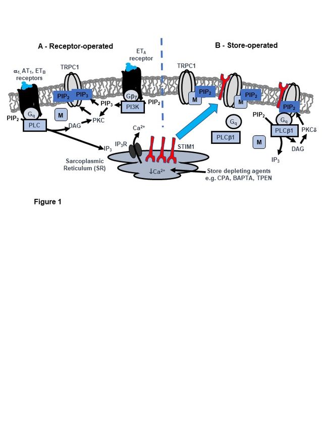
Figure 1. Proposed model of TRPC1-based store-operated channels (SOCs) in native contractile
vascular smooth muscle cells (VSMCs). A, Receptor stimulation of distinct Gq-PLC and Gβγ-PI3K-PIP3
pathways lead to PKC-dependent phosphorylation of TRPC1 and channel opening by PIP2 and PIP3
respectively. Local levels of PIP2 involved in channel activation is controlled by MARCKS (M). B, Store
depletion by receptor-mediated IP3 generation and store depleting agents such as CPA, BAPTA and
TPEN activate TRPC1-based SOCs through a STIM1-TRPC1-mediated pathway. In unstimulated cells,
SR Ca2+ stores are full and TRPC1-based SOCs are in a closed state. In this configuration, channels
are associated with MARCKS which buffers local PIP2 levels and do not interact with Gq, PLCβ1 or
STIM1. Following SR Ca2+ depletion, STIM1 (red) is activated and translocates from the SR to the
plasma membrane where it interacts with TRPC1. Formation of STIM1-TRPC1 interactions enable
binding and activation of Gq and PLCβ1 activity, PIP2 hydrolysis, DAG generation, stimulation PKCδ
and PKC-dependent phosphorylation of TRPC1. This leads to dissociation of MARCKS from TRPC1
and release of PIP2 (previously protected from PIP2 hydrolysis) into the local environment where it
acts as the activating ligand.
Author Contributions: All authors were involved in production of this manuscript and have discussed the final
version of the manuscript.
Funding: This work was supported by Biotechnology and Biological Sciences Research Council grants (BB/J007226/1
and BB/M018350/1 to APA).
Conflicts of Interest: To the best of our knowledge, the authors declare no conflict of interest.
References
1. Albert, A.P.; Large, W.A. Store-operated Ca2+ -permeable non-selective cation channels in smooth muscle
cells. Cell Calcium 2003, 33, 345–356. [CrossRef]
2. Albert, A.P.; Saleh, S.N.; Peppiatt-Wildman, C.M.; Large, W.A. Multiple activation mechanisms of
store-operated TRPC channels in smooth muscle cells. J. Physiol. 2007, 583, 25–36. [CrossRef] [PubMed]
3. Abramowitz, J.; Birnbaumer, L. Physiology and pathophysiology of canonical transient receptor potential
channels. FASEB J. 2009, 23, 297–328. [CrossRef] [PubMed]
4. Beech, D.J. Characteristics of transient receptor potential canonical calcium-permeable channels and their
relevance to vascular physiology and disease. Circ. J. 2013, 77, 570–579. [CrossRef]
5. Earley, S.; Brayden, J.E. Transient receptor potential channels in the vasculature. Physiol. Rev. 2015, 95,
645–690. [CrossRef]
6. Large, W.A. Receptor-operated Ca2+ -permeable nonselective cation channels in vascular smooth muscle: A
physiologic perspective. J. Cardiovasc. Electrophysiol. 2002, 13, 493–501. [CrossRef]
7. Albert, A.P.; Saleh, S.N.; Large, W.A. Identification of canonical transient receptor potential (TRPC) channel
proteins in native vascular smooth muscle cells. Curr. Med. Chem. 2009, 16, 1158–1165. [CrossRef]Cells 2020, 9, 179 10 of 13
8. Albert, A.P.; Large, W.A. Activation of store-operated channels by noradrenaline via protein kinase C in
rabbit portal vein myocytes. J. Physiol. 2002, 544, 113–125. [CrossRef]
9. Saleh, S.N.; Albert, A.P.; Peppiatt, C.M.; Large, W.A. Angiotensin II activates two cation conductances with
distinct TRPC1 and TRPC6 channel properties in rabbit mesenteric artery myocytes. J. Physiol. 2006, 577,
479–495. [CrossRef]
10. Shi, J.; Ju, M.; Saleh, S.N.; Albert, A.P.; Large, W.A. TRPC6 channels stimulated by angiotensin II are inhibited
by TRPC1/C5 channel activity through a Ca2+ - and PKC-dependent mechanism in native vascular myocytes.
J. Physiol. 2010, 588, 3671–3682. [CrossRef]
11. Shi, J.; Ju, M.; Large, W.A.; Albert, A.P. Pharmacological profile of phosphatidylinositol 3-kinases and related
phosphatidylinositols mediating endothelin(A) receptor-operated native TRPC channels in rabbit coronary
artery myocytes. Br. J. Pharmacol. 2012, 166, 2161–2175. [CrossRef]
12. Prakriya, M.; Lewis, R.S. Store-operated calcium channels. Physiol. Rev. 2015, 95, 1383–1436. [CrossRef]
13. Cheng, K.T.; Ong, H.L.; Liu, X.; Ambudkar, I.S. Contribution and regulation of TRPC channels in
store-operated Ca2+ entry. Curr. Top. Membr. 2013, 71, 149–179.
14. Liao, Y.; Abramowitz, J.; Birnbaumer, L. The TRPC family of TRP channels: Roles inferred (mostly) from
knockout mice and relationship to ORAI proteins. Handb. Exp. Pharmacol. 2014, 223, 1055–1075.
15. Worley, P.F.; Zeng, W.; Huang, G.N.; Yuan, J.P.; Kim, J.Y.; Lee, M.G.; Muallem, S. TRPC channels as
STIM1-regulated store-operated channels. Cell Calcium 2007, 42, 205–211. [CrossRef]
16. Yuan, J.P.; Kim, M.S.; Zeng, W.; Shin, D.M.; Huang, G.; Worley, P.F.; Muallem, S. TRPC channels as
STIM1-regulated SOCs. Channels 2009, 3, 221–225. [CrossRef]
17. Lee, K.P.; Choi, S.; Hong, J.H.; Ahuja, M.; Graham, S.; Ma, R.; So, I.; Shin, D.M.; Muallem, S.; Yuan, J.P.
Molecular determinants mediating gating of transient receptor potential canonical (TRPC) channels by
stromal interaction molecule 1 (STIM1). J. Biol. Chem. 2014, 289, 6372–6382. [CrossRef]
18. Asanov, A.; Sampieri, A.; Moreno, C.; Pacheco, J.; Salgado, A.; Sherry, R.; Vaca, L. Combined single channel
and single molecule detection identifies subunit composition of STIM1-activated transient receptor potential
canonical (TRPC) channels. Cell Calcium 2015, 57, 1–13. [CrossRef]
19. Ambudkar, I.S.; de Souza, L.B.; Ong, H.L. TRPC1, Orai1, and STIM1 in SOCE: Friends in tight spaces.
Cell Calcium 2017, 63, 33–39. [CrossRef]
20. Trepakova, E.S.; Gericke, M.; Hirakawa, Y.; Weisbrod, R.M.; Cohen, R.A.; Bolotina, V.M. Properties of a native
cation channel activated by Ca2+ store depletion in vascular smooth muscle cells. J. Biol. Chem. 2001, 276,
7782–7790. [CrossRef]
21. Albert, A.P.; Large, W.A. A Ca2+ -permeable non-selective cation channel activated by depletion of internal
Ca2+ stores in single rabbit portal vein myocytes. J. Physiol. 2002, 538, 717–728. [CrossRef] [PubMed]
22. Saleh, S.N.; Albert, A.P.; Peppiatt-Wildman, C.M.; Large, W.A. Diverse properties of store-operated TRPC
channels activated by protein kinase C in vascular myocytes. J. Physiol. 2008, 586, 2463–2476. [CrossRef]
23. Large, W.A.; Saleh, S.N.; Albert, A.P. Role of phosphoinositol 4,5-bisphosphate and diacylycerol in regulating
native TRPC channel proteins in vascular smooth muscle. Cell Calcium 2009, 45, 574–582. [CrossRef]
24. Albert, A.P. Gating mechanisms of canonical transient receptor potential channel proteins: Role of
phosphoinositols and diacylglycerol. Adv. Exp. Med. Biol. 2011, 704, 391–411.
25. Shi, J.; Ju, M.; Abramowitz, J.; Large, W.A.; Birnbaumer, L.; Albert, A.P. TRPC1 proteins confer PKC
and phosphoinositol activation on native heteromeric TRPC1/C5 channels in vascular smooth muscle:
Comparative study of wild-type and TRPC1−/− mice. FASEB J. 2012, 26, 409–419. [CrossRef]
26. Shi, J.; Birnbaumer, L.; Large, W.A.; Albert, A.P. Myristoylated alanine-rich C kinase substrate coordinates
native TRPC1 channel activation by phosphatidylinositol 4,5-bisphosphate and protein kinase C in vascular
smooth muscle. FASEB J. 2014, 28, 244–255. [CrossRef]
27. Shi, J.; Miralles, F.; Birnbaumer, L.; Large, W.A.; Albert, A.P. Store depletion induces Gαq-mediated PLCβ1
activity to stimulate TRPC1 channels in vascular smooth muscle cells. FASEB J. 2016, 30, 702–715. [CrossRef]
28. Shi, J.; Miralles, F.; Birnbaumer, L.; Large, W.A.; Albert, A.P. Store-operated interactions between
plasmalemmal STIM1 and TRPC1 proteins stimulate PLCβ1 to induce TRPC1 channel activation in vascular
smooth muscle cells. J. Physiol. 2017, 595, 1039–1058. [CrossRef]
29. Shi, J.; Miralles, F.; Kinet, J.P.; Birnbaumer, L.; Large, W.A.; Albert, A.P. Evidence that Orai1 does not contribute
to store-operated TRPC1 channels in vascular smooth muscle cells. Channels 2017, 11, 329–339. [CrossRef]Cells 2020, 9, 179 11 of 13
30. Rubaiy, H.N.; Ludlow, M.J.; Henrot, M.; Gaunt, H.J.; Miteva, K.; Cheung, S.Y.; Tanahashi, Y.; Hamzah, N.;
Musialowski, K.E.; Blythe, N.M.; et al. Picomolar, selective, and subtype-specific small-molecule inhibition
of TRPC1/4/5 channels. J. Biol. Chem. 2017, 292, 8158–8173. [CrossRef]
31. Martin-Aragon Baudel, M.; Shi, J.; Jahan, K.S.; Large, W.A.; Albert, A.P. Obligatory role for PKCδ in
PIP2-mediated activation of store-operated TRPC1 channels in vascular smooth muscle cells (manuscript in
preparation). (manuscript in preparation).
32. Xu, S.Z.; Beech, D.J. TrpC1 is a membrane-spanning subunit of store-operated Ca2+ channels in native
vascular smooth muscle cells. Circ. Res. 2001, 88, 84–87. [CrossRef] [PubMed]
33. Xu, S.Z.; Boulay, G.; Flemming, R.; Beech, D.J. E3-targeted anti-TRPC5 antibody inhibits store-operated
calcium entry in freshly isolated pial arterioles. Am. J. Physiol. Heart Circ. Physiol. 2006, 291, H2653–H2659.
[CrossRef]
34. Liu, M.; Albert, A.P.; Large, W.A. Facilitatory effect of Ins(1,4,5)P3 on store-operated Ca2+ -permeable cation
channels in rabbit portal vein myocytes. J. Physiol. 2005, 566, 161–171. [CrossRef]
35. Berra-Romani, R.; Mazzocco-Spezzia, A.; Pulina, M.V.; Golovina, V.A. Ca2+ handling is altered when arterial
myocytes progress from a contractile to a proliferative phenotype in culture. Am. J. Physiol. Cell Physiol.
2008, 295, C779–C790. [CrossRef]
36. Potier, M.; Gonzalez, J.C.; Motiani, R.K.; Abdullaev, I.F.; Bisaillon, J.M.; Singer, H.A.; Trebak, M. Evidence for
STIM1- and Orai1-dependent store-operated calcium influx through ICRAC in vascular smooth muscle cells:
Role in proliferation and migration. FASEB J. 2009, 23, 2425–2437. [CrossRef]
37. Beech, D.J. Orai1 calcium channels in the vasculature. Pflug. Arch. 2012, 463, 635–647. [CrossRef]
38. Trebak, M. STIM/Orai signalling complexes in vascular smooth muscle. J. Physiol. 2012, 590, 4201–4208.
[CrossRef]
39. Li, J.; Sukumar, P.; Milligan, C.J.; Kumar, B.; Ma, Z.Y.; Munsch, C.M.; Jiang, L.H.; Porter, K.E.; Beech, D.J.
Interactions, functions, and independence of plasma membrane STIM1 and TRPC1 in vascular smooth
muscle cells. Circ. Res. 2008, 103, 97–104. [CrossRef]
40. Ng, L.C.; O0 Neill, K.G.; French, D.; Airey, J.A.; Singer, C.A.; Tian, H.; Shen, X.M.; Hume, J.R. TRPC1 and
STIM1 mediate capacitative Ca2+ entry in mouse pulmonary arterial smooth muscle cells. J. Physiol. 2009,
587, 2429–2442. [CrossRef]
41. Ng, L.C.; Ramduny, D.; Airey, J.A.; Singer, C.A.; Keller, P.S.; Shen, X.M.; Tian, H.; Valencik, M.; Hume, J.R.
Orai1 interacts with STIM1 and mediates capacitative Ca2+ entry in mouse pulmonary arterial smooth
muscle cells. Am. J. Physiol. Cell Physiol. 2010, 299, C1079–C1090. [CrossRef] [PubMed]
42. Li, J.; McKeown, L.; Ojelabi, O.; Stacey, M.; Foster, R.; O0 Regan, D.; Porter, K.E.; Beech, D.J. Nanomolar
potency and selectivity of a Ca2+ release-activated Ca2+ channel inhibitor against store-operated Ca2+ entry
and migration of vascular smooth muscle cells. Br. J. Pharmacol. 2011, 164, 382–393. [CrossRef] [PubMed]
43. Dietrich, A.; Kalwa, H.; Storch, U.; Mederos y Schnitzler, M.; Salannova, B.; Pinkenburg, O.; Dubrovska, G.;
Essin, K.; Gollasch, M.; Birnbaumer, L.; et al. Pressure-induced and store-operated cation influx in vascular
smooth muscle cells is independent of TRPC1. Pflug. Arch. 2007, 455, 465–477. [CrossRef] [PubMed]
44. Saleh, S.N.; Albert, A.P.; Large, W.A. Obligatory role for phosphatidylinositol 4,5-bisphosphate in activation
of native TRPC1 store-operated channels in vascular myocytes. J. Physiol. 2009, 587, 531–540. [CrossRef]
45. Albert, A.P.; Saleh, S.N.; Large, W.A. Inhibition of native TRPC6 channel activity by phosphatidylinositol
4,5-bisphosphate in mesenteric artery myocytes. J. Physiol. 2008, 586, 3087–3095. [CrossRef]
46. Imai, Y.; Itsuki, K.; Okamura, Y.; Inoue, R.; Mori, M.X. A self-limiting regulation of vasoconstrictor-activated
TRPC3/C6/C7 channels coupled to PI(4,5)P2-diacylglycerol signalling. J. Physiol. 2012, 590, 1101–1119.
[CrossRef]
47. Salamanca, D.A.; Khalil, R.A. Protein kinase C isoforms as specific targets for modulation of vascular smooth
muscle function in hypertension. Biochem. Pharmacol. 2005, 70, 1537–1547. [CrossRef]
48. Kwon, Y.; Hofmann, T.; Montell, C. Integration of phosphoinositide- and calmodulin-mediated regulation of
TRPC6. Mol. Cell 2007, 25, 491–503. [CrossRef]
49. Liu, M.; Large, W.A.; Albert, A.P. Stimulation of beta-adrenoceptors inhibits store-operated channel currents
via a cAMP-dependent protein kinase mechanism in rabbit portal vein myocytes. J. Physiol. 2005, 562,
395–406. [CrossRef]
50. Albert, A.P.; Liu, M.; Large, W.A. Dual effect of calmodulin on store-operated Ca2+ -permeable cation
channels in rabbit portal vein myocytes. Br. J. Pharmacol. 2006, 148, 1001–1011. [CrossRef]Cells 2020, 9, 179 12 of 13
51. Chen, I.S.; Dai, Z.K.; Welsh, D.G.; Chen, I.J.; Wu, B.N. Protein kinases modulate store-operated channels in
pulmonary artery smooth muscle cells. J. Biomed. Sci. 2011, 18, 2. [CrossRef] [PubMed]
52. Logothetis, D.E.; Petrou, V.I.; Zhang, M.; Mahajan, R.; Meng, X.Y.; Adney, S.K.; Cui, M.; Baki, L.
Phosphoinositide control of membrane protein function: A frontier led by studies on ion channels.
Annu. Rev. Physiol. 2015, 77, 81–104. [CrossRef] [PubMed]
53. McLaughlin, S.; Wang, J.; Gambhir, A.; Murray, D. PIP2 and proteins: Interactions, organization, and
information flow. Annu. Rev. Biophys. Biomol. Struct. 2002, 31, 151–175. [CrossRef] [PubMed]
54. Gamper, N.; Shapiro, M.S. Target-specific PIP2 signalling: How might it work? J. Physiol. 2007, 582, 967–975.
[CrossRef] [PubMed]
55. Gallant, C.; You, J.Y.; Sasaki, Y.; Grabarek, Z.; Morgan, K.G. MARCKS is a major PKC-dependent regulator of
calmodulin targeting in smooth muscle. J. Cell Sci. 2005, 118, 3595–3605. [CrossRef] [PubMed]
56. Quinn, K.V.; Behe, P.; Tinker, A. Monitoring changes in membrane phosphatidyylinositol 4,5-bisphoshate in
living cells using a domain from the transcription factor tubby. J. Physiol. 2008, 586, 2855–2871. [CrossRef]
[PubMed]
57. Balla, T.; Vamai, P. Visualization of cellular phosphoinositide pools with GFP-fused protein-domains.
Curr. Protoc. Cell. Biol. 2009, 42. [CrossRef]
58. Szentpetery, Z.; Balla, A.; Kim, Y.J.; Lemmon, M.A.; Balla, T. Live cell imaging with protein domains vapable
of recognizing phosphatidylinositol 4,5-bisphosphate; a comparative study. BMC Cell Biol. 2009, 10, 67.
[CrossRef]
59. Balla, T. Phosphoinositides: Tiny lipids with giant impact on cell regulation. Physiol. Rev. 2013, 93, 1019–1137.
[CrossRef]
60. Park, C.Y.; Shcheglovitov, A.; Dolmetsch, R. The CRAC channel activator binds and inhibits L-type
voltage-gated calcium channels. Science 2010, 330, 101–105. [CrossRef]
61. Wang, Y.; Deng, X.; Mancarella, S.; Hendron, E.; Eguchi, S.; Soboloff, J.; Tang, X.D.; Gill, D.L. The calcium
store sensor, STIM1, reciprocally controls Orai1 and CaV1.2 channels. Science 2010, 330, 105–109. [CrossRef]
[PubMed]
62. Jousset, H.; Rrieden, M.; Demaurex, N. STIM1 knockdown reveals that store-operated Ca2+ channels
located close to sarco/endoplasmic Ca2+ -ATPases (SECRA) pumps silently refull the endoplasmic tecticulum.
J. Biol. Chem. 2007, 282, 11456–11464. [CrossRef] [PubMed]
63. Ritchie, M.F.; Samakai, E.; Soboloff, J. STIM1 is required for attenuation of PMCA-mediated Ca2+ clearance
during T-cell activation. EMBO J. 2012, 31, 1123–1133. [CrossRef] [PubMed]
64. Lefkimmiatis, K.; Srikanthan, M.; Maiellaro, I.; Moyer, M.P.; Curci, S.; Hofer, A.M. Store-operated cyclic AMP
signalling mediated by STIM1. Nat. Cell Biol. 2009, 11, 433–442. [CrossRef] [PubMed]
65. Spassova, M.A.; Soboloff, J.; He, L.P.; Xu, W.; Dziadek, M.A.; Gill, D.L. STIM1 has a plasma membrane role in
the activation of store-operated Ca2+ channels. Proc. Natl. Acad. Sci. USA 2006, 103, 4040–4045. [CrossRef]
[PubMed]
66. Thompson, J.L.; Shuttleworth, T.J. Exploring the unique features of the ARC channel a store-operated Orai
channel. Channels 2013, 7, 364–373. [CrossRef]
67. Yang, B.; Gwozdz, T.; Dutko-Gwozdz, J.; Bolotina, V.M. Orai1 and Ca2+ -independent phospholipase A2
are required for store-operated Icat-SOC current, Ca2+ entry, and proliferation of primary vascular smooth
muscle cells. Am. J. Physiol. Cell Physiol. 2012, 302, C748–C756. [CrossRef]
68. Bolotina, V.M. Orai, STIM1 and iPLA2 beta: A view from a different perspective. J. Physiol. 2008, 586,
3035–3042. [CrossRef]
69. Avila-Medina, J.; Calderón-Sánchez, E.; González-Rodríguez, P.; Monje-Quiroga, F.; Rosado, J.A.;
Castellano, A.; Ordonez, A.; Smani, T. Orai1 and TRPC1 Proteins Co-localize with CaV1.2 Channels
to Form a Signal Complex in Vascular Smooth Muscle Cells. J. Biol. Chem. 2016, 291, 21148–21159. [CrossRef]
70. Avila-Medina, J.; Mayoral-Gonzalez, I.; Dominguez-Rodriguez, A.; Gallardo-Castillo, I.; Ribas, J.; Ordonez, A.;
Rosado, J.A.; Smani, T. The Complex Role of Store Operated Calcium Entry Pathways and Related Proteins in
the Function of Cardiac, Skeletal and Vascular Smooth Muscle Cells. Front. Physiol. 2018, 9, 257. [CrossRef]
71. Chakraborty, S.; Berwick, Z.C.; Bartlett, P.J.; Kumar, S.; Thomas, A.P.; Sturek, M.; Tune, J.D.; Obukhov, A.G.
Bromoenol lactone inhibits voltage-gated Ca2+ and transient receptor potential canonical channels.
J. Pharmacol. Exp. Ther. 2011, 339, 329–340. [CrossRef] [PubMed]Cells 2020, 9, 179 13 of 13
72. Saleh, S.N.; Albert, A.P.; Large, W.A. Activation of native TRPC1/C5/C6 channels by endothelin-1 is mediated
by both PIP3 and PIP2 in rabbit coronary artery myocytes. J. Physiol. 2009, 587, 5361–5375. [CrossRef]
[PubMed]
© 2020 by the authors. Licensee MDPI, Basel, Switzerland. This article is an open access
article distributed under the terms and conditions of the Creative Commons Attribution
(CC BY) license (http://creativecommons.org/licenses/by/4.0/).You can also read