Demystifying Excess Immune Response in COVID-19 to Reposition an Orphan Drug for Down-Regulation of NF-κB: A Systematic Review
←
→
Page content transcription
If your browser does not render page correctly, please read the page content below
Review

Demystifying Excess Immune Response in COVID-19 to
Reposition an Orphan Drug for Down-Regulation of NF-κB:
A Systematic Review
Apparao Peddapalli 1,†, Manish Gehani 2,†, Arunasree M. Kalle 3, Siva R. Peddapalli 4, Angela E. Peter 5 and
Shashwat Sharad 6,7,*

                                          1 Department of Microbiology, King George Hospital, Visakhapatnam 531011, Andhra Pradesh, India;
                                            vatavaapparao@gmail.com
                                          2 Department of Biological Sciences, Birla Institute of Technology and Science, Pilani-Hyderabad Campus,

                                            Hyderabad 500078, Telangana, India; dr.manishgehani@gmail.com
                                          3 Department of Animal Biology, School of Life Sciences, University of Hyderabad, Hyderabad 500046,

                                            Telangana, India; arunasreemk@uohyd.ac.in
                                          4 Department of Biological Sciences-Biotechnology, Florida Institute of Technology, Melbourne, FL 32901,

                                            USA; peddapalli.rasagnya@gmail.com
                                          5 Department of Biotechnology, College of Science & Technology, Andhra University, Visakhapatnam

                                            530003, Andhra Pradesh, India; angelapeter.728@gmail.com
                                          6 Center for Prostate Disease Research, John P. Murtha Cancer Center Research Program, Department of

                                            Surgery, Uniformed Services University of the Health Sciences and the Walter Reed National Military
                                            Medical Center, Bethesda, MD 20817, USA
                                          7 Henry M. Jackson Foundation for the Advancement of Military Medicine, Bethesda, MD 20817, USA

Citation: Peddapalli, A.; Gehani, M.;     * Correspondence: ssharad@cpdr.org; Tel.: +1-240-694-8931
                                          † These authors contributed equally to this work.
Kalle, A.M.; Peddapalli, S.R.; Peter,
A.E.; Sharad, S. Demystifying Excess
Immune Response in COVID-19 to
                                          Abstract: The immunological findings from autopsies, biopsies, and various studies in COVID-19
Reposition an Orphan Drug for             patients show that the major cause of morbidity and mortality in COVID-19 is excess immune re-
Down-Regulation of NF-κB: A               sponse resulting in hyper-inflammation. With the objective to review various mechanisms of excess
Systematic Review. Viruses 2021, 13,      immune response in adult COVID-19 patients, Pubmed was searched for free full articles not related
378. https://doi.org/10.3390/             to therapeutics or co-morbid sub-groups, published in English until 27.10.2020, irrespective of type
v13030378                                 of article, country, or region. Joanna Briggs Institute's design-specific checklists were used to assess
                                          the risk of bias. Out of 122 records screened for eligibility, 42 articles were included in the final
Academic Editor: Byram W. Bridle          review. The review found that eventually, most mechanisms result in cytokine excess and up-reg-
                                          ulation of Nuclear Factor-κB (NF-κB) signaling as a common pathway of excess immune response.
Received: 19 January 2021
                                          Molecules blocking NF-κB or targeting downstream effectors like Tumour Necrosis Factor α (TNFα)
Accepted: 23 February 2021
                                          are either undergoing clinical trials or lack specificity and cause unwanted side effects. Neutraliza-
Published: 27 February 2021
                                          tion of upstream histamine by histamine-conjugated normal human immunoglobulin has been
Publisher’s Note: MDPI stays neu-
                                          demonstrated to inhibit the nuclear translocation of NF-κB, thereby preventing the release of pro-
tral with regard to jurisdictional        inflammatory cytokines Interleukin (IL) 1β, TNF-α, and IL-6 and IL-10 in a safer manner. The au-
claims in published maps and insti-       thors recommend repositioning it in COVID-19.
tutional affiliations.
                                          Keywords: COVID-19; cytokine storm; SARS-CoV-2; repositioning; histamine-conjugated-normal
                                          human immunoglobulin; excess immune response; hyper-inflammation

Copyright: © 2021 by the authors. Li-
censee MDPI, Basel, Switzerland.
This article is an open access article    1. Introduction
distributed under the terms and con-
                                               Severe acute respiratory syndrome coronavirus 2 (SARS-CoV-2) belongs to the fam-
ditions of the Creative Commons At-
tribution (CC BY) license (http://crea-
                                          ily Coronaviridae of the Nidovirales order. Its genome is a positive‐sense single-stranded
tivecommons.org/licenses/by/4.0/).
                                          ribonucleic acid (RNA). It enters the body with the help of its spike protein (S protein) by
                                          infecting airway epithelium cells, alveolar epithelial cells, vascular endothelial cells, and

Viruses 2021, 13, 378. https://doi.org/10.3390/v13030378                                                              www.mdpi.com/journal/viruses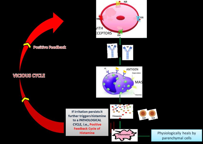
Viruses 2021, 13, 378 2 of 24
macrophages in the lung through human angiotensin-converting enzyme 2 (ACE2) recep-
tors [1–3]. The virus enters the cells mainly through endocytosis, with the help of phos-
phatidylinositol 3-phosphate 5-kinase (PIKfyve), two-pore channels 2 (TPC2), and cathep-
sin L [3,4]. Transmembrane serine protease 2 (TMPRSS2) helps in priming the S protein
[5]. The virus entry reduces ACE2 expression, which results in a dysfunction of the renin–
angiotensin system (RAS). This influences blood pressure and fluid–electrolyte balance
while causing inflammation and enhancing vascular permeability in the airways [6]. After
the entry, it starts replicating actively at the site of infection. It is also reported to directly
invade other organs and proliferate in the gut with the help of ACE2 receptors in these
organs [7–13]. Hamming et al. studied the expression of ACE2 in various human organs
and found that it was expressed on lung alveolar epithelial cells; enterocytes of the small
intestine; and arterial and venous endothelial cells and arterial smooth muscle cells in oral
and nasal mucosa, nasopharynx, lung, stomach, small intestine, colon, skin, lymph nodes,
thymus, bone marrow, spleen, liver, kidney, and brain [14]. The clearance of viral RNA in
stools of patients is more delayed as compared to the respiratory system [13].
1.1. Normal Response of the Human Immune System to SARS-CoV-2 Infection
Exposure to the virus results in variable severity of disease ranging from asympto-
matic or mild disease to severe disease. In a normal scenario, the human immune system
elicits a regulated immune response to SARS-CoV-2 infection. The release of the virus
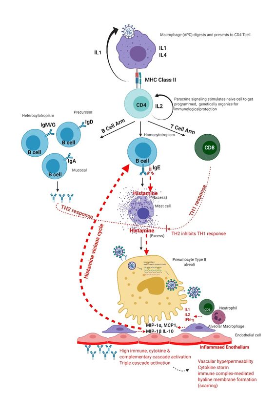
causes the host cells to undergo a phenomenon known as pyroptosis (Figure 1) [6], which
releases molecular patterns and triggers innate immunity. The innate immunity causes
the creation of barrier, secretion of mucus, and shredding of epithelial layer containing
virus. Neutrophils engulf the enemy by phagocytosis and produce enzymes to kill it [15].
Natural killer (NK) cells cause apoptosis of rogue self-cells containing the pathogens [16].
Expression of pro-inflammatory genes is induced with the help of factors like Nuclear
Factor-κB (NF-κB) [17]; as a result, pro-inflammatory cytokines and chemokines are re-
leased, which attract monocytes, macrophages, and T cells to the site of the insult [6]. Type
I interferons (IFN) IFN-α, and IFN-β block viral replication and augment antiviral effector
mechanisms. SARS-CoV-1 and likely the homologous SARS-CoV-2 expresses proteins
that inhibit type I IFN production [18]. This delays the antiviral response and facilitates
rapid viral replication and extensive virus-induced direct cytopathic effects in the early
stages of disease [18].
Along with triggering of the innate system, the adaptive immune system is also
started. Timing of adaptive immune response is vital for efficient viral clearance by innate
immunity [19]. Macrophages or dendritic cells engulf the viruses and function as antigen-
presenting cells. With the help of Interleukin (IL)-1, these antigen-presenting cells present
small epitope antigens to the immature cluster of differentiation (CD)4+T cells by major
histocompatibility complex (MHC) class 2 receptors in the presence of a co-signal mole-
cule called CD27 and form mature helper T cells [20]. IL-2 acts in an autocrine manner and
determines which foreign particles are extracellular and need an antibody response, and
which are intracellular and need T-cell response [20,21].
Depending upon the pattern of cytokine release, T helper (Th) cells are differentiated
into either Th1 or Th2 effector cells, and histamine regulates the balance of both [22]. Th1
type helper cells promote cell-mediated immunity by activating macrophages, while Th2
type helper cells activate B-cell proliferation [21]. Similarly, antigen is presented to imma-
ture CD8+T cells through MHC 1 to form mature cytotoxic T cells [20], which break open
the infected cells, and the foreign organisms are killed once they come out of the broken
cell [23].
The T cells and B cells meet in the secondary lymphoid organs and exchange infor-
mation, due to which naïve B cells are activated to become effector B cells, which secrete
disease-antigen-specific antibodies or immunoglobulins (Ig) of five types—IgD, IgA, IgE,
IgG, and IgM. IgD aids the activation of naïve B-cells. IgA acts locally on the site of insult.
Initially IgM and later IgG target foreign particles by hetero-cytotrophism [24]. IgE targets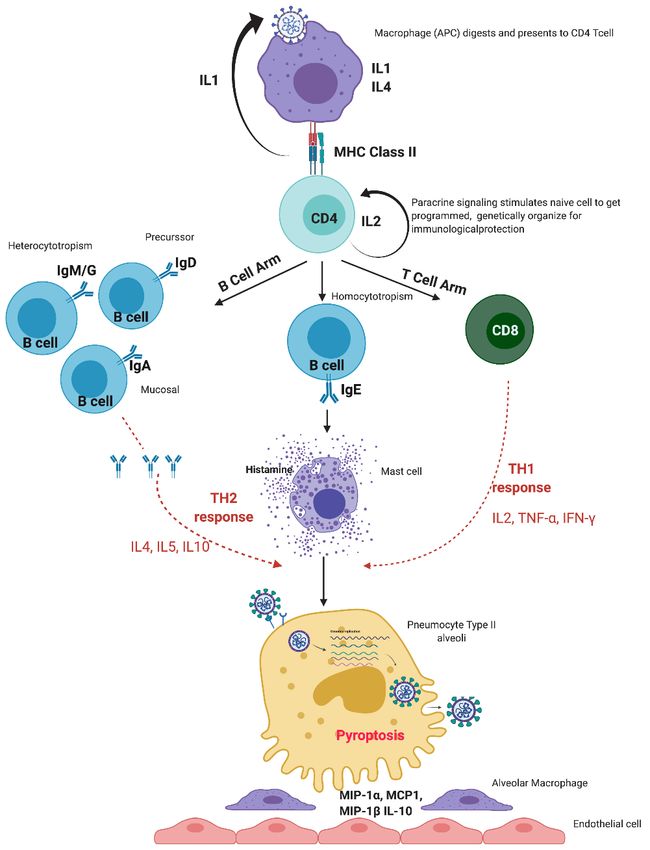
Viruses 2021, 13, 378                                                                                          3 of 24

                        the body’s own mast cells by homo-cytotrophism, causing degranulation and histamine
                        release [24].
                              Studies have found that histamine plays a key role in coronavirus disease 2019
                        (COVID-19) due to its immunomodulatory actions on mast cell histamine–cytokine cross-
                        talk [25]. Histamine acts through four receptors H1, H2, H3, and H4. The H1 receptor is
                        ubiquitous and is activated through guanine nucleotide-binding protein (G) αq; H2 is
                        highly expressed in B-cells, T-cells, gastric parietal cells and is coupled to Gαs; H3 is ex-
                        clusively expressed in neurons and is coupled to Gαi/o; and H4 is found on immune cells,
                        lung, central nervous system (CNS), spleen and is coupled to Gi/o [26]. Histamine acti-
                        vates other mediator biomolecules, such as serotonin, arachidonic acid metabolites, plate-
                        let-activating-factor, complement mediator substances Complement (C) 3a and C5a, and
                        cytokines. All these biomolecules, in return, can trigger histamine. In addition, the cumu-
                        lative action of angiotensin II and vasopressin via Gq pathways contribute to inflamma-
                        tion [27–30].
                              The secreted histamine promotes vascular smooth muscle contraction and diapedesis
                        of all immune cells, antibodies, and mediators into the site of insult [31]. In a normal sce-
                        nario, these immune entities neutralize and kill all the foreign particles, and macrophages
                        do scavenging. The body subdues what is not needed out of the immune molecules, fol-
                        lowing the law of conservation of mass. Histamine is neutralized by its biological antago-
                        nist adrenaline after the insult is resolved [32]. The serum also has histamine neutralizing
                        ability, which takes care of the residual histamine. This histamine homeostasis is im-
                        portant for immune modulation and regulation of inflammation in the body [22].
Viruses 2021, 13, 378 4 of 24
Figure 1. Normal immune response of the human immune system to SARS-CoV-2 infection. MIP:
Macrophage inflammatory protein-1 alpha, MCP: monocyte chemoattractant protein.
1.2. Need for Reviewing Excess Immune Response of Human Immune System to SARS-CoV-2
Infection in this Pandemic Situation
Nearly 20% of COVID-19 patients develop serious complications due to excess im-
mune response of the human immune system [33], resulting in pneumonitis, Acute Res-
piratory Distress Syndrome (ARDS), encephalopathy, hypercoagulability, pulmonary em-
bolism, deep vein thrombosis, ischemic stroke, myocardial infarction, systemic arterial
embolism, disseminated intravascular coagulation, virus-activated cytokine storm syn-
drome, fulminant myocarditis, septic shock, mimicry of vasculitis, endothelium damage,
and multiple organ failure in humans [7,34–38]. Immune-mediated damage due to
COVID-19 occurs in various organs. Lungs of patients of COVID-19 show marked alveo-
lar inflammatory cell infiltrate, diffuse alveolar damage, formation of hyaline membranes,
and diffuse thickening of the alveolar wall [35]. The spleen shows atrophy, and lymph
nodes show necrosis with reduction of lymphocytes in lymphoid organs [35,36]. OlderViruses 2021, 13, 378 5 of 24
age, male gender, underlying co-morbidities, and secondary infections are reportedly as-
sociated with high fatality [39].
Many authors have described a multitude of mechanisms of the excess immune re-
sponse of the human immune system to SARS-CoV-2, but to date, the complex and heter-
ogeneous nature of hyper-inflammation in COVID-19 is not fully understood. The objec-
tive of this study was to review the reported extracellular and intracellular mechanisms
in the patients showing excess immune response to SARS-CoV-2 infection in published
articles, to suggest life-saving solutions in this pandemic, and to propose further research.
2. Materials and Methods
A systematic review was conducted to synthesize a narrative of mechanisms of ex-
cess immune response in patients of COVID-19. The protocol of conducting the review
was registered in advance in the PROSPERO register with registration number
CRD42020214230.
2.1. Search Strategy
MEDLINE database was searched through Pubmed using keyword:
(COVID-19 OR SARS-CoV-2 OR nCoV OR "Novel Corona") AND ("excess immune
response" OR "exaggerated immune response" OR "excessive immune response" OR "ex-
cess immunity" OR "exaggerated immunity" OR "excessive immunity" OR "hyperinflam-
mation" OR "excess inflammation" OR "exaggerated inflammation" OR "excessive inflam-
mation") NOT ("clinical trials" OR "clinical trial" OR "trial")
There was no restriction related to the type of article, country, or region, so as to
include findings in a broad population studied at varied timings during the pandemic.
Articles published in English, related to human species, and available as free full text were
included. The last search was run on 27.10.2020. The search was not rerun prior to the final
analysis and no unpublished studies were sought.
As Pubmed provides clinical literature focused on human species, it suited the study
question of the review regarding the excess immune response of the human immune sys-
tem to SARS-CoV-2 virus. Although Pubmed yielded a sufficient number of articles that
were made available free of cost during the pandemic, authors admit that searching only
one portal and including articles published only in English may have introduced publica-
tion bias.
2.2. Selection of Studies
Studies describing mechanisms of the excess immune response of the human im-
mune system to SARS-CoV-2 and the intra-cellular pathways among the reported mech-
anisms were studied. Study participants were COVID-19 patients eliciting excess immune
response. Exposure was excess immune response in severe COVID-19 resulting in hyper-
inflammation, as compared to normal immune response of the human immune system to
SARS-CoV-2 infection. Two reviewers screened titles, abstracts, and full papers in an un-
blinded standardized manner and assessed the eligibility of retrieved articles based on the
following criteria.
2.2.1. Inclusion Criteria
1. The articles describing immunology in COVID-19 patients especially in the context
of excess immune response;
2. Articles pertaining to adult population showing excess immune response in COVID-
19.
2.2.2. Exclusion Criteria
1. Articles related to drug repositioning, therapeutics, target drugs, therapies, treat-
ments, and vaccines;Viruses 2021, 13, 378 6 of 24
2. Articles focusing on a sub-group of patients suffering from a particular co-morbidity;
3. Articles with study design as clinical trial.
Articles utilizing a drug as a material to elicit a phenomenon or to describe immuno-
logical concepts or explaining drugs or pathways or therapeutic targets just as a sugges-
tion were not excluded.
The reviewers were blinded to each other's decisions. The screening process was val-
idated, and disagreements were resolved by other co-authors of the paper and were rec-
orded in excel sheets.
2.3. Data Extraction
Extraction was done by one reviewer and was checked and verified by the other re-
viewer. Disagreements were resolved by mutual consensus. Data regarding study design,
type of article, month of publication, participant demographics, mechanism of excess im-
mune response, and mechanism of normal immune response were extracted from the
studies in an Excel-based extraction sheet. Description of mechanisms of excess immune
response in patients was compiled from the included articles and synthesized into a nar-
rative. The extracted data were validated by all authors of this article.
2.4. Assessment of Risk of Bias and Quality of Studies
Quality assessment was done by two reviewers independently, and disagreements
were resolved by consultation with each other. Assessment was done at the study level.
Characteristics to be assessed depended upon the study design of the screened record.
Joanna Briggs Institute's (JBI) critical appraisal tools [40] for respective study design of
articles were used. For reviews, the initial five criteria of the JBI checklist were considered
critical, and the remaining six criteria were planned to be assessed only when the initial
five were satisfied. For narrative reviews, which could not fulfill the initial five criteria of
the JBI tool, the Scale for the quality Assessment of Narrative Review Articles (SANRA)
tool [41] was used. For research papers with cross-sectional design and secondary data
analysis, all criteria of the JBI tool for analytical cross-sectional studies were considered
essential to be fulfilled for inclusion in the review. Similarly, for remaining articles like
editorials, viewpoints, or comments, all criteria of the JBI tool for text and opinion were
considered essential to be fulfilled for inclusion in the review. The data of only those stud-
ies were included in the final narrative synthesis, which fulfilled the criteria of quality
assessment.
2.5. Strategy for Data Synthesis
A formal narrative synthesis was planned. A minimum of 10 studies was considered
to be required for synthesis. Qualitative data aggregated in an Excel sheet was synthesized
for presenting common mechanisms of excess immune response of COVID-19 patients
and the intracellular pathways reported in the studies using the formal method "meta-
study", including meta-theory, meta-method, and meta-data-analysis. Indication of inter-
pretation of results was for deriving common mechanisms and exploring their common
links or pathways at the intracellular level. Two researchers were involved in data syn-
thesis, and the discrepancies were resolved by consultation among all co-authors.
2.6. Primary Outcome
Description of the mechanism of excess immune response of human immune system
to SARS-CoV-2 and the intracellular pathways involved was the outcome of interest,
where the immune response was measured based on the presence of immune cells, anti-
bodies, and mediators of innate and/or acquired immunity found by any of the following
analyses: immunological findings on autopsies, analysis of biomarkers, broncho-alveolarViruses 2021, 13, 378 7 of 24
lavage fluid analysis, histopathological analysis, electron microscopy, and immunostain-
ing. The measure of effect was the mortality rate due to various reported mechanisms for
excess immune response.
3. Results
3.1. Flow of Studies through the Review Process
Out of a total 122 records screened for eligibility, 42 articles were included in the final
review. The flow of the studies through the review process is depicted in the Preferred
Reporting Items for Systematic Reviews and Meta-Analyses (PRISMA) flow diagram pro-
vided below (Figure 2). All 42 included articles passed quality assessment as per their
respective study design or article type. The details of the characteristics of the included
studies are mentioned in Table 1. The studies included in the review were conducted in
small geographies or limited patient populations and may not be generalizable over all of
humankind.
Authors reported their findings based on different procedures like autopsies, analy-
sis of biomarkers, broncho-alveolar lavage fluid analysis, histopathological analysis, elec-
tron microscopy, and immunostaining.
Figure 2. Preferred Reporting Items for Systematic Reviews and Meta-Analyses (PRISMA) dia-
gram showing selection of studies for inclusion in the review.Viruses 2021, 13, 378 8 of 24
Table 1. Characteristics of the studies included in the review.
Characteristic 1 Number of Studies
Month of publication N = 42
April 2020 6
May 2020 8
June 2020 12
July 2020 8
August 2020 3
September 2020 3
October 2020 2
Article type N = 42
Research Paper 8
Systematic Review 1
Narrative Review 22
Editorial 1
Commentary 2
Comment 3
Hypothesis 2
Viewpoint/perspective 2
Updating article 1
Outcome (Mechanism of excess immune response) N = 42
Extracellular mechanisms 2
Cytokine storm 12
Neutrophil-related mechanism (NETosis, neutrophilia, and HMGB1 induced inflammation) 6
Lymphocyte-related mechanisms 4
Secondary haemophagocytic lymphohistocytosis 3
Direct injury 3
Immunothrombosis-related mechanisms 3
Hyperferritinemia 2
Macrophage activation syndrome (including Galactin-3 up-regulation) 2
Hypoxia-induced dysregulated immune response 1
Cannabinoid receptor-mediated immune suppression 1
Quasi-programmed aging 1
IL-6 attenuated HLA-DR expression 1
Intracellular mechanisms
NLRP3 inflammasome activation 2
Dysfunction of platelet mitochondria 1
PROS1 signalling 1
NF-κB pathway 1
ACE2/bradykinin B1R/DABK axis involvement 1
1 All included articles were published in the English language and pertained to adult patients with severe COVID-19 with
hyper-inflammation. 2 Two studies described both cytokine storm and direct invasion, while one study described both
cytokine storm and secondary haemophagocytic lymphohistocytosis. Abbreviations: ACE2—angiotensin-converting en-
zyme 2; DABK—[des-Arg9]-bradykinin; HLA-DR—Human Leukocyte Antigen–DR isotype; HMGB1—high-mobility
group box 1; NET—neutrophil extracellular traps; NF-κB—Nuclear Factor-κB; NLRP3—Nod-like receptor family, pyrin
domain-containing 3.
3.2. Mortality Rate due to Excess Immune Response in COVID-19
The authors set out to measure the mortality rate in COVID-19 attributable to excess
immune response. Efforts were made to measure mortality due to the individual reported
mechanisms.
Based on the articles included in the review, the deaths attributable to excess immune
response and its individual mechanisms as reported above could not be quantified due to
a paucity of data. Mortality due to severe respiratory failure was reported to be up to 60%
[42]. In another study, respiratory failure was found to be the leading cause of COVID-19Viruses 2021, 13, 378 9 of 24
death in 69.5% of patients [43]. These estimates are close to the findings of Wu et al., who
had found the mortality due to ARDS to be 52.4% [44]. The mortality rate among patients
with disseminated intravascular coagulation (DIC) in COVID-19 was reported to be 71.4%
[45].
The overall case fatality rate of COVID-19 was reported to range from 1% to 10% [46].
The mortality rate in severe disease was reported to be 4% to 15% [47]. The case fatality
rates for COVID-19 in various co-morbidities were documented to be 10.5% in patients
with cardiovascular disease, 7.3% in patients with diabetes, and 6.0% in those with hyper-
tension [8]. The case fatality rate in patients who did not have any co-morbidity varied
from 3% to 4% [8]. Deaths in females ranged from 29% to 85% of total deaths, the latter
being in older females [48]. While several lab investigations indicated the severity of dis-
ease, authors reported that a D-dimer level of more than 1 mg/liter predicted an 18-fold
increase in the risk of death [49]. Another study reported a 10% increased risk of mortality
with every 10% increase in the level of D-dimer or IL-6 [50].
3.3. Reporting of Mechanisms of Excess Immune Response
Since hyper-inflammation has been found predominantly in patients in severe
COVID-19 [51], the mechanisms reported by the review have been observed primarily in
severe COVID-19, unlike the initial stage of the infection. The mechanisms of excess im-
mune response shown by the human immune system to SARS-CoV-2 were reported dif-
ferently by various authors in the articles included in the review. A total of 14 different
extracellular mechanisms were suggested by authors, while another four articles ex-
plained receptor-level activity. Another three articles exclusively explained intracellular
mechanisms. Very few articles described intracellular continuum of extracellular mecha-
nisms.
4. Discussion
We evaluated various mechanisms proposed or demonstrated by the authors in the
articles included in the review. The same are discussed and summarized hereunder. We
also tried to find out the common pathways among the reported mechanisms as dis-
cussed.
4.1. Mechanisms of Excess Immune Response of the Human Immune System Due to
SARS-CoV-2
4.1.1. Extracellular Mechanisms
Cytokine storm and Related Mechanisms
Cytokines are a family of small molecular proteins secreted by immune cells and tis-
sues. These include IL, colony-stimulating factor (CSF), IFN, tumor necrosis factor (TNF),
growth factor (GF), and chemokines [43]. Cytokines take part in immune responses
through the activation of many signaling pathways like Janus kinase (JAK) signal trans-
ducer and activator of transcription protein (STAT), TNF receptor-associated factor
(TRAF)-NF-κB, TRAF-activator protein 1 (AP-1), and interleukin-1 receptor-associated ki-
nase (IRAK)-NF-κB [43]. The cytokine storm is described as a systemic acute inflamma-
tory manifestation during viral infections in which the cytokine level rises in the body
[46]. The cytokine storm observed in COVID-19 is more complex than other conditions
due to the heterogeneous nature of hyper-inflammation at various stages of the disease
[52] and due to a complex interaction of various components and axes [53]. It has been
proposed to present in various forms such as Macrophage Activation Syndrome (MAS)
or secondary haemophagocytic lymphohistiocytosis (sHLH), which are hyperferritinemic
conditions [54]. The other mechanisms like immune-thrombosis or neutrophil extracellu-
lar traps (NET) are also associated with excess cytokine release. Delayed but elevated lev-Viruses 2021, 13, 378 10 of 24
els of pro-inflammatory cytokines and chemokines and delayed production of viro-pro-
tective IFNs lead to dysregulated immune response and cytokine storm with inefficient
viral clearance [55].
Hyperferritinemia
Alunno et al. expressed that COVID-19 can be another hyperferritinemic condition
like sHLH and MAS, which is characterized by persistent IFNγ-dependent stimulation of
toll-like receptors (TLRs), antigen-presenting cells, and T-cell-uncontrolled activation,
leading to cytokine storm [52]. The excess ferritin circulating in the body may originate
either due to active secretion by macrophages and hepatocytes or due to the death of the
cells. The circulating ferritin shows pro-inflammatory activity. Moreover, free iron re-
leased from ferritin leads to oxidative stress on red blood cells (RBC) and fibrin, which
induces coagulation [56] and triggers multiple chain reactions [57]. RBCs are also dam-
aged due to the tropism of the virus [58]. Damaged RBCs further release iron in circulation
due to the disruption of hemoglobin [58]. Heavy-molecular-weight ferritin is shown to
regulate an iron-independent signaling pathway, which eventually activates NF-κB [59]
and causes secretion of excess cytokines. Removal of excess iron is done by the phagocytic
system [60].
MAS and sHLH
Excessive cytokines result in activation of macrophages, which in turn increases the
secretion of more cytokines like IL-6 and IL-10 [45] and contributes to lung damage [61].
McGonagle et al. elaborated that MAS in COVID-19 is atypical and that MAS-like lung
inflammation and coagulopathy are more centered on the lung, which is aggravated by
virus-induced immune suppression [62]. Galectin 3 is a carbohydrate-binding protein ex-
pressed by macrophages, epithelial cells, and alveolar cells, which drives macrophage-
related hyper-inflammation, mediates viral adhesion, and promotes lung fibrosis [63]. Gi-
rija et al. proposed distinct mechanisms of three phases of cytokine storm [46]. Initial in-
filtration of the airway is induced by IFN-αβ and IFN-γ through mechanisms involving
Fas–Fas ligand (FasL) or TNF-related apoptosis-inducing ligand (TRAIL)–death receptor
5 (DR5) [55]. This causes the apoptosis of airway and alveolar epithelial cells and damage
to the pulmonary microvasculature, leading to vascular leakage, alveolar edema, and hy-
poxia [55]. In the next phase, TNF-mediated T-cell apoptosis occurs [46]. Activated mac-
rophages accumulate in lung tissues through the abrogation of myeloid-specific STAT-1
signaling [46]. In the final phase, ARDS occurs due to IL-6, chemokine (C-X-C motif) lig-
and (CXCL)8, IL-1β, and Granulocyte-macrophage colony-stimulating factor (GM-CSF),
chemokine (C-C motif) ligand (CCL) 2, CCL5, IFN-γ inducible protein (IP) -10, and
CCL3 [46].
The role of IL-6 in the distinct MAS-like lung inflammation remained unclear to
McGonagle et al. [62]. Another study demonstrated that lung inflammation and sustained
cytokine production was due to the fact that IL-6 attenuates Human Leukocyte Antigen–
DR isotype (HLA-DR) membrane expression in CD14 monocytes and decreases the pro-
duction of IFN-γ by CD4 cells [42]. This leads to defective antigen presentation and lym-
phopenia, which causes the defective function of lymphoid cells [42,64], whereas mono-
cytes remain potent and keep producing TNF-α and IL-6 [42].
The presentation of severe COVID-19 resembles sHLH, due to aberrant activation of
T cells, NK cells, and macrophages, causing overproduction of inflammatory cytokines
and hemophagocytosis [65].
Neutrophil-related mechanisms
Excess neutrophils sustain inflammation in COVID-19 [66]. Due to the need to pro-
duce more neutrophils, bone marrow can produce fewer cells of other types [67]. NETs
are extracellular webs secreted by activated neutrophils under the influence of inflamma-
tory cytokines to prevent the spread of pathogens and facilitate the accumulation of anti-Viruses 2021, 13, 378 11 of 24
microbial factors. The release of NETs is induced by damage-associated molecular pat-
terns (DAMPs), particularly the high-mobility group box 1 (HMGB1) [68]. NETs are of
two types: suicidal NETs and vital NETs. Suicidal NETs require the generation of reactive
oxygen species (ROS) and activation of Raf/ mitogen-activated protein kinases ERK
(MERK)/extracellular signal-regulated kinases (ERK) pathway, while vital NETs sustain
longer [68]. Laforge et al. postulated that excess ROS generated due to neutrophils leads
to oxidative damage and excess immune response [69]. Inhibition of nuclear factor
erythroid 2-related factor (NRF) 2-mediated pathways responsible for antioxidant de-
fenses, and activation of NF-κB signaling can promote inflammation and oxidative dam-
age during respiratory infections [69]. Small pathogens lead to excessive NETosis [68].
Excessive NET formation tends to aggravate secretion of more pro-inflammatory cyto-
kines and formation of microvascular thrombosis by stimulating the intrinsic pathway
through activation of Factor XII and propagating a pro-coagulant state [18,70]. NETosis in
lung tissue also results in an insufficient anti-viral response by negatively regulating T
cells and NK cells [47]. At high levels of HMGB1, pulmonary expression of the receptor
for glycated end products (RAGE) is reported to cause detrimental inflammasome activa-
tion [71]. Misbalance in neutrophil serine cascade activator proteases and their inhibitors
cause a proteolytic storm, which advances to a cytokine storm [72].
Immunothrombosis
Immunothrombosis or thromboinflammation is a process of formation of blood clots
due to the interaction of platelets, coagulation factors, and innate immune effector systems
like monocytes/macrophages, polymorphonuclear neutrophils, and the complement sys-
tem [18]. COVID-19-induced coagulopathy results from platelet hyper-reactivity, hyper-
coagulability, hypo-fibrinolysis due to an imbalance between tissue plasminogen activa-
tor (tPA)/urokinase plasminogen activator (uPA) and plasminogen activator inhibitor-1
(PAI-1), complement overactivation, and renin–angiotensin aldosterone system (RAAS)
derangement in the presence of underlying inflammatory-induced endothelial dysfunc-
tion [18]. Prolonged prothrombin time (PT) and minimally affected activated partial
thromboplastin time (aPTT) in COVID-19 suggest a predominant tissue factor-factor VIIa
(TF-F VIIa)-mediated activation of the extrinsic coagulation pathway. COVID-19-associ-
ated coagulopathy has been proposed to be pulmonary-specific intravascular coagulopa-
thy or local DIC [73]. A bidirectional interaction between inflammation and coagulation
has been observed. Innate immunity, pro-inflammatory cytokines, chemokines, adhesion
molecules, tissue factor expression, platelet and endothelial activation, and micro-parti-
cles promote coagulation [73]. In turn, the activated coagulation products, including
thrombin, Factor Xa, fibrin, and the TF–FVIIa complex through activating protease-acti-
vated receptors (PARs), induce secretion of pro-inflammatory cytokines [73]. Platelet-de-
pendent protection of endothelial barrier integrity warrants differentiation of pro-throm-
botic and pro-inflammatory mechanisms of platelets [74]. Anticoagulant protein S is en-
coded by PROS1 gene and is released due to rupture of the tissue containing it [49]. Lemke
et al. hypothesized that excessive blood clotting and excess immune response are linked
by consumption and exhaustion of protein S by the growing clot, which leads to inactiva-
tion of the immunosuppressive Mer receptor tyrosine kinase (MERTK) on macrophages
and leads to the secretion of excess cytokines [49].
Other Mechanisms
Lymphocyte-Related Mechanisms
Song et al. demonstrated that despite a decrease in the absolute counts of CD8+ T
cells, over-activation of these cells increased T-cell inhibitory molecules expression and
increased multiple cytotoxic granules expression result in excess acute inflammation [75].
An increase in inhibitory receptors T-cell immunoglobulin mucin-3 (TIM-3) and Lympho-
cyte-activation gene-3 (LAG-3) on T effector cells modulates pro-inflammatory T cell re-
sponses [76]. Lymphopenia was found to be due to activation of apoptosis and the P53-
signalling pathway in lymphocytes [77]. Exhaustion of lymphocytes causes the secretionViruses 2021, 13, 378 12 of 24
of excessive inflammatory cytokines as a compensatory mechanism [54,78]. Compromised
mechanisms of innate immune response pave the way for cytokine storm. Delayed re-
sponse of type I IFNs (IFN α and β) hampers innate immune response against viruses
through JAK-STAT signal transduction pathway. TLRs recruit signal transfer proteins and
eventually activate the NF-κB pathway to secrete cytokines [79]. Cavalli et al. demon-
strated predominant B-cell activation, which depends on a mammalian target of rapamy-
cin (mTOR) pathway and can lead to hyper-inflammation [80]. A cross-referenced article
also proposed antibody-dependent enhancement as a cause of cytokine storm [81].
Multiple Organ Damage
An et al. review described that pattern recognition receptors (PRR) recognize patho-
gen-associated molecular patterns (PAMP) during viral infections and activate IFN regu-
latory factor (IRF) and NF-κB, which promotes the release of pro-inflammatory cytokines
and chemokines from infected local cells in the lungs [43]. The pro-inflammatory cyto-
kines and chemokines are spilled over into the circulatory system, causing systemic cyto-
kine storm and multiple organ damage [43]. Mechanisms of cardiac injury include direct
myocardial injury by the virus through ACE2 entry, which is mediated by thrombaxane
A2 [82], hypoxia-induced myocardial injury, microvascular damage and endothelial shed-
ding, and cytokine/inflammation-mediated damage [8,83]. We support the view that the
toxin effect of histamine on H1 and H2 receptors in myocardium causes cardiotoxicity
[84]. Systemic hyper-inflammation leads to CNS complications [7]. Rare direct neuro-in-
vasion causing CNS complications remains unproven to date [7]. Similarly, other organs
also suffer damage due to hypoxia and deregulation of the control mechanism of inflam-
mation [85].
A feature of COVID-19 is that the acute condition goes into chronic condition ab-
ruptly over a small period [86]. Due to the ongoing chronic inflammation, we are of the
opinion that all organs predisposed to COVID-19-related damage will be affected even in
the post-COVID phase. The post-COVID sequelae may include interstitial lung disease,
dilated cardiomyopathy, chronic renal failure, cirrhosis of liver, other fibrotic conditions
of organs, vasculopathies, psychiatric disorders, neuropathies, and arthropathies [87–92].
Hyper-inflammation in sub-groups of patients
NETs increase due to diabetes, obesity, increasing age, and male gender. All these
conditions also show a shift from Th1 to Th2 cytokine response [68]. Testosterone has been
found to induce DAMP release, resulting in increased TLR4 signaling in males [68].
Giaglis et al. proposed that Progesterone in females protects them from NETosis [93]. On
the other hand, Rossi et al. proposed that estrogen in women stimulates cannabinoid re-
ceptor type 2, which leads to a limit of the release of pro-inflammatory cytokines, a shift
of the macrophage phenotype towards the anti-inflammatory M2 type, and an enhance-
ment of the immune-modulating properties of mesenchymal stromal cells [94]. O’Brien et
al. proposed that the regulation of expression and extracellular release of Heat Shock Pro-
tein-HSP27 by estrogens is responsible for the relative protection of females from severe
COVID-19 [48]. Blagosklonny et al. proposed that COVID-19 is an age-dependent syn-
drome associated with inflammaging and immunosenescence, hyperinflammation, hy-
perthrombosis, and cytokine storms as explained by the hyper-function theory of quasi-
programmed aging [95].
4.1.2. Intra-Cellular Mechanisms
It is proposed that rapid and excessive stimulation of the innate immune response
triggers activation of the Nod-like receptor family, pyrin domain-containing 3 (NLRP3)
inflammasome pathway in response to recognition of PAMPs or DAMPs like endogenous
or exogenous adenosine triphosphate (ATP), ROS, or lysosomal proteases [48,96]. This
activates PRRs such as TLRs, which bind to the virus [48], or nucleotide-binding oligomer-
ization domain-containing protein 2 (NOD2). Eventually, NF-κB is activated, leading toViruses 2021, 13, 378 13 of 24
the release of pro-inflammatory cytokines, causing acute lung injury [96]. NLRP3 activa-
tion leads to pyroptosis of infected cells, which is an inflammatory programmed cell death
pathway of T lymphocytes mediated by IL-1β and IL-18 [96].
According to Saleh et al., the cytokine storm, oxidative stress, microbiota dysregula-
tion, iron overload, and accumulation of ROS cause intra- and extra-mitochondrial dys-
function [50]. Dysfunction of the platelet mitochondria leads to coagulopathy [50].
4.1.3. Signaling Pathways in an Excess Immune Response
Cytokine storm is characterized by ACE2 receptor-mediated inflammatory response,
cell pyroptosis, delayed IFN α and β response by blocking STAT1 phosphorylation, and
Anti S IgG-mediated lung injury [79]. The IL-6/JAK/STAT signaling pathway is reported
to transduce extracellular signals transmitted by many pro-inflammatory factors [97].
Mahmudpour et al. explained that excess inflammatory cytokines are released due to
down-regulation of ACE2 through dysregulation of the renin–angiotensin–aldosterone
system (ACE/angiotensin II/angiotensin II type 1 receptor-AT1R axis) [53], which results
in overproduction of angiotensin II, thereby enhancing IL-6 production via JAK/STAT
pathway, and ultimately results in exacerbation of vascular and lung injuries [97]. Down-
regulation of ACE2 leads to hyper-activation of NF-κB by IL-6 STATs axis; attenuation of
Mas receptor (ACE2/MasR axis); production of Angiotensin-(1–7) that reduces the expres-
sion of p38 mitogen-activated protein kinase mitogen-activated protein kinase (MAPK)
and NF-κB and inflammatory factors such as IL-6, TNFα, and IL-8; increased activation of
[des-Arg9]-bradykinin (DABK) (ACE2/bradykinin B1R/DABK axis); and activation of the
complement system including C5a and C5b-9 components [53]. Further, the angiotensin
II/AT1 receptor axis activates a disintegrin and metalloproteinase (ADAM) 17, which
cleaves and inactivates ACE2 and enhances angiotensin II retention [97]. For the complete
induction of NF-κB pathway, the activation of STAT3 is reportedly required [97].
The sphingosine-1-phosphate (S1P)/sphingosine-1-phosphate receptor 1 (S1PR1) axis
has been demonstrated to regulate the migration of numerous types of immune cells, in-
cluding T and B lymphocytes, NK cells, and dendritic cells, and to inhibit the pathological
damage induced by the host innate and adaptive immune responses [97].
The S1PR1 pathway agonism is documented to suppress cytokine and chemokine
production, independently of TLR3 and TLR7 signaling or other endosome and cytosolic
innate pathogen-sensing pathways, by targeting myeloid differentiation primary re-
sponse gene 88 (MyD88)/TIR (Toll/interleukin-1 receptor)-domain-containing adapter-in-
ducing IFN-β (TRIF) signaling, which are common actors with the NF-κB pathway [98].
NF-κB is a family of inducible transcription factors and a central mediator of induc-
tion of pro-inflammatory genes [17]. Exacerbation of NF-κB activation is implicated as the
underlying mechanism in lung inflammatory pathology induced by respiratory viruses
including SARS-CoV [97]. Moreover, SARS-CoV spike protein is documented to be asso-
ciated with an increase in I-κBα degradation, which leads to activation of NF-κB pathway
[97]. NF-κB levels were found to be higher in SARS-CoV-2-infected lungs, and suppres-
sion of this pathway enhanced IFN-mediated antiviral immunity and improved the infec-
tion outcome [99,100].
Based on the findings of the review, we observed a common pattern in the proposed
mechanisms by various authors. Most of the described mechanisms interact bi-direction-
ally with inflammatory cytokines by being induced by them and in return secreting more
pro-inflammatory cytokines. Eventually, most mechanisms result in cytokine excess or
storm. At the intra-cellular level, up-regulation of NF-κB was documented to play a major
role in most of the mechanisms of excess immune response, as explained by the authors,
including macrophage activation with release of cytokines, T-cell activation, and regula-
tion of inflammasome particularly of NLRP3 variety [17]. However, we admit the limita-
tion that not all studies explained the proposed mechanism up to the signaling at the intra-
cellular level.Viruses 2021, 13, 378 14 of 24
4.2. Role of Histamine and NF-κB in Excess Immune Response in COVID-19
Conti et al. showed that histamine increases IL-1 levels, causing hyper-inflammation
in COVID-19 and cytokine storm [101]. Types I, II, and III reactions of the B-cell arm pro-
duce Th2 subset cytokines, while Th1 subset cytokines are produced by cell-mediated type
IV reaction of T-cell arm [102–108]. The homeostasis between Th1 subset cytokines and
Th2 subset cytokines is lost and excess Th2 cytokines arrest the activity of Th1 subset cy-
tokines [109–111]. The Th2 subset cytokines give positive feedback to the IgE-producing
B-cells and result in increased production of IgE [112–116]. Bax et al. showed that excess
IgE causes excess histamine release from mast cells [111]. Any viral product remaining in
the body can prolong the phase of fighting the insult and can exaggerate the process of
inflammation [117,118]. Remnants of microorganisms like SARS-CoV-2 try to give an im-
pression that there is still a persistent foreign particle [119,120]. Due to a prolonged fight
against the insult, adrenaline gets exhausted and is unable to neutralize excess histamine,
as the body has higher reserves of histamine since the mast cells are greater in number
and are strategically placed all over the body. This un-neutralized histamine causes hista-
mine imbalance and leads to the release of more pro-inflammatory Th2 cytokines
[110,111,121,122]. In this way, an IgE-mediated positive feedback vicious cycle (Figure 3)
is established, which results in excessive inflammation [109–111].
Figure 3. Positive feedback cycle of histamine.
Excess histamine causes excess diapedesis of all immune cells, antibodies, and medi-
ators through vascular endothelium into the alveoli [6,23,123–126]. To prevent the excess
diapedesis, a serous gel-like fluid is secreted at the site, which eventually thickens. This
gel is made by three cascade reactions—complement, kinin, and coagulation. Ghebrehi-
wet et al. showed that the three cascades are linked with one another by Hageman factor
(factor XII) [127], and when coagulation starts, all other cascades also start [127]. Comple-
ment cascade makes membrane attack complex (MAC) to kill the viruses [128]. Kinin
pathway does the autonomic regulation [129]. Chaudhry and Babiker postulated that co-
agulation triggers the fibrinogen pathway and arrests the viruses from going anywhere
[130]. Further, Nayak et al. showed that SARS-CoV-2 infects type II pneumocytes, leading
to reduced production of surfactant (Figure 4) [131]. This causes a collapse of the alveoli,
retention of fluid in lungs, and progressive hypoxemic respiratory failure [131,132].Viruses 2021, 13, 378 15 of 24
Figure 4. Excess immune response of the human immune system to SARS-CoV-2 infection.
Holden et al. showed that the dysregulated immune reaction is enhanced by excess
histamine results due to the potentiation of NF-κB-dependent transcription and release of
pro-inflammatory factors at the intracellular level [133]. Ayoub et al. explained that NF-
κB exists in an inactive state in the cytoplasm of cells bound to its inhibitory protein I-κB.
Phosphorylation of I-κB by IκB kinase (IKK) enzyme leads to its degradation. This results
in translocation of NF-κB to the nucleus, where it promotes transcription of IL-10 and
many pro-inflammatory factors like TNF-α, IL-1β, and IL-6 [134].
4.3. Drugs Acting on NF-κB Signaling
Various possible mechanisms of attenuation of hyper-inflammation include antago-
nism of IL6 receptors, inhibition of the JAK/STAT signaling pathway, agonism of S1PR1,
blockade of TNFα, and down-regulation of NF- κB, either directly or indirectly by neu-Viruses 2021, 13, 378 16 of 24
tralization of excess histamine [97,133]. The findings of this review provide enough evi-
dence of the benefit of targeting NF-κB down-regulation in the attenuation of the excess
immune response to COVID-19. We researched various drugs and therapeutic approaches
that can serve this purpose. NF-κB is required for normal immune response and survival
of the cell. Hence, global inhibition of NF-κB signaling may affect the normal functioning
of the cells due to the complexity of intrinsic pathways [17,97]. Liu et al. expressed that
the development of drugs for selectively down-regulating NF-κB for clinical use has been
a challenge for current science [17]. Various drugs have shown the ability to down-regu-
late NF-κB by acting upon different steps of its activity. Selective IKK inhibitors block the
IKK-dependant phosphorylation of I-κB; proteasome inhibitors such as Bortezomib block
I-κBα degradation, tacrolimus, and I-κBα super-repressor block nuclear translocation of
NF-κB; and glucocorticoids and Peroxisome proliferator-activated receptors (PPAR) ago-
nists block binding of NF-κB to deoxyribonucleic acid (DNA) [17]. Caffeic acid phenethyl
ester (CAPE), Bay 11–7082, and parthenolide have been shown to inhibit NF-κB activation
and reduce inflammation [97,99]. Hiscott et al. highlighted that molecules blocking NF-
κB either are undergoing clinical trials or lack specificity and may cause unwanted side
effects like broad suppression of innate immunity [135]. Hence, direct targeting of down-
stream effectors like TNFα by monoclonal antibodies like infliximab and adalimumab,
which block TNFα, has been attempted to attenuate hyper-inflammation in several im-
mune-mediated disorders [97]. These monoclonal antibodies are expensive, and drugs
like adalimumab are under investigation in COVID-19 patients [136]. Galloway et al. re-
ported that the major issue with TNFα-blockers has been an increased risk of bacterial and
fungal superinfections [137], which can be a cause of concern in COVID-19 patients, who
as documented by Zhou et al., mostly suffer superadded lung infections due to pre-exist-
ing foci [138]. Zhou et al. reported that at least one in seven COVID-19 patients encounter
a secondary bacterial infection resulting in 50% of the fatalities due to untreated or un-
treatable secondary bacterial infections, occurring mostly in the lung [138].
Since histamine potentiates pro-inflammatory effects of NF-κB, targeting the up-
stream mediator histamine is a highly potential therapeutic approach in current times [22].
This approach not only helps in down-regulating NF-κB effectively and safely but also
helps in reducing other effects of excess histamine.
4.4. Neutralization of Excess Histamine and Down-Regulation of NF-κB
The need to neutralize histamine by some exogenous agent to prevent excess immune
response was perceived by researchers in the past for various diseases. One approach,
which is widely used in clinical practice, is the use of anti-histaminic drugs, which work
by attaching to histamine receptors, but the persistent histamine molecules in the milieu
behave like a toxin and cause toxin-mediated damage as reported by Comas-Basté et al.
[139]. Moreover, histamine receptors are widely expressed in the body and are different
in distribution between genders and age groups. This warrants caution in blocking hista-
mine receptors and requires using a mixture of agonists and antagonists to avoid delete-
rious side effects [22]. Another approach based on the discussion presented above is the
administration of exogenous adrenaline, but it cannot be tolerated by the body beyond a
limit.
In 1951, Parrot and Laborde developed a new treatment method of co-administration
of histamine and human serum gamma-globulin to restore the histamine-neutralizing
ability of the body (histaminopexy). Immunoglobulin provides a larger co-molecule, and
the complex of both the drugs behaves as a foreign body to produce antibodies within the
body [140,141].
Haruo Yoshii and Yuriko Fukata administered intramuscular Histamine-conjugated-
normal human immunoglobulin (Histamine dihydrochloride + Human normal immuno-
globulin) for producing antihistamine antibodies within the human body [142–145]. This
formulation reaches the lymph nodes and stimulates B cells to produce IgG antihistamineViruses 2021, 13, 378 17 of 24
antibodies. The antibodies in turn reach the inflammatory site and neutralize the hista-
mine directly, thereby enhancing the histamine-neutralizing ability of the plasma.
The inventors recommended its use in the treatment of diseases associated with any
abnormal immune response, including infectious diseases, parasitic diseases, respiratory
diseases, and autoimmune diseases. A radioimmunoassay using a Histamine kit detected
no residual histamine after administration of Histamine-conjugated-normal human im-
munoglobulin. An in vivo experiment in Bagg albino mice (BALB/c) for producing mouse-
histamine-added gamma-globulins specific to trinitrophenyl showed significant promot-
ing action upon IgG and IgM antibody production and potent immune-modulatory ac-
tion. In an experimental allergic encephalomyelitis model of Female Lewis rats, the drug
showed immune suppression comparative to cyclosporine A, proving the theory of posi-
tive feedback vicious cycle of hyper-inflammation due to histamine excess [142–145]. Ad-
ministration of Histamine-conjugated-normal human immunoglobulin has shown re-
markable clinical improvement in patients of diseases with an underlying excess immune
response like urticarial, eosinophilia caused by a malignant tumor, chronic articular rheu-
matism, systemic lupus erythematosus, multiple sclerosis, and several diseases associated
with hyper-eosinophilia [142–146].
Neutralization of histamine by Histamine-conjugated-normal human immunoglobu-
lin has been demonstrated to inhibit the nuclear translocation of NF-κB and release of pro-
inflammatory cytokines IL-1β, TNF-α, IL-6, and IL-10 [134]. The NF-kB independent effect
of Histamine-conjugated-normal human immunoglobulin includes neutralization of his-
tamine to reduce its toxin-like effects. Thus, Histamine-conjugated-normal human immu-
noglobulin has a great potential to reduce the excess immune response or hyper-inflam-
mation encountered in diseases like COVID-19 without affecting the ability of the body to
clear the virus, especially while specific and selective drugs for down-regulation of NF-
κB are not available in current clinical practice.
In the wake of the COVID-19 pandemic, as a new drug takes around 12 to 15 years
from its discovery to its use in patients, the only option left with the researchers and cli-
nicians is to repurpose the existing therapeutics for use in COVID-19 [147]. In order to
figure out which candidate molecules have a therapeutic effect against SARS-COV-2 and
are suitable for repositioning studies, researchers have deployed various approaches to
screen commercially available drugs. A few of these approaches are Computer-Aided
Drug Design (CADD), molecular docking, re-trained multi-task deep model, homology
modeling, virtual high-throughput drug screening, drug-likeness profiling, and docking
scores [148–154]. Most of the approaches tried by other researchers are not based on hu-
man biology and the actual interaction of the virus with the human body. Hence, the
drugs identified in such ways may not provide the expected therapeutic effect in clinical
trials, which may cost lives due to delay in discovering ideal therapeutic candidates. The
evidence generated by this review is based on the immunological mechanism of excess
immune response in COVID-19 patients and can be useful in conducting further research
and formulating guidelines for clinical practice.
5. Translational Value and Future Research Direction
To our best knowledge, this review is a state-of-art explanation of varied mechanisms
of excess immune response in COVID-19, and it successfully brings out the common pat-
tern of “cytokine excess and dysregulated cytokine response” among them. This review
can form the basis for further research to understand the complex nature of hyper-inflam-
mation in COVID-19 and can save significant suffering and loss of human life resulting
from it. The review also critically appraises the available drugs for down-regulating NF-
κB. Our approach of basing our proposition on real biological findings instead of com-
puter-based modeling can provide a more real-world solution for the human body to cope
with SARS-CoV-2 infection. The therapeutic candidate suggested by the review needs val-
idation in clinical trials in COVID-19 patients.Viruses 2021, 13, 378 18 of 24
6. Conclusions
The immunological findings in COVID-19 patients indicate that hyper-inflammation
is the main culprit of morbidity and mortality in COVID-19. Histamine-conjugated-nor-
mal human immunoglobulin has been proved to be effective and safe in such situations
based on the past data, due to its effect on down-regulation of NF-κB without directly
blocking the NF-κB signaling pathway. It is already approved for use in humans and is
available in the market for the last 30 years as an orphan drug for allergies. From an eco-
nomic point of view, this treatment is inexpensive as compared to the methods currently
used. The authors recommend clinical trials and further analysis of available data for re-
positioning of Histamine-conjugated-normal human immunoglobulin in COVID-19 for
saving lives of the patients eliciting excess immune response.
Author Contributions: Conceptualization, A.P. and M.G.; methodology, M.G.; software, A.M.K.
and S.R.P.; validation, A.P., S.S., and M.G.; formal analysis, M.G.; investigation, S.R.P., A.M.K., and
A.E.P.; resources, A.P.; data curation, M.G.; writing—original draft preparation, M.G. and S.S.; writ-
ing—review and editing, S.S.; visualization, A.M.K., M.G., and A.E.P.; supervision, A.P.; project ad-
ministration, M.G.; funding acquisition, A.P. All authors have read and agreed to the published
version of the manuscript.
Funding: This research received no external funding.
Institutional Review Board Statement: Not applicable.
Informed Consent Statement: Not applicable.
Data Availability Statement: The data presented in this study are openly available in Harvard
Dataverse at https://doi.org/10.7910/DVN/EQXE0E (accessed on 1 January 2021), reference number
UNF:6:0kKzeLUxwDWCaZfoIrPIkg = = [fileUNF] [155].
Acknowledgments: We acknowledge V.V. Shailaja, Associate Professor (Microbiology), Gandhi
Medical Hospital, Hyderabad, India; and Mahwish Jawaid, Registrar (Microbiology), Maternity and
Children Hospital, Alkharj, Riyadh, Saudi Arabia, for their contribution towards editing the scien-
tific content of the article. BioRender Figure 1; Figure 4 were prepared by Arunasree Maarasanapalli
Kalle, Figure 2 was contributed by Manish Gehani, and Figure 3 was prepared by Angela
Elisabeth Peter.
Conflicts of Interest: The authors declare no conflict of interest. No external funding was received.
Pre-Registration: The review was prospectively registered in PROSPERO register with registration
number CRD42020214230.
References and Note
1. Yan, R.; Zhang, Y.; Li, Y.; Xia, L.; Guo, Y.; Zhou, Q. Structural basis for the recognition of SARS-CoV-2 by full-length human
ACE2. Science 2020, 367, 1444–1448, doi:10.1126/science.abb2762.
2. Li, W.; Moore, M.J.; Vasllieva, N.; Sui, J.; Wong, S.K.; Berne, M.A.; Somasundaran, M.; Sullivan, J.L.; Luzuriaga, K.; Greeneugh,
T.C.; et al. Angiotensin-converting enzyme 2 is a functional receptor for the SARS coronavirus. Nature 2003, 426, 450–454,
doi:10.1038/nature02145.
3. Ou, X.; Liu, Y.; Lei, X.; Li, P.; Mi, D.; Ren, L.; Guo, L.; Guo, R.; Chen, T.; Hu, J.; et al. Characterization of spike glycoprotein of
SARS-CoV-2 on virus entry and its immune cross-reactivity with SARS-CoV. Nat. Commun. 2020, 11, doi:10.1038/s41467-020-
15562-9.
4. Lu, R.; Zhao, X.; Li, J.; Niu, P.; Yang, B.; Wu, H.; Wang, W.; Song, H.; Huang, B.; Zhu, N.; et al. Genomic characterisation and
epidemiology of 2019 novel coronavirus: Implications for virus origins and receptor binding. Lancet 2020, 395, 565–574,
doi:10.1016/S0140-6736(20)30251-8.
5. Hoffmann, M.; Kleine-Weber, H.; Schroeder, S.; Krüger, N.; Herrler, T.; Erichsen, S.; Schiergens, T.S.; Herrler, G.; Wu, N.H.;
Nitsche, A.; et al. SARS-CoV-2 Cell Entry Depends on ACE2 and TMPRSS2 and Is Blocked by a Clinically Proven Protease
Inhibitor. Cell 2020, 181, 271–280, doi:10.1016/j.cell.2020.02.052.
6. Tay, M.Z.; Poh, C.M.; Rénia, L.; MacAry, P.A.; Ng, L.F.P. The trinity of COVID-19: Immunity, inflammation and intervention.
Nat. Rev. Immunol. 2020, 20, 363–374.
7. Najjar, S.; Najjar, A.; Chong, D.J.; Pramanik, B.K.; Kirsch, C.; Kuzniecky, R.I.; Pacia, S.V.; Azhar, S. Central nervous system
complications associated with SARS-CoV-2 infection: Integrative concepts of pathophysiology and case reports. J. Neuroinflam-
mation 2020, 17, 231.You can also read