Animal Models of Metabolic Epilepsy and Epilepsy Associated Metabolic Dysfunction: A Systematic Review - MDPI
←
→
Page content transcription
If your browser does not render page correctly, please read the page content below
pharmaceuticals

Review
Animal Models of Metabolic Epilepsy and Epilepsy
Associated Metabolic Dysfunction: A
Systematic Review
Uday Praful Kundap 1,2 , Yam Nath Paudel 1              and Mohd. Farooq Shaikh 2, *
 1   Research Center of the University of Montreal Hospital Center (CRCHUM), Department of Neurosciences,
     Université de Montréal, Montréal, QC H2X 0A9, Canada; uday.kundap@umontreal.ca (U.P.K.);
     yam.paudel@monash.edu (Y.N.P.)
 2   Neuropharmacology Research Strength, Jeffrey Cheah School of Medicine and Health Sciences,
     Monash University Malaysia, Selangor 47500, Malaysia
 *   Correspondence: farooq.shaikh@monash.edu; Tel.: +60-3-551-44-483

 Received: 8 May 2020; Accepted: 23 May 2020; Published: 26 May 2020

 Abstract: Epilepsy is a serious neurological disorder affecting around 70 million people globally
 and is characterized by spontaneous recurrent seizures. Recent evidence indicates that dysfunction
 in metabolic processes can lead to the alteration of neuronal and network excitability, thereby
 contributing to epileptogenesis. Developing a suitable animal model that can recapitulate all the
 clinical phenotypes of human metabolic epilepsy (ME) is crucial yet challenging. The specific
 environment of many symptoms as well as the primary state of the applicable neurobiology, genetics,
 and lack of valid biomarkers/diagnostic tests are the key factors that hinder the process of developing
 a suitable animal model. The present systematic review summarizes the current state of available
 animal models of metabolic dysfunction associated with epileptic disorders. A systematic search
 was performed by using the Preferred Reporting Items for Systematic Reviews and Meta-Analyses
 (PRISMA) model. A range of electronic databases, including google scholar, Springer, PubMed,
 ScienceDirect, and Scopus, were scanned between January 2000 and April 2020. Based on the selection
 criteria, 23 eligible articles were chosen and are discussed in the current review. Critical analysis of
 the selected literature delineated several available approaches that have been modeled into metabolic
 epilepsy and pointed out several drawbacks associated with the currently available models. The result
 describes available models of metabolic dysfunction associated with epileptic disorder, such as
 mitochondrial respiration deficits, Lafora disease (LD) model-altered glycogen metabolism, causing
 epilepsy, glucose transporter 1 (GLUT1) deficiency, adiponectin responsive seizures, phospholipid
 dysfunction, glutaric aciduria, mitochondrial disorders, pyruvate dehydrogenase (PDH) α-subunit
 gene (PDHA1), pyridoxine dependent epilepsy (PDE), BCL2-associated agonist of cell death (BAD),
 Kcna1 knock out (KO), and long noncoding RNAs (lncRNA) cancer susceptibility candidate 2 (lncRNA
 CASC2). Finally, the review highlights certain focus areas that may increase the possibilities of
 developing more suitable animal models and underscores the importance of the rationalization of
 animal models and evaluation methods for studying ME. The review also suggests the pressing need
 of developing precise robust animal models and evaluation methods for investigating ME.

 Keywords: metabolic epilepsy; animal model; mitochondrial dysfunction; metabolic genes;
 translational research

1. Introduction
     Metabolic abnormalities (ME) causing high brain activity are associated with an increased risk
of epilepsy development in affected individuals. ME is caused by an array of toxic or metabolic

Pharmaceuticals 2020, 13, 106; doi:10.3390/ph13060106                   www.mdpi.com/journal/pharmaceuticals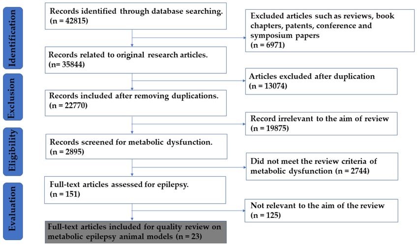
Pharmaceuticals 2020, 13, 106                                                                       2 of 35

diseases, such as mitochondrial dysfunction, alteration of intracellular osmolytes, accumulation of
toxic substances, and a decrease of substrates that are crucial for internal membrane function or cellular
metabolism [1]. All these factors combined result in a compromised efficacy to supply energy in the
brain area, leading to excitability of the neuronal cells and producing epileptic seizures [2]. Moreover,
the novelty of this research would be the study of how metabolic dysfunction can contribute to seizures
and exacerbate related sequalae such as neuronal loss and related complications [3].
     According to the International League Against Epilepsy (ILAE), ME is classified based on deficiency
syndrome and disorder related to mitochondria or metabolism [4] (Figure 1). Biotinidase deficiency
or “Holocarboxylase synthetase deficiency” is a condition whereby the body is not able to utilize
biotin properly [5]. The impairment of certain enzymes that are biotin dependent are categorized
under a group of disorders known as “multiple carboxylase deficiencies” [6]. Cerebral folate deficiency
(CFD) is a neurological syndrome associated with a low cerebrospinal fluid (CSF) concentration
of 5-methyltetrahydrofolate (5MTHF) in the presence of normal peripheral folate metabolism [7].
Moreover, CFD might result in cerebellar ataxia, epilepsy, dyskinesia, psychomotor retardation,
and spastic diplegia [8]. Disturbances in folate transport, which might be due to increased folate
turnover within the central nervous system (CNS), may also lead to CFD [9]. In a majority of the CFD
cases, the etiology remains elusive, however there is an increased understanding about the key role of
mutation in folate receptor 1 (FOLR1) gene in CFD [10]. Further, folate receptor auto-antibodies suggests
that CFD may be caused by the blocking of folic acid transport into CSF [8]. Creatine disorders are
comprised of three defects, namely reduced creatine production in guanidinoacetate methyltransferase
(GAMT), deficiencies of arginine glycine amidino transferase (AGAT), and decreased transport of
creatine into the brain [11]. Epilepsy is associated with GAMT deficiency, which positively responds to
the treatment which is substitutive with creatine monohydrate [12]. Folinic acid responsive seizures
are diagnosed as an increase in monoamine metabolite in CSF, however, their genetic cause remains
elusive [13]. Neonatal epileptic encephalopathy might be a cause of folinic acid responsive seizures
and is treatable as the patients with this type of seizures respond well to pyridoxine therapy [14].
     Mutations in solute carrier family 2 member1 (SLC2A1) gene are the cause of glucose transporter 1
(GLUT1) deficiency syndrome and results in the improper transportation of glucose into the brain [15].
A common inborn error of energy metabolism are a mitochondrial respiratory chain disorder. Tissues
with a high energy requirement are usually affected by these disorders, which are frequently observed
in childhood cerebral involvement and often leads to seizures [16]. Prominent myoclonic seizures are a
common characteristic feature of numeral mitochondrial disorders together with Alpher’s syndrome,
myoclonic epilepsy with ragged red fibers (MERRF), mitochondrial encephalopathy with lactic acidosis,
and stroke-like episodes (MELAS) [17,18]. A group of inherited diseases where either peroxisomal
function or one or more peroxisome biogenesis functions are disrupted are known as a peroxisomal
disorder [19]. Pyridoxal 50 -phosphate is the naturally active form of pyridoxine which is converted
by a series of enzymes involving pyridoxamine phosphate oxidase (PNPO) [20]. Decreased levels of
pyridoxal 50 -phosphate in the CSF along with epilepsy are usually associated with PNPO [21].
     Human ME constitutes a ranges of clinical, electrical, and behavioral demonstrations [22].
The selection or development of an animal model system is determined by several crucial factors,
such as the type of epilepsy to be modelled, including the reason to be studied, acquaintance, and
suitability, because of the large variety of pathological mechanisms involved in ME [23].
Pharmaceuticals 2020, 13, 106 3 of 35
Figure 1. Types of Metabolic Epilepsy.Pharmaceuticals 2020, 13, 106 4 of 35
Overall, 23 animal model studies that were mainly focused on different aspects of ME are reviewed
in the current systematic review. Current study discusses the metabolic alterations studied in animal
models that are associated with epilepsy. The major types of models are described with the use of
animals to study metabolic disorders causing epilepsy or related disorders of CNS, and are further
classified as follows: biotinidase and holocarboxylase synthase deficiency, CFD, creatine disorders,
GLUT1 deficiency, folinic acid responsive seizures, mitochondrial disorders, peroxisomal disorders
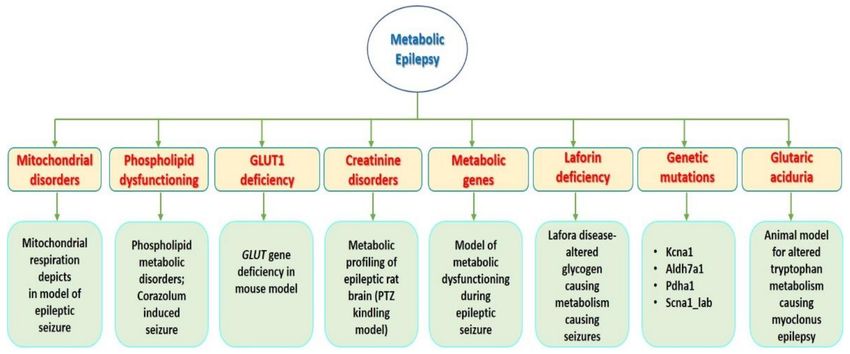
and pyridoxine-dependent epilepsy, and genetic knock out (KO) models (Figure 2).
Figure 2. Animal models of ME and epilepsy associated metabolic dysfunctions: This figure
demonstrates the overall studies of animal model related to ME with different multi approach
considered in the current review.
Developing a suitable animal model that can recapitulate the clinical features of human ME is
challenging as well as interesting [24]. The development of an animal model is complicated due
to the specific nature of various symptoms, lack of validated biomarkers and objective diagnostic
tests, the primary level of the specific neurobiology, and genetic factors [25]. Developing suitable
animal models of ME will open a window of opportunity in this domain as well as strengthen our
knowledge of the complex pathophysiology, mechanism, and therapy for the treatment of ME [26].
Herein, we systematically review the current state of several available pre-clinical models of ME and
other related alterations. With respect to the reviewed literature, the authors consider the probable
areas of interest that might intensify the likelihood of generating more valuable models, at least for
ME with related symptoms, and for explicit methods where animal models are used [27]. To the best
of our knowledge, no earlier reviews have systemically considered the available animal models that
are used to investigate ME and associated alterations. Hence, the current review in a systemic way
provides readers with a precise summary of available animal models that have been used to investigate
several aspects of metabolic alterations associated with, but not necessarily responsible for epileptic
disorders. In addition to a single read, this systematic review would strengthen the understanding of
readers of the importance of developing animal models of human ME. Moreover, the current systematic
review encourages that ME can also be modeled in a range of species, including zebrafish, tilapia, and
drosophila [28,29].
2. Results and Discussion
Exploring the selected database with the specific key words mentioned above in the methodology
yielded 42,815 records. After screening, the total number of articles excluded were 42,792, and thereof:
(a) 6971 abstracts, books, chapters, patents, symposiums, oral and poster presentations, and conferences;
(b) 13,074 duplicate articles were excluded; (c) 22,619 articles that did not meet review criteria, review
articles, and articles irrelevant to the aim of the review were also excluded. A further 23 articles were
compiled, included, and described in Table 1 and discussed in the systematic review forum (Figure 3).Pharmaceuticals 2020, 13, 106 5 of 35
Figure 3. Study evaluation PRISMA flow chart for selection of articles.
The 23 articles discussed herein consist of original research articles, pre-clinical studies, and research
reports which provide information of the available animal models for ME. In addition, the number
of articles found were limited to the type of metabolic study conducted using specific animal
species. During our analysis, we observed that, to date, only mice and rats were used to study ME.
However, a few human studies have also been recorded. As the precise mechanism underlying how
metabolic dysfunction can lead to epilepsy is still unknown, it is very challenging to mimic the same
clinical conditions in animal settings. We attempted to summarize all the article and describe the
main significant feature of the study to highlight the role of metabolic disorder in epileptogenesis.
Nevertheless, there are very few articles available. Moreover, we tried to classify them according to
the available classification scheme mentioned by ILAE. Books with different chapters on “epilepsy in
children, newborns, and inborn errors of metabolism” [30–32], as shown in Table 1, Figures 1 and 2,
and research reports and retrospective studies were included despite their limitations and scope [32].Pharmaceuticals 2020, 13, 106 6 of 35
Table 1. Tabular representation of animal models in ME.
Animal/Subject A Drug or Technique Used
No. of
S.N. Model Type Study Type Study Sample Used for Model for Modeling Metabolic Outcomes References
Citations
Design Epilepsy (ME)
• Abcc8 and Kcnj11 gene
significantly different
from control group.
• Hsd11b1 and Nr3c1 gene
n = 45 Groups—24 h SD rats, 30–35 4% Pilocarpine hydrochloride increase in fold change
Metabolic gene significantly different
Pre-clinical (n = 5), 10 days days old (350 mg/kg in saline, i.p.),
1 responsible for from control group. 6 [33]
(animal model) (n = 5), 1month (n = 5) (125–150 g), methyl-scopolamine prior to
epilepsy in obese rat
and 2 months (n = 5). male rats. Pilocarpine Injection. • Metabolic dysfunction
causes repeated
reoccurrence of
seizures leading to
chronic epilepsy.
• Increase in metabolites
like Myo-inositol, lactate,
creatine, phosphocholine,
Metabolic profiling GPC, Succinate.
of epileptic rat brain • Clear metabolic changes
Male
(PTZ kindling in the cerebellum
Wistar rats,
induced seizures) PTZ (37 mg/kg body and hippocampus of
group (a and b)
Type: Creatine Pre-clinical weight) every 48 h or every kindled rats.
2 n = 10 were 4 weeks 25 [34]
disorders and (animal model) 72 h at the weekend • Indicating increase
old and group
Succinic over a 5-week period in energy demand,
(c) were 9
semialdehyde altered neurotransmitters,
weeks.
dehydrogenase increase neuron loss
deficiency and gliosis.
• Technique used 1H
NMR spectroscopy.
Mutation of • E6-AP protein can affect
ubiquitin ligase the metabolism of p53 in
(Ube3a phenotype) • postmitotic neurons.
causes Angelman • n = 6 LTP UBE3Atm1Alb/J • Ube3a mice at maternal
syndrome in • n = 11 LTP null deficiency closely
mice—a rare genetic Pre-clinical • Maternal mutation 8 and 12 weeks of age mimic the phenotype
3 608 [35,36]
epileptic (animal model) deficiency (AS) mice, 12–16 weeks of age. of human AS
neurodegeneration. (n = 12) • UPD mice • Decrease in
Type: disruption of • WT mice GABA/Glutamate
UBE3A, ratio following ketone
Mitochondrial ester administration.
disordersPharmaceuticals 2020, 13, 106 7 of 35
Table 1. Cont.
Animal/Subject A Drug or Technique Used
No. of
S.N. Model Type Study Type Study Sample Used for Model for Modeling Metabolic Outcomes References
Citations
Design Epilepsy (ME)
• In immature rates,
threshold of 2nd seizure
was lower than for the 1st
seizure. I was vice versa
Immature for the mature rats.
Metabolic features • Immature animal showed
animals: SD rat • Flurothyl was used
in repetitive more c-fos mRNA
(n = 7 pups (P15) to induce repetitive
seizures. Pre-clinical expression in the regions
4 immature; n = 6 Mature animals: seizures in immature and 10 [23]
Type: (animal model) of the CNS.
mature) male mature rats.
Mitochondrial • Consequences of
Sprague–Dawley
disorders repetitive seizures
rats (P60).
in immature animal
is more related to
metabolic disorder then
mature animals.
• G1D antisense mice
represents similar features
that closely resemble
human phenotype.
• Glutamine and its
synthetase expression
Glut1 gene were preserved in
G1D transgenic
deficiency in mouse G1D mice.
Pre-clinical antisense mice 3 G1D gene knockdown to
5 model. n = 104 • TCA cycle intermediate, 16 [37]
(animal model) and 5 months produce Glut1 deficiency
Type: GLUT-1 amino acid and
of age
deficiency neurotransmitter contents
were normal, so there
is no basis to suspect
that in G1D mice, TCA
cycle is responsible for
energy failure.Pharmaceuticals 2020, 13, 106 8 of 35
Table 1. Cont.
Animal/Subject A Drug or Technique Used
No. of
S.N. Model Type Study Type Study Sample Used for Model for Modeling Metabolic Outcomes References
Citations
Design Epilepsy (ME)
• Mitochondrial
respiration deficits
occur in experimental.
Mitochondrial • Novel methodology
respiration deficits for assessing
in rat epilepsy cellular metabolism.
Pre-clinical Adult male SD
6 model n = 4–8 in each group KA (11 mg/kg, s.c.). • Increased steady-state 16 [38]
(animal model) rats (300–350 g)
Type: ROS in mice and deletion
Mitochondrial of manganese superoxide
disorders dismutase results in
deficits in mitochondrial
oxygen consumption.
• Metabolic connectivity
• Pre-treated with was confirmed by
lithium chloride magnetic resonance
(127 mg/kg, i.p.) & imaging (fMRI) based on
methylscopolamine-bromide blood-oxygen-dependent
Abnormal (1 mg/kg, i.p) 24 h (BOLD) signals.
metabolic function and 30 min before • The technique is
in the Adult male SD Pilocarpine administration. used provide vital
Pilocarpine-induced Pre-clinical rats (7 weeks • Pilocarpine hydrochloride information on various
7 (30 mg/kg, i.p.) was brain disorders and 13 [39]
epilepsy rat model. (animal model) old), weighing
Type: 180–200 g injected and repeated functional connectivity
Mitochondrial doses (10 mg/kg) were abnormalities in diseases
disorders then administered every such as epilepsy.
30 min until stage 4 • Complex brain
seizures developed connectivity, abnormal
according to the brain metabolism and
Racine scale topologic features were
theoretically measured.Pharmaceuticals 2020, 13, 106 9 of 35
Table 1. Cont.
Animal/Subject A Drug or Technique Used
No. of
S.N. Model Type Study Type Study Sample Used for Model for Modeling Metabolic Outcomes References
Citations
Design Epilepsy (ME)
• Greater fat
accumulation due to
Metabolic • C57BL/6J adiponectin deficiency.
dysfunction via mice and • Low dose of
adiponectin • (n = 17 per ADP-KO mice. intrahippocampal
deficiency. Pre-clinical genotype) • Control KA resulted in severe
8 • (n = 10 per WT and KA-Induced Seizure neuronal damage and 26 [40]
Type: adiponectin (animal model)
-responsive seizures genotype) • gliosis in ADP-KO mice.
(Mitochondrial Adiponectin- • Clonic seizures (seizure
disorders) deficient mice score of 3+) occurred
in 50% of HFD-fed
ADP-KO mice.
• Tryptophan metabolism
along 5-HT and
kynurenine (KYN)
• The wild pathways are disrupted
Myoclonus type mice
Epilepsy: in EPM1.
(129SvJ
impairment of • CSTB-deficient animals
Pre-clinical n = 4 (mice) strain) and
serotonin (5HT) and showed no change in
(animal model) Unverricht- heterozygous Valporic acid induced
3-Hydroxyanthranilic tryptophan concentration.
9 and Clinical Lundborg type for CSTB metabolic disturbances in 8 [41]
Acid metabolism. mice age • In humans’ patients with
study (Human (EPM1) diagnosed myclonus epilepsy sodium valproate has
Type: adiponectin- 4 months.
Subjects) human patients n = 2 been shown to reduce
Responsive seizures • Male and
(Mitochondrial serum tryptophan level.
female age
disorders) 35 ± 5 years • Reduced absorption of
tryptophan from GI tract
is been observed with
valproate treatment.Pharmaceuticals 2020, 13, 106 10 of 35
Table 1. Cont.
Animal/Subject A Drug or Technique Used
No. of
S.N. Model Type Study Type Study Sample Used for Model for Modeling Metabolic Outcomes References
Citations
Design Epilepsy (ME)
• Characterized metabolic
and mitochondrial
functions between acute
hippocampal slices from
Model for metabolic • Pilocarpine hydrochloride epileptic rat’s brain and
dysfunction during (320 mg/kg, i.p.; 30 min pharmaco-resistant TLE
Pre-clinical after pre-treatment patients was done.
epileptic seizure in • Male
(animal model) with scopolamine • NADPH transients were
Pilocarpine treated • Rats n = 6 Wistar rats
10 and Clinical hydrobromide (1 mg/kg, observed in dentate gyrus, 104 [42]
rats (115–130 g)
study (Human s.c.; CA3, CA1 of rat’s brain.
Type:
subjects) • Pharmaco-resistant TLE • The metabolic dysfunction
Mitochondrial
disorders involved patients. elicited in each neuron
of AHS tissues,
represents a negative
activation-dependent
mitochondrial depolarization.
Lafora • Epm2a−/− • Laforin or malin
LKO mice Genetic knock down deficiency causes
disease—altered
model Epm2a−/− /Gys1+/+ are labeled C6 hyperphosphorylation.
glycogen Pre-clinical
(mixed as LKO mice model and
11 metabolism causing (animal model) n = 3–8 genotype • Malformed long-chained 34 [43]
C57BL/6J Epm2a−/− /Gys1+/+ knock glycogen gest collected
epilepsy. In-vitro study
and down are labelled as DKO in many brain cells
Type: Laforin or
129Sv/J) experimental mice causing epilepsy.
malin deficency
• Decrease in t
phosphatidylcholine
Single intramuscularinjections and elevation in
Phospholipid lysophosphatidylcholines
of corazolum (dose, 8–9 mg
metabolic disorders- content was observed.
Male albino rats peranimal), sodium thiosulfate
corazolum- induced Pre-clinical
12 n = 50 weighing (1 mg per animal), and vitamin • Cardiolipins and 0 [44]
seizures. (animal model) phosphatidylserines
180–200 g, E (0.4 mg per animal) to
Type: Phospholipid were significantly
produce corazolum- induced
dysfunctioning upregulated at the
seizures.
point of development of
epileptic seizures.Pharmaceuticals 2020, 13, 106 11 of 35
Table 1. Cont.
Animal/Subject A Drug or Technique Used
No. of
S.N. Model Type Study Type Study Sample Used for Model for Modeling Metabolic Outcomes References
Citations
Design Epilepsy (ME)
• The CSTB-deficient
mice had constitutively
increased TRP, 5HT, and
Animal model for 5-month-old 5-hydroxyindole acetic
altered tryptophan mice acid (5HIAA) levels
metabolismin homozygous. • Increased levels of KYN in
Pre-clinical
13 causing myoclonus n=3 for a disruption By disruption in the Cstb gene the cerebellum. 14 [45]
(animal model)
Epilepsy. in the Cstb gene • CSTB metabolic gene
Type: Glutaric (Cstb−/− , deficiency in specific
Aciduria 129SvJ strain brain regions, may
be responsible for the
myoclonic/seizure.
• Astrocytes activation is
inhibited by LncRNA
CASC2 in epileptic rats.
long noncoding • Adenosine metabolism is
RNAs cancer inhibited by LncRNA
susceptibility CASC2 in the epileptic
candidate 2 rat’s hippocampus.
Pre-clinical Male SD rats LncRNA CASC2 suppression
14 (lncRNA CASC2) 5 group n = 12 • Adenosine 1 [46]
(animal model) (200−220 g). in PTZ induced rats.
inhibits astrocytic metabolism-related
activation and proteins p-P38, ENT1 and
adenosine ADK were also found to
metabolism be reduced in PTZ treated
rats, which were increased
by lncRNA CASC2.
• Translocation of HMGB1
from nuclear to cytosol to
HMGB1 modulates Primary rat extracellular space.
Neuronal cell culture
glutamate Pre-clinical neural cells • HMGB1 contributed
15 plate—Cells (4 × 10⁴ KA—10 µM 13 [47]
metabolism in KA (animal model) (PRNCs)—BrainBit protein that triggers
cells/well)
induced seizures (E18 rat cortex) tissue damage and
inflammatory response.Pharmaceuticals 2020, 13, 106 12 of 35
Table 1. Cont.
Animal/Subject A Drug or Technique Used
No. of
S.N. Model Type Study Type Study Sample Used for Model for Modeling Metabolic Outcomes References
Citations
Design Epilepsy (ME)
• Lipid peroxidation causes
selective alteration in
cell signaling, protein
and DNA damage
and cytotoxicity in
damaged brain.
Lipid metabolism Six months old • Free iron radicals
2 groups (n = 10; n Fe2+ /Fe3+ from damaged
altered in Pre-clinical male Wistar rats, ferric chloride (FeCl3) to cause
16 represents the hemoglobin induces 4 [48]
post-traumatic (animal model) weighing post-traumatic epilepsy (PTE).
number) inflammatory mechanism
epileptic rat model 350–400 g
at the accident sight.
• Oxidative-stress causes
peroxidation of lipids
which induced damage
or destruction of lipid
components in the brain.
• Scn1Lab mutant zebrafish
showed a decrease in
baseline glycolytic rate
Altered glycolysis and oxygen consumption
and mitochondrial Scn1Lab mutant voltage-gated sodium rate (OCR)
Pre-clinical • Glucose and
17 respiration in a 96 plate well zebrafish channel-1A_Lab mutation 24 [49]
(animal model) mitochondrial
zebrafish model of (HM/WT), 5dfp (SCN1A_Lab)
Dravet Syndrome hypometabolism
contribute to the
pathophysiology of
Dravet Syndrome.
• The metabolic dysfunction
Alterations in such as glycolysis, the
cytosolic and TCA cycle and electron
mitochondrial transport trigger epilepsy.
Pre-clinical Pilocarpine induced status
18 [U-13C] glucose n = 10–12 group -2 Male CD1 mice • Impairment to oxidative 9 [50]
(animal model) epilepticus (SE) model glucose metabolism along
metabolism in a
chronic epilepsy with TCA cycle enzymes
mouse model deactivation is observed in
epileptic brain.Pharmaceuticals 2020, 13, 106 13 of 35
Table 1. Cont.
Animal/Subject A Drug or Technique Used
No. of
S.N. Model Type Study Type Study Sample Used for Model for Modeling Metabolic Outcomes References
Citations
Design Epilepsy (ME)
• BAD KO increases
longevity and decreases
seizure severity in
BAD KO provides Male and female Kcna1−/− mice.
metabolic seizure Kcna1−/− (n = 29; 10 • Kcna1 −/− Bad −/− mice
resistance in a Pre-clinical female, 19 male) BCL2-associated agonist of cell outlived Kcna1−/− mice by
19 Kcna1−/− mice 6 [51]
genetic model of (animal model) and Kcna1−/− Bad−/− death (BAD)—Kcna1−/− mice approximately 2 weeks.
epilepsy with (n = 15; 10 female, 5
• Kcna1−/− Bad−/− mice
SUDEP male) mice
also spent significantly
less time in seizure than
Kcna1−/− mice on P24.
• To identify the total
metabolic changes linked
with the administration
of ketogenic diet
(medium-chain TAG
diet) MCTD in dogs.
Metabolic • Various techniques like
perturbations ultra-performance liquid
Male n = 10 and
associated with the 21 dogs with chromatography-MS
female n = 6 dogs
consumption of a Pre-clinical idiopathic (UPLC-MS) were used
20 Avg. weight 29.3 kg Idiopathic epilepsy in dogs 8 [52]
ketogenic (animal model) epilepsy of to collect metabolic and
Avg. year
medium-chain TAG different breed lipidomic profiles.
4.59 years old
diet in dogs with • The study also suggests
idiopathic epilepsy that MCT consumption
improves administration
of ketogenic diet for
neurological diseases but
also offers new strategy
for research.Pharmaceuticals 2020, 13, 106 14 of 35
Table 1. Cont.
Animal/Subject A Drug or Technique Used
No. of
S.N. Model Type Study Type Study Sample Used for Model for Modeling Metabolic Outcomes References
Citations
Design Epilepsy (ME)
• Study tries to report
the new metabolic
based phenotypic drug
screening model that can
uncover novel targeted
A novel therapy relevant for future
metabolism-based 5–7dpf 96 plate drug design.
zebrafish model to well, • They found consistency in
uncovers HDACs 1 Pre-clinical Zebrafish larvae, wild-type Kcna1-null mice, phenotype resulted from
21 pharmacological-induction 15 [53]
and 3 as a potential (animal model) Kcna1-null mice zebrafish (TL PTZ induced zebrafish model.
combined strain) and targeted KO model.
anti-seizure drug Kcna1-null mice • They screened 870
target: compounds and identified
Vorinostat as a potent
anti-seizure drug and
showed to have a
selective HDAC1 and
HDAC3 inhibition.
• The paper describes
the new technique
of clustered regularly
interspaced short
palindromic repeat
(CRISPR)/CAS9
gene editing.
• Aldh7a1 loss-of-function
cause accumulation of
Pyridoxine-dependent toxic PDE biomarkers,
epilepsy in Pre-clinical Zebrafish larvae Aldh7a1-null mutation, recurrent spontaneous
22 Zebrafish larvae seizures from day 10 32 [54]
zebrafish caused by (animal model) 5–14dpf pyridoxin dependent epilepsy
Aldh7a1 deficiency post-fertilization (dpf)
and premature death at
day 14.
• The analysis technique
like mass spectrometry
(MS) of untreated aldh7a1
mutated fish identified
number of alterations in
amino acid levels, lysine
metabolism pathway.Pharmaceuticals 2020, 13, 106 15 of 35
Table 1. Cont.
Animal/Subject A Drug or Technique Used
No. of
S.N. Model Type Study Type Study Sample Used for Model for Modeling Metabolic Outcomes References
Citations
Design Epilepsy (ME)
• PDHD mice
exhibited decreased
cerebral glutamate
Pyruvate Zebrafish larvae concentration but normal
Pre-clinical Human blood sample GABA content.
dehydrogenase and Pdha1 KO Pdha1 knockdown
23 (animal model) mouse model of • EEG recordings from 3 [55]
deficiency in mouse mouse 2–3 mouse model (PDHD)
Clinical data (PDHD) the mice and patients
model months old
with PDHD confirmed
globally decreased basal
electrical activity.Pharmaceuticals 2020, 13, 106 16 of 35
2.1. Metabolic Genes Responsible for Epilepsy in the Obese Rat
Mounting evidence has suggested that obesity may be associated with disorders of the neural
pathway [56]. Obesity is highly comorbid with neurological disorders [57]. In a clinical trial
of pediatric populations diagnosed with epilepsy, body mass index (BMI) has been found to be
elevated in epileptic populations as compared to normal control [58]. Anti-epileptic drugs (AEDs)
associated weight gain have also been reported in many studies. In an experimental study, lithium
and pilocarpine induced status epilepticus (SE) in female Wistar rats, causing weight gain and
obesity [59]. Moreover, findings are emerging which report that patients under valproic-acid therapy
have greater chances of the progression of metabolic disorders [60]. Various genes associated with
metabolic dysfunction and the neuroendocrine regulation of obesity might contribute to neurological
disorders like epilepsy [61]. Intracellular glucocorticoid metabolism in the brain has been regulated by
11β-hydroxysteroid dehydrogenase Type 1 (Hsd11b1) [62]. In rodents and humans, the upregulation
of Hsd11b1 gene in adipose tissue and the brain is associated with obesity, metabolic dysfunction,
and neuronal degeneration [63]. Glucocorticoid receptors (Nr3c1) have an affinity towards cortisol,
which is present in several brain regions and peripheral tissues, and play a crucial role in regulating
negative feedback mechanisms of the metabolic function, hypothalamus-pituitary-adrenal axis, and
many other physiological processes [33]. In spite of the fact that the comorbidity of obesity and epilepsy
has been recently studied [58], a couple of studies have shown the basic mechanisms relating to the
association between weight gain and epilepsy [59].
The genes linked with neuroendocrine function or metabolism that regulates obesity (Nr3c1,
Hsd11b1, Kcnj11, Abcc8, Drd2, NPY, Mc4r, Lepr, and brain derived neurotropic factor (BDNF) were
determined [64]. The expression levels of Hsd11 were significantly upregulated in animals with
epilepsy at 24 h post-SE, and the decrease in the expression level takes place at 10 days and one
month. As the Hsd11b1 level increases (through a negative feedback mechanism of increased levels of
intracellular cortisone), the level of Nr3c1 is down-regulated [65]. It has been reported that, during
epileptogenesis, glucocorticoid metabolism is changed, and this might be due to alterations in metabolic
gene expression of glucocorticoid, [33].
Downregulation of BDNF has a detrimental effect on glucocorticoids. It was observed that
Abcc8 mRNA levels were downregulated and Kcnj11 levels tend to increase by two months post-SE.
The study reported no significant difference in the hippocampal mRNA expression level of some of
the genes like MC4r, Drd2, NPY, or BDNF in the control and epileptic animal. The results of Lepr
gene mRNA levels were too low in the hippocampus to perform the analysis. After Pilocarpine
administration in rats, they become rigorously obese and demonstrated substantial differences in the
hippocampal expression level of genes that are involved in energy metabolism and glucocorticoid
regulation. Herein, authors hypothesized that feedback loops regulating energy metabolism and
dysregulation of neuroendocrine mechanisms in the hippocampus might be due to epileptogenesis [33].
The animal model utilized herein can be further used to study various parameters related to gene
expression of metabolic dysregulation in epilepsy with obesity.
2.2. Metabolic Profiling of Epileptic Rat Brain in PTZ Kindling Model
Globally, over 70 million people suffer from epilepsy [66,67], and up to one third of people with
epilepsy do not respond to mainstream AEDs [68]. The PTZ kindling model is among the most
frequently used models to induce epilepsy and is characterized by an increased susceptibility to
seizures [34,69]. Seizures are known to increase blood flow, overall brain metabolic rate, flux through
glycolysis, and the tricarboxylic acid (TCA) cycle [70,71]. These metabolic alterations are thought to
be a result of the increased adenosine triphosphate (ATP) demand during a seizure [70]. Over the
years, very few animal model prototypes have been developed to study the basic mechanisms of
metabolic alteration leading to epilepsy. During seizures increase in ATP demand is thought to be
due to metabolic alterations [72]. Amino acid metabolism has been widely studied in recent years,
and studies suggest that PTZ kindling favored the uptake of specific amino acids, namely leucine,Pharmaceuticals 2020, 13, 106 17 of 35
but demolished the process of transamination reaction, resulting into glutamate production [73].
An earlier reported study performed proton nuclear magnetic resonance (1 H NMR) spectroscopy
in combination with multivariate data analysis on cellular extracts from four brain regions with
the aim to identify metabolic changes that occur following a seizure. This metabolomics approach
has been successfully applied in order to the study several neurological disorders, such as Batten
disease, Huntington’s disease, and spinocerebellar ataxia [34]. This study utilizes unique approach in
order to get deep insights into the changes in global metabolic rate of glutamate and other essentials
transmitters after seizures [74]. The study also reported decreased levels of N-acetyl aspartate (NAA)
in PTZ-kindled animals, and also reduced in diseases where neuronal loss takes place [75]. The same
result was found in kainic acid-induced seizures and related alterations [76,77].
2.3. Mutation of Ubiquitin Ligase (Ube3a Phenotype) Causes Angelman Syndrome in Mice-a Rare Genetic
Epileptic Neurodegeneration
Angelman syndrome (AS) is an uncommon neurodegenerative disorder characterized by seizure
disorder with a specific electroencephalogram (EEG) [78]. AS is detected in one out of every
12,000–20,000 population. Severe delays in developmental and childhood epilepsy are involved in AS
which is considered as one of the important genetic syndromes [79]. Moreover, the disorder exhibits
some specific features, such as difficulty in learning, ataxia, and subtle dysmorphic facial features [80].
Moreover, the disorder is triggered due to multiple genetic abnormalities, including in the chromosome
15q11-13 region [81]. Patients with AS demonstrate problems in controlling epileptic seizures and are
often noncompliant to many prescribed medications involving several seizure types [35]. AS occurrs in
humans during the maternal deficiency of ligase protein and it was observed that an E6-AP ubiquitin
ligase (mouse gene UBE3A/humane/ube3a) supports the deprivation of p53. The study conducted by
Jiang et al. in 1998 reported that mutation in Ubiquitin-protein ligase E3A (Ube3a) phenotype of mice
with maternal insufficiency (m−/p+) replicates to human AS with induced seizures and demonstrates
dysfunction in motor coordination and deficits in learning [36]. Long-term potentiation (LTP) was
significantly decreased in mutant mice which suggests that it might be abnormal in AS. To confirm
the model for AS, the cytoplasmic accumulation of p53 was reported to be upregulated in postmitotic
neurons in mice with AS. Mice demonstrated abnormal behavior and ataxia according to EEG. As well
as the size of skeletal bone, brain weight was observed to be reduced, and these features are similar
with human AS patients. One of the major metabolic disorders observed was the late onset of obesity
which might be due to an increase in the cytoplasmic profusion of p53. The study highlighted that
null mutation for Ube3a mice at maternal deficiency clearly recapitulates the phenotype of human AS.
E6-AP protein might disturb the metabolism of p53 in postmitotic neurons, which may upregulate the
concentration of p53. The study also suggested that the development of a mouse model will allow the
opportunity to categorize other protein targets to E6-AP, which are supposed to regulate metabolic
disorders that might lead to AS and epilepsy [36].
2.4. Metabolic Features in Repetitive Seizures
According to ILAE, repetitive seizures are those exhibiting continuous seizure episodes observed
within 24 h [4]. The prevalence of seizures is comparatively higher in children as compared to the
young and adult population. There is an increased understanding that early life seizures might trigger
long-lasting changes [82]. Childhood seizures, either repetitive or prolonged, might lead to damage
to the neuronal metabolic pathway as well as problems in learning and memory like Alzheimer
disease [83]. Flurothyl (FL) was used to induce repetitive brief seizures in mature and immature
rats. The rationale behind this study was to suggest metabolic activity (2-deoxyglucose labeling) and
markers of neuronal activity (c-fos mRNA expression) in repetitive epileptic immature and mature
animals. This study sheds light on the extent of damage to metabolic activity and gene expression
that occurred during repetitive seizures in the brain. The study also well reported that c-fos mRNA
expression level was highly expressed in the major areas of immature rats as compared to the adult rats.Pharmaceuticals 2020, 13, 106 18 of 35
Repetitive seizures resulted in lower 2-deoxyglucose labeling in many regions of the brain. Neuronal
activity patterns and seizure behavior are significantly observed in the immature rats as compared to
the mature rats. It is worth noting that the 2-deoxyglucose labeling technique discussed in the study is
not sensitive enough to measure metabolic activity that could unravel the distinct FL seizure-related
changes [23]. The study clearly highlighted the importance of developing a suitable animal model that
would allow to determine the effects and metabolism in animals of repetitive epilepsy and to correlate
these variations to those in the adult animals.
2.5. Epilepsy and Metabolic Dysfunction in a Mouse Model-Glut Deficiency (G1D)
Glucose has a profound role in brain growth and neural excitation as glucose is the major source
of carbon and energy for the brain [84]. Most of the carbon required is supplied by the brain to
generate acetyl-coenzyme A (acetyl-CoA), which is one of the vital steps in myelin synthesis [85].
Further metabolism of acetyl-CoA is responsible for neurotransmitter production via the TCA cycle.
Impairment of glucose transportation into the brain is caused due to GLUT1 deficiency syndrome.
It is related more with hyper-excitability rather than hypo-excitability which leads to seizure activity
in patients [86]. Despite this, our knowledge of brain metabolism during Glut deficiency (G1D)
remains limited. Currently available mouse models of G1D have broadened the knowledge and
allowed to investigate the energy failure or metabolism of the brain during the disease. This study has
provided a novel aspect of cerebral metabolism and hyper-excitable circuit during GLUT1 deficiency
by identifying key metabolic and electrophysiological features of transgenic mice, i.e., antisense
GLUT1 [37]. Moreover, this study clearly described the somatic metabolic features of antisense mice,
general phenotype, and electrophysiological changes of GLUT1 deficiency syndrome causing epilepsy
like disorders. The study also attempts to replicate the most common clinical phenotypes of human
including seizures and predominantly movement disorders. The results generated herein provided
the novel perspectives related to fatty acid synthesis, flux from fatty acids to triglyceride, anaerobic
glycolysis, and cholesterol esters in GLUT1 deficiency as these mechanisms are plausibly responsible
for epileptogenesis [37].
2.6. Mitochondrial Respiration Deficits in Rat Epilepsy Model
Metabolism is simply described as the biochemical progressions taking place within the living
organism in order to sustain life [16]. Impairment of metabolic function accounts for the cause of
several neurological disorders. Among the several known functions, the generation of ATP is the
prime function of mitochondria [87]. The primary purpose of mitochondria is to generate reactive
oxygen species (ROS) and the production of ATP, but they also exhibit an ability to induce seizures.
This is mainly because the energy requirement during the seizure and mitochondrial sensitivity might
lead to oxidative damage [88]. A wide variety of mutations in nuclear genes or mitochondrial DNA
progressing to the impairment of mitochondrial respiratory chain or synthesis of mitochondrial ATP
have been associated with epileptic disorders [3]. In addition, mitochondrial malfunction leads to
neuronal cell death, which is an important feature of TLE [87]. In the model of temporal lobe epilepsy
(TLE), it was reported that mitochondrial respiration might be impaired by oxidative damage to
electron transport chain (ETC) enzymes, leading to the increased generation of mitochondrial ROS.
The aim of this study was to determine if variations in cellular bioenergetics occur using the real-time
analysis of depletion in oxygen level in the mitochondria and glycolytic rates in an animal. The study
hypothesized that epileptogenic injury initiated the amplified steady-state levels of ROS which might
result in impaired mitochondrial respiration [38]. The study validated that ROS mediated metabolic
problems exists in experimental TLE. It is well reported that respiration defects in mitochondria occur
during TLE and ROS experimental models, which might mechanistically contribute to these defects.
The finding of the study provides deep insights about the novel perspectives for evaluating cellular
metabolism during the complete time frame of disease progression [38].Pharmaceuticals 2020, 13, 106 19 of 35
Oxidative stress is a well-known player in the pathogenesis of neurological disorders [89].
The mitochondria are considered as the prime source of ROS causing oxidative stress. Earlier
study has demonstrated the precise role of NADPH oxidase (NOX) enzymes in generating ROS [90].
The cellular function by NOX enzymes in CNS has a pathological effect on many cell types, but the
precise mechanism is not yet well understood [91]. The systematic review of the effect of antioxidant
compounds on neuropathological alterations represents psychotic-like symptoms resembling the
human first psychotic episode. Oxidative stress and redox dysregulation have a negative impact on the
CNS that might initiate the progression of healthy mental status to a psychotic state [27]. An increase
in substantial levels of oxidative stress biomarkers and reduced levels of antioxidants are reported
to be present in epileptic subjects. The increase in the generation of ROS has been documented to be
responsible for inducing epilepsy by recurrent seizures as well as by mitochondrial dysfunctions [28].
Autophagy is a catalytic process which plays a role in maintaining cellular homeostasis by the
degradation of cytoplasmic macromolecules and organelles. Cell death is reported to be associated
with excessive autophagy [29]. Further, a huge number of studies have reported that autophagy might
play a crucial role in several neurological disorders. There are very few studies describing the putative
role of autophagy in epilepsy [92]. NOX-induced oxidative stress plays a major role in inducing
autophagy, but the role of NADPH-mediated autophagy in epilepsy is still unknown. The study
suggests that PTZ kindling induces the production of ROS and peroxidation of lipids, which causes
mitochondrial injuries and the activation of NOX [93].
2.7. A Rat Model of Pilocarpine-Induced Epilepsy with an Abnormality in Metabolic Connectivity
Functional connectivity (FC) between brain regions is mapped using functional magnetic
resonance imaging (fMRI) on the basis of blood-oxygen-dependent (BOLD) signals that oscillate
synchronously [94]. The activation of membrane phospholipases, proteases, and nucleases which
cause debasement of membrane phospholipids, proteolysis of cytoskeleton proteins, and protein
phosphorylation is the main feature of long term seizure progression [95]. This alteration marked the
major liberation of free fatty acids (FFA), mainly free arachidonic acid, lipid peroxides, and radical free
ions [96,97]. Some models of PTZ kindles have stated the changes in oxidative defense mechanism in
frontal cortex, hippocampus causing increase in FFA, glutathione peroxidase, and superoxide dismutase
increase [98]. The over utilization of brain glucose increases after long term seizure activity, which
causes neuronal damage [99]. Information regarding human brain metabolism and disease state is
provided by the rationality of signals among the brain. fMRI alone cannot accurately assess the effective
connectivity (EC) across the brain [39]. The study proposed an innovative way to measure EC which is
labeled as metabolic connectivity mapping (MCM), that assimilates one-directed FC with local energy
metabolism from fMRI and positron emission tomography (PET) data acquired simultaneously [39].
The graphical approaches of theoretic means were used to evaluate complex brain connectivity and to
measure regional abnormal brain metabolism and topologic features. The connectivity of abnormal
metabolism was demonstrated in the rat model of pilocarpine-induced epilepsy [100]. It was reported
that the functional correlation was significantly different along with graphical theoretic properties
between the epileptic rats and control groups. The involvement was found particularly in the amygdala
and entorhinal cortex. The study also unraveled the connections that are abnormal and involved in
the left insular cortex as well as in the left amygdala by using threshold-free network modeling [101].
The topological properties and brain network modeling can provide vital information about various
brain disorders and functional connectivity abnormalities in diseases such as epilepsy. Furthermore,
the results obtained from rodents and other models indicate that metabolic functional brain network
analysis can be a useful tools for pre-clinical research using rat brain models that can produce the
hallmarks of different human brain dysfunctions like epilepsy [39].Pharmaceuticals 2020, 13, 106 20 of 35
2.8. Epilepsy Due to Metabolic Dysfunction via Adiponectin Deficiency
Genetic diseases concerning disorders of metabolism are mainly present in the form of inborn
errors. Genetic diseases normally occur in the newborn and in infants but can even occur during
adulthood [102]. Metabolic syndromes have a detrimental effect on the CNS, and recent findings suggest
that obesity rates are higher in children with metabolic epilepsy. Adipose tissues are responsible for
the secretion of adiponectin which regulates lipid and glucose metabolism in the brain and peripheral
organs of our body [103]. A frequent symptom of seizures, no specific EEG pattern, or seizure types
constitute the main characteristic features of inborn errors of metabolism [104]. Insufficiency of
adiponectin leads to metabolic symptoms which are characterized by hyperlipidemia, impaired glucose
tolerance, obesity, cardiovascular morbidity, insulin resistance, and neurodegenerative disorders [105].
Adiponectin can act as a protective agent against ischemic brain injury and other neurological disorders
via interfering with inflammatory pathways and endothelial functions [106].
This study mainly focused on whether seizures associated with brain injury would aggravate
metabolic syndrome due to adiponectin insufficiency. To study the relationship between epileptic
seizures and metabolic syndrome, adiponectin KO mice and wild-type C57BL/6J were fed a high-fat diet
(HFD). The lowest dose of kainic acid (KA) treatment was administered to induce seizures. The greater
fat accumulation was reported in mice fed with HFD having adiponectin deficiency. This resulted
in the accumulation of fats, impaired glucose tolerance, increased seizure severity, as well as led to
hyperlipidemia and increased hippocampal pathology. The study attempted to develop an animal
model replicating features of metabolic dysfunction leading to epilepsy via adiponectin deficiency.
The incidence of clonic seizures was reported in 50% of HFD-fed adiponectin-KO mice. A low dose of
intra-hippocampal KA resulted in severe neuronal damage and gliosis in adiponectin-KO mice [40].
There was no increment in seizure sensitivity in the normal chow-fed mice with adiponectin deficiency.
This result highlighted that adiponectin deficiency might stimulate brain pathology and seizure activity
due to changes in metabolic parameters. A better understanding of the precise mechanisms of how
adiponectin deficiency stimulates brain pathology and seizure activity would offer a broader horizon
for further model development that could assist in the prevention and treatment of epilepsy linked
with metabolic syndrome [40].
2.9. Myoclonus Epilepsy Model: Impairment of Serotonin (5HT) and 3-Hydroxyanthranilic Acid Metabolism
Myoclonus epilepsy is characterized by continuous convulsive frequent febrile seizures, followed
by non-febrile seizures, mainly unilateral and clonic of frequent SE and long duration [107]. The loss
of function in mutation genes takes place inside the gene encoding cystatin B (CSTB) and might
cause several neurodegenerative diseases. The cathepsin family of proteases is known as a cysteine
protease inhibitor [108]. The goal of the study was to assess the amount of tryptophan and its
metabolites in the brain. As well as the study evaluates the metabolism of tryptophan in serum of
mice deficient with CSTB (a model system for EPM1) and progressive myoclonus epilepsy (EPM1)
patients along 5-HT and kynurenine (KYN) pathways. The results demonstrate that the metabolism
of tryptophan was disturbed in EPM1 along 5-HT and KYN pathways [41]. The metabolism of
tryptophan by 5-HT and KYN pathways are well established, whereby tryptophan is converted
to 5-hydroxytryptophan via Tryptophan-5-hydroxylase enzyme. These metabolites are further
decarboxylated to form 5-hydroxy-tryptamine [109]. Homozygous mice mimicking the disruption
in CSTB gene is now available and the same transgenic mice display the conditions similar to EPM1
patients pre-clinically [110]. The study also outlined that tryptophan metabolism along with 5-HT and
KYN pathways are disturbed in EPM1. Disturbances of 5-HT metabolism in the brain are due to a
mutation in the CSTB gene and they are not related to a decrease in the level of L-tryptophan (a 5-HT
precursor). KYN pathway is well known for tryptophan metabolism and reported to be abnormal in
EPM1 patients and CSTB-deficient mice [41].Pharmaceuticals 2020, 13, 106 21 of 35
2.10. Model for Metabolic Dysfunction during an Epileptic Seizure in Pilocarpine-Treated Rats
TLE includes all seizure types or electric firing occurring in the temporal lobe region of the brain,
irrespective of the pathology and the location of the initiation of seizures [111]. It depicts that seizures
are specifically arising in the mesial structure of the entorhinal cortex, hippocampus, and amygdala.
TLE is the common form of epilepsy prevailing from the focal region and is recurrently unaffected
by anticonvulsants, whereas in a few patients, it is progressive in nature [111]. The mechanism
underlying the pathogenesis of TLE still remains poorly understood. However, there is a notion
that metabolic dysfunction contributes to the pathogenesis of TLE. Functional neuroimaging studies
of hippocampal tissue from TLE patients displayed a reduction in glucose consumption in seizure
foci and corresponding brain structures in epileptic patients. This might be due to the disruption of
oxidation or glycolytic energy metabolism. However, the study did not report any detailed record
investigating the cause metabolic or mitochondrial dysfunction during neuronal activation.
On a positive note, the next study characterized mitochondrial and metabolic functions in
Pilocarpine-treated hippocampal slices of chronic epileptic rat [42]. The results suggested that the
NADPH recording under fluorescence light indicted the conditions of cellular energy metabolism
during neuronal activation in several areas of acute hippocampal slices. In control rats, the electrical
stimulations described by a brief early drop and followed by prolonged overshoot indicated an
increase in NADP+ decline. Whereas in epileptic rats, overshoots were significantly smaller in the
hippocampal cornu ammonis 1 (CA1) area. However, In TLE patients who were classified as two
groups, namely Ammon’s horn sclerosis (AHS) and non-AHS, a large drop and very small overshoot
were observed in the CA3, dentate gyrus, CA1, and subiculum. The metabolic dysfunction elicited in
each neuron of AHS tissues represents a negative activation-dependent mitochondrial depolarization.
These findings were established by applying confocal laser scanning microscopy in specific neurons
of AHS tissue, indicating a negative action potential and mitochondrial depolarization-dependent
activation. The neurons and glial cells in metabolic dysfunction might affect ATP homeostasis
significantly and intrinsic anti-oxidative mechanisms as well. Under specific situations, this turbulence
might favor the manifestation of seizures and neuronal vulnerability along with status epilepticus [42].
These findings indicated a severe dysfunction in the metabolic process during epileptic seizures
in the hippocampus in humans and chronic epileptic rats. Thus, the findings suggest that cellular
hypometabolism in the epileptic cells in the brain indicate mitochondrial enzyme defects in TLE.
2.11. Lafora Disease Model-Altered Glycogen Metabolism Causing Epilepsy
LD is caused by an interaction between two enzymes, laforin and malin (ubiquitin E3 ligase). It is
characterized by the formation of weak branched polymer like glycogen (polyglucosan), also known as
lafora bodies. These lafora bodies accumulate in the liver, various parts of muscular structures, neurons,
and other tissues [112]. In the brain, the neuronal dendrites are overtaken by these accumulated
lafora bodies and initiate neuronal degeneration followed by fatal seizures. Disrupted long-chained
glycogen is accumulated in many brain cells, causing epilepsy [113]. About half of the LD cases results
from mutations in the epilepsy progressive myoclonus type 2A (EPM2A) gene, which encodes laforin,
a member of the dual specificity protein phosphatase family capable to release the small quantity of
covalent phosphate normally present in glycogen [112].
In the study discussed herein, authors attempted to genetically block the synthesis of brain
glycogen in LD mice. The mouse model of LD in this research is described as Epm2a−/− LKO model
(mixed C57BL/6J and 129Sv/J). Offspring with these two genetic knockdown Epm2a−/− /Gys1+/+ are
labeled as LKO mice model whereas Epm2a−/− /Gys1+/+ knockdown are labeled as DKO experimental
mice. LKO mice are characterized by increased astrocytes and gliosis. The genetic modulation resulted
in the long-term prevention of formation of laforin bodies, neurodegeneration, and seizure onset.
The study postulated that glycogen synthesis is necessary for lafora body formation, leading to LD,
which in turn results in progressive myoclonic epilepsy. The findings of the study also point out that
lafora bodies were found to be pathogenic and the main cause of neuronal degeneration and fetal deathPharmaceuticals 2020, 13, 106 22 of 35
within 10 years of the first epileptic episode. In addition, the study suggests that the animal model
utilized herein is suitable for the study of metabolic epilepsy where the inhibition of glycogen synthesis
prevents the formation of pathogenic lafora bodies, thus preventing epilepsy. These findings opens a
novel avenues for the treatment of LD with known small molecule glycogen synthesis inhibitors [43].
2.12. Animal Model for Phospholipid Metabolic Disorders: Corazolum Induced Seizures
Phospholipids are the two parallel layers arranged together and lined up to form a phospholipid
bilayer. Cell membranes are built up by phospholipid bilayer and play a vital role in cell function [114].
Phospholipid plays an important role in cell metabolism, cell structure, as well as functional and
physicochemical characteristics [115]. Breakdown in the phospholipid membrane has been implicated
in neurodegeneration [116].
One of the earlier reported studies well investigated how experimental epileptoid seizures
induced by Corazolum in rodents leads to quantitative and qualitative disruptions in phospholipids,
affecting the proportion between phospholipids, and changing the quotient K-the ratio between
total neutral phospholipids (lysophosphatidylcholines, sphingomyelins, phosphatidylcholines,
and phosphatidylethanolamines) and total acid phospholipids (monophosphoinositides,
phosphatidylserines [44]. In an experimental investigation, Corazolum was injected intramuscularly
to induce Corazolum-induced epileptoid seizures in rats. This was associated with a significant
decrease in the phosphatidylcholine and elevation in lysophosphatidylcholines content. Blood samples
were collected, centrifuged, and treated to remove the purified phospholipids by one-dimensional
thin-layer chromatography. It was reported that cardiolipins and phosphatidylserines were significantly
upregulated at the point of development of epileptic seizures, thereby confirming the essential level
of respiratory potential at which the process of oxidation became much less rigorous in the ischemic
brain. The study further determined the role of ultralow concentrations of vitamin E and sodium
thiosulfate which were injected prior to Corazolum and observed that it had a mobilizing effect on the
endogenous antiradical defense system of the cell. Disturbances in metabolism by the administration
of Corazolum in phospholipids were induced in intact animals. A special feature observed during
study of Corazolum-induced epileptic seizures was long-lasting benefits of lysophosphatidylcholines
over the content of lysophosphatidylcholines in the control animals [44].
2.13. An Animal Model for Altered Tryptophan Metabolism Causing Myoclonus Epilepsy
Unverricht-Lundborg disease (ULD), also known as EPM1, is the most common form of
progressive myoclonus epilepsy (PMEs) [117]. CSTB mutation is supposed to be the underlying
cause of a majority of ULD cases which might ultimately lead to altered tryptophan metabolism
followed by PME [117]. However, a new clinical and molecular form of ULD without mutation in
CSTB gene has been reported [118]. The main features of EPM1 includes spontaneous, as well as
stimulus-sensitive myoclonus, generalized tonic-clonic seizures, intention tremor, ataxia, incoordination,
and dysarthria. However patients may progress to additional motor disabilities and even rapidly
progressing dementia [119].
CSTB KO mice displays similar phenotypes of progressive neurodegeneration, ataxia and
myoclonic seizures in a mammalian model of EPM1 mutation [120]. The study aimed to examine
the tryptophan metabolism and the 5HT and KYN pathway in the brain of CSTB deficient mice in
relation to their plausible involvement in seizure phenotype. The results showed that cerebral cortex
and cerebellum of CSTB-deficient animal had elevated levels of 5HT, tryptophan, and 5-hydroxyindole
acetic acid (5HIAA), as detected by high-pressure liquid chromatography (HPLC) assay. It was
also reported that the level of KYN was increased in the cerebellum of CSTB-deficient mice. These
neurotransmitter changes were associated with ataxia and myoclonic phenotype of epileptic seizures.
The levels were increased due to deregulated tryptophan metabolism along the 5-HT and KYN
pathways in the cerebellum of CSTB−/− mice. The authors highlighted that CSTB mice provide a
secondary enhancement for tryptophan metabolism in CNS which may contribute to epileptic-likeYou can also read