Clinical and genetic heterogeneity of primary ciliopathies (Review)
←
→
Page content transcription
If your browser does not render page correctly, please read the page content below
INTERNATIONAL JOURNAL OF MOlecular medicine 48: 176, 2021
Clinical and genetic heterogeneity of primary ciliopathies (Review)
INA OFELIA FOCȘA1, MAGDALENA BUDIȘTEANU2‑4 and MIHAELA BĂLGRĂDEAN5,6
1
Department of Medical Genetics, University of Medicine and Pharmacy ‘Carol Davila’, 021901 Bucharest;
2
Department of Pediatric Neurology, ‘Prof. Dr. Alexandru Obregia’ Clinical Hospital of Psychiatry, 041914 Bucharest;
3
Medical Genetic Laboratory, ‘Victor Babeș’ National Institute of Pathology, 050096 Bucharest;
4
Department of Medical Genetics, Titu Maiorescu University, 040441 Bucharest;
5
Department of Pediatrics and Pediatric Nephrology, Emergency Clinical Hospital for Children ‘Maria Skłodowska Curie’;
6
Department of Pediatrics, University of Medicine and Pharmacy ‘Carol Davila’, 077120 Bucharest, Romania
Received April 11, 2021; Accepted June 28, 2021
DOI: 10.3892/ijmm.2021.5009
Abstract. Ciliopathies comprise a group of complex disorders, by abnormal cilia biogenesis (1,2). The two main subcatego‑
with involvement of the majority of organs and systems. In ries, namely motile and immotile/primary ciliopathies, both
total, >180 causal genes have been identified and, in addition involve disruption of the cilium, and also share several causal
to Mendelian inheritance, oligogenicity, genetic modifications, genes (3‑5). However, clinically, they are quite different;
epistatic interactions and retrotransposon insertions have all while motile ciliopathies (Kartagener syndrome and primary
been described when defining the ciliopathic phenotype. It is ciliary dyskinesia) are characterized by pulmonary disease,
remarkable how the structural and functional impairment of infertility, situs inversus or reversal of organ laterality (6),
a single, minuscule organelle may lead to the pathogenesis of primary ciliopathies include a wide class of diseases that range
highly pleiotropic diseases. Thus, combined efforts have been from organ‑specific disorders to pleiotropic syndromes with
made to identify the genetic substratum and to determine the multiorgan involvement. These distinct phenotypes may be
pathophysiological mechanism underlying the clinical presen‑ explained through the structural differences between primary
tation, in order to diagnose and classify ciliopathies. Yet, and motile cilia, as well as their distinct functions (7).
predicting the phenotype, given the intricacy of the genetic The aim of the present review was to comprehensively
cause and overlapping clinical characteristics, represents a describe the primary ciliopathies, focusing on genetic hetero‑
major challenge. In the future, advances in proteomics, cell geneity, diagnosis and clinical aspects, with a brief overview
biology and model organisms may provide new insights that of their biological basis.
could remodel the field of ciliopathies.
2. Cilium
Contents Structure. Motile cilia have been observed in protozoa since
the early microscopy era (8). Unlike motile cilia, which are
1. Introduction concentrated in clusters and line the respiratory tract, fallopian
2. Cilium tubes, the efferent ductules of the testis and brain ventricles (9),
3. Ciliopathies the primary cilium is a single hair‑like organelle, with variable
4. Conclusions length (1‑9 µm) (10), projecting from the apical surface of
almost all types of cells, with certain exceptions (lymphocytes,
granulocytes, hepatocytes and acinar cells) (11). Primary cilia
1. Introduction are dynamic organelles that are assembled in the G 0/G1 cell
cycle stage and become disassembled with the onset of cell
Ciliopathies comprise a heterogeneous group of genetic disor‑ division (12).
ders caused by structural or functional disruption of cilia, or Both types of cilia are structurally composed of a micro‑
tubule backbone, termed the axoneme, surrounded by matrix
and covered by the ciliary membrane, which is continuous with
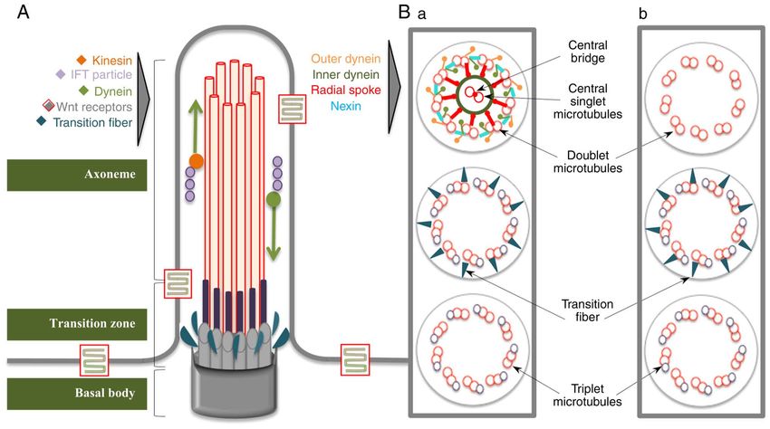
the plasma membrane (Fig. 1). At the base of this ensemble, a
Correspondence to: Dr Ina Ofelia Focșa, Department of Medical specialized centriole, referred to as the basal body (BB), docks
Genetics, University of Medicine and Pharmacy ‘Carol Davila’, the cilium to the cell (13). The axoneme of the primary cilium
3‑5 Pierre de Coubertin Boulevard, 021901 Bucharest, Romania consists of 9 outer doublet microtubules, (9 + 0 type), while
E‑mail: ina.focsa@umfcd.ro motile cilia possess an extra inner pair of microtubules, rein‑
forced by nexin bridges (9 + 2 type) and an accessory structure
Key words: cilia, ciliopathies, signaling, oligogenic, modifier,
involved in motility, formed of dynein arms and radial
epistasis, pleiotropy
spokes (14). Each doublet contains a complete microtubule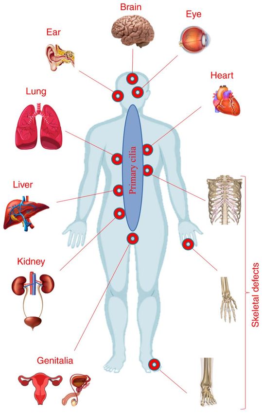
2                       FOCȘA et al: CLINICAL AND GENETIC HETEROGENEITY OF PRIMARY CILIOPATHIES

(A tubule) and one incomplete microtubule (B tubule), which         (GPCRs), Notch, transforming growth factor‑β (TGF‑β), mech‑
are composed of tubulin protofilaments and are attached to each     anistic target of rapamycin (mTOR) and Salvador‑Warts‑Hippo
other through tektins and Ca‑binding ribbon proteins (15). The      (SWH) signaling. In addition, other signaling pathways that
BB structure contains 9 radially arranged microtubule triplets      have been linked to primary cilia include extracellular matrix
(A, B and C) and no central pair. The A and B tubules expand        protein‑mediated signaling, transient receptor potential
into the proximal segment of the cilium and are connected to        channel‑mediated signaling, vasopressin signaling in renal
the ciliary membrane by Y fibers, constituting a distinctive        epithelial cells, somatostatin, serotonin and melanin‑concen‑
subcompartment known as the transition zone. Proximal to            trating hormone signaling (37,38).
the Y fibers are the transitional fibers, which help to anchor          The Wnt signaling pathway comprises a large family of
the BB to the plasma membrane (16). The BB is responsible for       secreted, cysteine‑rich proteins, acting as a network of signal
the configuration of the microtubule scaffold and coordinates       transduction pathways that are responsible for embryonic
the ciliary trafficking pathway; thus, it is involved, together     development, as well as tissue homeostasis and regeneration
with transitional fibers, in ciliogenesis (17). Surrounding the     in adults (39). At least three signaling pathways have been
transitional fibers are numerous strings of particles, acting as    described: The canonical Wnt pathway (or Wnt/β ‑catenin
a selective filter for intraflagellar transport (IFT) molecules,    pathway), and the non‑canonical planar cell polarity (PCP)
known as the ciliary necklace (18,19). In the distal region of      and Wnt/Ca 2+ pathways. The primary cilium and BB were
the cilium, the backbone contains a single microtubule fiber        found to be required for the regulation of both canonical
(A tubule), which delimits the ciliary tip, a proteic zone with     and non‑canonical Wnt signaling pathways. Canonical Wnt
cell type‑specific structure and function (20,21). In addition to   signaling acts through its end effectors as co‑transcriptional
this classical structure of the cilia, there is evidence showing    factors, together with the T‑cell factor/lymphoid enhancer
the existence of motile 9 + 0 cilia, covering the node, which       factor 1 family of proteins, and co‑activates the expression
are responsible for left‑right asymmetry, or sensory 9 + 2 cilia,   of Wnt target genes to modulate the cell cycle, leading to cell
which are observed in the inner ear cells (22‑26).                  differentiation, proliferation, adhesion and migration, and
                                                                    tissue development (40). Canonical Wnt signaling appears
Function of the primary cilium. Since their discovery in            to be directly or indirectly implicated in the formation of
the kidneys and the thyroid gland by Zimmermann in 1898,            almost all organ systems during embryogenesis; it has been
primary cilia have been considered as vestigial organelles,         shown to be involved in anterior head fold formation and
without a specific function, due to their lack of motility and      neuroectodermal patterning, in controlling further posterior
their absence in several cells during mitosis (27). In 1975,        patterning, as well as in the genesis and development of the
Webber and Lee (28) raised the hypothesis of a possible             heart, lungs, kidney, eyes, skin, blood cells and bone (41,42).
sensory role of mammalian nephron cilia, by comparing them          In addition, the essential role of the Wnt pathway in stem
to those in sensory tissues. This hypothesis was confirmed          cell renewal has been highlighted (42,43). The non‑canonical
in 2000 in a study by Pazour et al (29), which presented            PCP pathway appears to act independently on transcription
experimental evidence showing the physiological function            and plays a key role in the modification and rearrangement of
of the primary cilium. Once the implication of primary cilia        the actin cytoskeleton. Moreover, the molecular constituents
in human diseases was demonstrated (30), the awareness of           of this pathway were shown to randomize the orientation of
the significance of this organelle increased. Subsequently, a       polarized epithelial cells and to coordinate the morphology
number of studies demonstrated the complex roles of primary         and convergent extension of dorsal mesodermal and ecto‑
cilia as mechanoreceptors, chemoreceptors and osmosen‑              dermal cells during gastrulation and neural tube closure (44).
sors (31‑34).                                                       Yet, the role of cilia in canonical Wnt signal transduction is
    A highly specialized process occurring in the ciliary           controversial, although several studies have suggested the
compartment is the IFT: A bidirectional movement during             importance of primary cilia in the decrease of canonical
which a protein complex (IFT particle) is shuttled along the        Wnt signaling (45‑47). By contrast, the contribution of the
microtubule backbone from the BB to the tip of the cilium           integrity of primary cilia to the non‑canonical PCP Wnt
(through kinesin‑anterograde transport) and back (facilitated       pathway is well established. Movement of the BB to the
by dynein‑retrograde transport) (35). As the synthesis of           apical cell surface and centriolar position are essential for the
proteins essential for the development of cilia is not possible     establishment of cell polarity; thus, defects in ciliary proteins
inside the ciliary compartment and proteins are carried             implicated in ciliogenesis and BB migration lead to various
through IFT, the importance of IFT in ciliogenesis must be          PCP errors (48). The Wnt/Ca2+ pathway shares a number of
emphasized, as well as its involvement in the delivery of           components with the PCP, but has been described as a sepa‑
signals from the cilium to the cell, higlighting its significant    rate pathway, which stimulates intracellular Ca2+ release from
role in cilia‑mediated signaling pathways (36).                     the endoplasmic reticulum. Ca 2+ waves are hypothesized to
    Furthermore, >25 receptors and ion channels have been           serve as a key modulator in early pattern formation during
localized to the ciliary membrane, where a growing number of        embryo gastrulation. The Wnt/Ca 2+ pathway regulates
extracellular signals are received and transduced by the ciliary    embryogenesis in a complex manner, including promoting
ensemble, facilitating certain signaling pathways that control      ventral cell fate, negative regulation of dorsal axis develop‑
the development of organs, as well as behavioral processes.         ment, regulation of tissue separation and convergent extension
Particularly important primary cilia‑related signaling path‑        movements during gastrulation, as well as heart formation.
ways include the following: Wingless (Wnt), Hedgehog (Hh),          The Wnt/Ca2+ pathway also functions as a critical modulator
receptor tyrosine kinase (RTK), G‑protein coupled receptors         of both the canonical and PCP pathways (44). Wnt signaling
INTERNATIONAL JOURNAL OF MOlecular medicine 48: 176, 2021 3
Figure 1. Schematic representation of the cilium structure. (A) Longitudinal section, (B‑a) transversal view of the motile cilium and (B‑b) transversal view of
the primary cilium. IFT, intraflagellar transport; Wnt, wingless.
also regulates a number of other signaling pathways that have signaling (56). PDGFRα, which is bound to the membrane of
not been yet completely elucidated, but appear to be linked the primary cilium, regulates cytoskeletal reorganization to
with myogenesis, axonal guidance, neuronal migration and drive directional migration of fibroblasts in wound healing.
synaptogenesis (49,50). Defects in primary cilia lead to abnormal wound healing.
Another key signaling pathway that was demonstrated Moreover, disassembly of cilia, which allows the centriole to
to be essential for a variety of developmental processes participate in mitosis during cell cycle progression, is modu‑
is the Hh signaling pathway, described through its three lated by PGFRα signaling (57). Along with this signaling,
Hh homologues: Desert Hh, Indian Hh (Ihh) and Sonic Hh other RTK signaling pathways have recently been described,
(Shh). The Shh pathway, the most extensively investigated including EGFR, which plays an important role in mechano‑
signaling pathway, functions due to the synergy of several sensation and the migration of kidney epithelial cells or airway
molecule/proteins acting as transmembrane receptors, namely smooth muscle cells, and insulin‑like growth factor receptor,
Patched homolog 1, Smoothened (SMO) and GLI transcription which is involved in preadipocyte differentiation (37).
factors, leading to the transcription of Hh target genes (51). GPCRs comprise a large family of transmembrane recep‑
The role of Shh proteins emerges during embryonic develop‑ tors divided into six classes (A‑F), for which >30 receptors
ment and morphogenesis, controlling left‑right asymmetry, belonging to the A (rhodopsin‑like receptors), B (secretin
dorso‑ventral axes and distal limb patterning. Moreover, receptor family) and F (frizzled/SMO‑component of Shh
proliferation of hematopoietic, retinal and neural stem cells, as signaling) classes are found on the ciliary membrane. Among
well as development of epithelial tissues during organogenesis, these receptors, opsin, olfactory, serotonin (HTR6), soma‑
appear to be modulated by Shh (52). Primary cilia are essen‑ tostatin (SSTR3), vasopressin (V2R), dopamine (D1R, D2R and
tial for the transduction of Hh signaling, playing a dual role D5R) and prostaglandin (EP4) receptors are involved in a wide
through positive and negative regulation. It has been shown spectrum of cellular and physiological processes, including
that abnormal cilia may lead to either loss‑of‑function Hh photoreception, olfactory sensation, feeding behavior, pain
phenotypes in the neural tube, or gain‑of‑function Hh pheno‑ sensation, osmotic function in kidney cells, physiological
types in the limbs, indicating that the Hh pathway may play an function in cardiac myocytes, neuronal processes and energy
important role in primary cilia biogenesis (38). homeostasis (58,59). GPCRs are involved in neuronal or
The migration, proliferation, differentiation and apoptosis retinal cilia function and control the length of primary cilia
of cells are also controlled by another cilia‑related pathway, or ciliogenesis. Conversely, absence or shortening of primary
the platelet‑derived growth factor receptor‑ α (PDGFRα) cilia may interfere with normal brain development, inter‑
pathway (53,54). PDGFRα belongs to the large family of neuron connectivity, gonadotropin hormone release at the
RTK transmembrane receptors and is required for activation nerve terminals or the sensory potential of cells (48).
of the Ras‑Mek1/2‑Erk1/2 pathway, thus causing axonemal In addition to these main signaling cascades, an increasing
reestablishment, cell cycle progression and chemotaxis (55). number of pathways have been associated with primary cilia.
The development of numerous cells and tissues, including It has been concluded that the Notch signaling is involved in
neurons, oligodendrocytes, astrocytes, alveolar smooth muscle the physiology of primary cilia. Notch3 receptor, which is
cells, cardiac fibroblasts and bone cells, relies on PDGFα localized in the ciliary membrane, is activated by presenilin 2,4 FOCȘA et al: CLINICAL AND GENETIC HETEROGENEITY OF PRIMARY CILIOPATHIES
a ciliary BB enzyme, thereby regulating epidermal cell
proliferation and differentiation (60). Loss of primary cilia or
knockdown of IFT molecules may result in diminished Notch
activation, leading to decreased cell proliferation and differen‑
tiation defects. In the neuroepithelia of the developing neural
tube, activation of Notch signaling leads to increased primary
cilium length, as well as accumulation of Smo molecules
within the primary cilium. This interplay between the Notch
and Shh pathways in primary cilia may specify ventral cell
fate in the developing neural tube (38).
TGF‑β signaling has been recently associated with cilia,
whereas TGF‑β1 and TGF‑β2 receptors are located on the
ciliary tip. Primary cilia use diverse methods to regulate
TGF‑β pathways, through SMAD2/3 and ERK1/2 activation
by TGF stimulation, modulating various cellular processes,
such as the differentiation of cardiomyocytes, osteocytes and
myofibroblasts. Moreover, endothelial primary cilia, which
act as flow sensors in the blood vessels, inhibit the endothe‑
lial‑to‑mesenchymal transition, and this process is related
to attenuation of the TGF‑β signaling. There is also strong
evidence regarding the impairment of mechanosensation and
maturation in human osteoblasts due to shortening of primary
ciliary length through TGF‑β signaling (61,62).
The SWH pathway controls organ size and cell prolifera‑
tion through a core of serine/threonine‑kinases that interact
with nephrocystin 4 or Crumbs 3 receptors located in the
cilium. One of the major components of SWH signaling,
MST1/2, which is localized to the BB, has been found to be
crucial for primary cilia biogenesis, with loss of MST1/2
leading to defects in ciliogenesis (63).
It has also been demonstrated that the primary cilium regu‑
lates mTOR signaling, which plays a pivotal role in metabolism Figure 2. Organ and system involvement in primary ciliopathies.
and cell proliferation, thereby determining cell size, through
the Lkb1 tumor suppressor, AMP‑activated protein kinase and
folliculin. In epithelial primary cilia, the mTOR pathway is polydactyly and cognitive problems with learning difficulties,
upregulated by polycystin‑1 through the tuberin protein, thus was reported in 1920 by Bardet and in 1922 by Biedl (70).
being involved in cyst formation (64,65).
Brain‑derived neurotrophic factor signaling, which is Clinical overlaps in primary ciliopathies. As a result of the
involved in neuronal development, synaptic plasticity, satiety presence of primary cilia in nearly all tissues and organs,
and weight control, has recently been proposed to be linked impairment of their structure or function may result in a vast
with the BBS4 protein and primary cilia (66). group of phenotypes, ranging from single organ impairment
to complex systemic disorders (Fig. 2). In addition to their
3. Ciliopathies isolated involvement, the kidney and the eyes (retina) are also
implicated in defining the heterogeneous pattern of primary
Given the notable complexity of interconnected signaling ciliopathies, with the participation of other organs, including
pathways in cilia, the role of the primary cilium as a cellular the brain, skeletal system and liver (71). Additional system
hub is becoming increasingly obvious; its clinical importance contributions in defining the ciliopathic clinical picture are
emerges from the consequences of its structural or functional summarized in Table I.
defects, which lead to a broad category of disorders, collec‑
tively termed as ciliopathies. Renal manifestations. Renal impairment is the most common
The term ‘ciliopathy’ is most likely attributed to immotile sign in primary ciliopathies, histologically characterized
or primary cilia‑related disorders, and it has been recently by renal cysts, a thickened and irregular tubular basement
allocated to certain conditions that have long been known membrane, and interstitial fibrosis. Clinically, two frequently
as separate clinical entities (67). The first ciliophathy ever observed categories have been defined: Polycystic kidney
defined was Bardet‑Biedl syndrome (BBS) in 2003 (68), disease (PKD) and nephronophthisis (NPHP). Both entities
although this disease had been known since 1866, when are characterized by a progressive decline in renal func‑
Laurence and Moon (69) first described the phenotype, tion, eventually leading to renal failure (72,73). The onset
including retinitis pigmentosa, mental retardation, hypogo‑ of the diseases varies. Some signs could be detected prena‑
nadism and spastic paraplegia, in four cases. Decades later, tally due to the presence of oligohydramnios and enlarged
a similar phenotype, consisting of obesity, retinal dystrophy, kidneys, or shortly after birth due to the occurrence of severeINTERNATIONAL JOURNAL OF MOlecular medicine 48: 176, 2021 5
Table I. Additional clinical features of ciliopathiesa.
Type of system Clinical feature
Cardiovascular Atrial or/and ventricular septal defects, dilated cardiomyopathy, hypertrophic cardiomyopathy and
valvular defects
Respiratory Breathing abnormalities, respiratory insufficiency, pulmonary hypoplasia, atelectatic lungs and intersti‑
tial fibrosis
Endocrine Panhypopituitarism, growth hormone deficiency, hypothyroidism, diabetes mellitus and hypogonadism
Genital Genital hypoplasia, micropenis and ambiguous genitalia
Pancreatic Pancreatic dysgenesis, pancreatic fibrosis and cystic pancreas
Aural Sensorial hearing loss
Features displayed in this table were collected after an overview analysis of OMIM clinical synopsis (www.omim.org). OMIM, Online
a
Mendelian Inheritance in Man.
hypertension or respiratory insufficiency (74,75). During of the fourth ventricle, thickened and horizontalized supe‑
childhood, the symptoms of renal disease are unspecific and rior cerebellar peduncles and a deepened interpeduncular
may include polydipsia, polyuria, secondary enuresis and fossa (87,88). Neurological abnormalities may also include
urinary concentration defects. Poor growth may occur due to Dandy‑Walker malformation (DWM), ventriculomegaly,
chronic dehydration. As a result of renal insufficiency and its periventricular nodular heterotopia, hydrocephalus, encepha‑
progression to end‑stage renal failure, new complications may locele/meningocele, polymicrogyria, absence of the pituitary
develop, including anemia, metabolic acidosis, anorexia and/or gland, corpus callosum defects and morphological brainstem
hypertension (76). Renal ultrasound examination shows large, abnormalities (83,89‑91). A wide range of clinical signs may
normal‑sized or small kidneys, with increased echogenicity, be observed, such as hypotonia, ataxia, developmental delay,
loss of corticomedullary differentiation and the presence of intellectual disability (ID), impaired or absent speech, behav‑
renal cysts (76,77). Dysplastic, lobulated or horseshoe kidneys, ioral disturbances such as hyperactivity and aggressiveness,
kidney malrotation and renal agenesis are less frequently and self‑mutilation (92,93).
encountered in ciliopathic disorders (78).
Skeletal manifestations. Clinical manifestations of the skeletal
Liver manifestations. Liver cysts, liver fibrosis and ductal plate system may vary from mild phenotypes, such as polydactyly,
malformation with abnormal bile ducts may be summarized as to severe deformities, possibly leading to death. Polydactyly
liver fibrocystic diseases, and they are often found, in addition of the hands and/or feet, which is usually post‑axial, but may
to PKD, in primary ciliopathies (79). The liver disease can also be pre‑axial and, in some cases, central or mesoaxial, is
remain asymptomatic, or it can lead to complications, particu‑ present in most individuals with cilia‑related disorders (94‑96).
larly portal hypertension and esophageal varices, cholangitis In addition to polydactyly, the hands and feet may be affected
or cholestasis (80). End‑stage hepatic disease requiring trans‑ to various degrees by oligodactyly, syndactyly, campto‑
plantation has also been reported in some patients (81,82). The dactyly, brachydactyly, carpal and tarsal shortening, short
cardinal symptom is hepatomegaly, which can be associated long bones, rhizomelic micromelia, fibular aplasia or limb
with elevated serum levels of hepatic enzymes, or liver hyper‑ agenesis (97,98). Truncal skeletal defects may include a
echogenicity on abdominal ultrasound (83). constrictive thoracic cage, with shortened and horizontalized
ribs, which may be life‑threatening in some cases; abnormal
Ocular manifestations. Retinal dystrophy (with both rod and or absent clavicles, small scapulae and scoliosis may also be
cone photoreceptor involvement) is commonly encountered in observed (99). Cranioskeletal characteristics include cranio‑
primary ciliopathic disorders. The clinical manifestations of synostosis, macro‑ or microcephaly, head shape anomalies,
visual impairment range from night blindness, color blindness frontal bossing, a prominent forehead, bitemporal narrowing,
and loss of peripheral vision, to progressive visual loss and cleft palate, zygomatic arch hypoplasia, maxillary hypoplasia
complete blindness (84). Disruptions of ocular motility, such and micrognathia (100).
as oculomotor apraxia and nystagmus, are also frequently
described (85,86). Additional ocular defects include stra‑ Diagnosis of primary ciliopathies
bismus, amblyopia, astigmatism, congenital cataracts and Clinical diagnosis. Given the numerous overlapping features
coloboma (78). and marked genetic heterogeneity, considerable efforts have
been made to diagnose and classify ciliopathies, in order to
Central nervous system (CNS) manifestations. The major optimize clinical management of the patients and improve the
neuroimaging finding, which characterizes a distinct group accuracy of genetic counseling.
of diseases referred to as Joubert syndrome (JS) and related For some of these diseases with severe phenotypes leading
disorders, is the ‘molar tooth sign’ (MTS), comprising to a high mortality rate in utero or during the perinatal
cerebellar vermis hypoplasia or aplasia, with enlargement period, a prenatal diagnosis is possible in the presence of6 FOCȘA et al: CLINICAL AND GENETIC HETEROGENEITY OF PRIMARY CILIOPATHIES
Figure 3. Diagram of the clinical diagnosis algorithm of primary ciliopathies. Adapted with permission from (107). MTS, molar tooth sign; PKD, polycystic
kydney disease; RP, retinitis pigmentosa.
pathognomonic ultrasonographic signs in conjunction with with recognized ciliopathies, of which >140 genes (Table SI)
α‑fetoprotein testing of the amniotic fluid and DNA testing are implicated in primary ciliopathies. Other candidate genes
of the fetus (101). Early in the pregnancy (weeks 8‑11), ultra‑ (>240), of which the protein products have been shown to be
sonographic screening can detect certain fetal malformations, associated with cilia function or structure, may be involved in
such as an enlarged cisterna magna or encephalocele (102). either new or confirmed primary or motile ciliopathies (110).
Between 11 and 14 weeks of pregnancy, enlarged polycystic Ciliopathies are considered to be Mendelian disorders (111),
kidneys or polydactyly may be detected (103), while later although a plethora of evidence has also indicated a non‑Mende‑
in the second trimester, other brain anomalies (e.g., DWM lian pattern of inheritance, or even environmental contribution
and hydrocephalus) (104) and severe skeletal anomalies to defining the phenotype. Genetic locus heterogeneity, copy
(e.g., rhizomelic shortening of the long bones and hypoplastic number variants (112,113), oligogenicity (114,115), multiple
thoracic cage) may be identified (105). Fetal MRI can detect allelism (116‑119) and transposon‑mediated mutagenesis (120)
MTS at 27 weeks of pregnancy (106). have been described, highlighting the marked complexity of
Postnatally, Beales and Kenny proposed a clinical diag‑ the genetic mechanisms responsible for ciliopathic pheno‑
nosis algorithm starting with the presence of renal and retinal types. Moreover, the severity or variability of the phenotypes
involvement and/or polydactyly (Fig. 3). Adding limb or rib is suggested to be modulated by the pattern of ciliary gene
abnormalities to this core of clinical manifestations may easily expression and its effect on protein function (null, truncating
direct the diagnosis to ciliary skeletal dysplasias. Furthermore, or hypomorphic) (121), by epistatic interactions (122,123) and
identification of ectodermal defects in this group suggests the by genetic modifiers or stochastic effects (45,111,124‑128).
diagnosis of oral‑facial‑digital syndrome (OFDS) or cranio‑
ectodermal dysplasia (CED), while their absence indicates Classification of primary ciliopathies. Several types of ciliop‑
the diagnosis of short‑rib polydactyly syndrome (SRPS). athies have been recognized, considering the level to which an
The detection of MTS and other CNS abnormalities should organ is affected for defining their phenotype.
raise the suspicion of JS or JS‑related disorders, whereas the
presence of obesity points towards the diagnosis of BBS or Retinal ciliopathies. Retinal ciliopathies include clinical enti‑
Alström syndrome (ALMS) (107). ties manifesting as retinal degeneration, and they are caused by
defective morphogenesis or dysfunction of specialized sensory
Genetics and molecular diagnosis. Since the description cilia from the retina that form the outer segment of photorecep‑
of the first ciliopathy gene, BBS6, by two distinct research tors. Proteins, such as rhodopsin or ambient lighting‑dependent
groups in 2000, due to the advances in genomic sequencing proteins, are trafficked along these specialized primary cilia
technologies, a number of genes have been associated with by means of IFT particles. Impairment of IFT leads to the
ciliary phenotypes (108,109). Only in the last 5 years, through accumulation of rhodopsin, defects in outer segment develop‑
the intensive use of specific gene panels, whole exome and ment and cell death, which result in the phenotype of retinal
whole genome sequencing >100 new ciliary genes have been degeneration (129,130). Among non‑syndromic retinal ciliopa‑
identified. At present, there are >190 known genes associated thies, the ocular phenotype ranges from the most commonINTERNATIONAL JOURNAL OF MOlecular medicine 48: 176, 2021 7
retinitis pigmentosa [Mendelian Inheritance in Man (MIM), ARPKD is more severe, with antenatal onset and diagnosis
26800] (47), which initially manifests as night blindness, during late pregnancy or at birth, leading to increased perinatal
followed by loss of peripheral vision, due to the impairment death rate (30‑50%). Death occurs as a consequence of respira‑
of rod photoreceptor function, and can progress to complete tory insufficiency due to pulmonary hypoplasia and thoracic
blindness (84,131) to the most severe congenital retinal compression by the extremely expanded kidneys (75). NPHP,
dystrophy, Leber congenital amaurosis (LCA; MIM, 204000), which is characterized by corticomedullary cysts, atrophy
which frequently results in blindness within the first year of and interstitial fibrosis resulting in nephron disintegration, is
life. Visual loss is usually accompanied by sensory nystagmus, the main cause of ESRD in children (140). The severity and,
amaurotic pupillary response and absent electroretinogram subsequently, the progression to ESRD, depend on the clinical
signs. Photophobia, high refractive errors, keratoconus and variant, namely the infantile, juvenile or adolescent variant. The
enophthalmos are often seen in LCA. Involvement of the retina infantile variant is the most severe, with prenatal manifestations
may range from normal, to retinal degeneration, retinal aplasia consisting of oligohydramnios and bilateral enlarged cystic
or biochemical dysfunction (dysplasia) (132). Overlapping kidneys. Thus, ESRD develops in the first year of life. The first
with these two disorders, other ocular dystrophies have also symptoms of the classical juvenile form, which is characterized
been described as retinal ciliopathies: Cone dystrophy (MIM, by renal interstitial fibrosis and inflammation, with progression
304020), characterized by visual loss and color vision defects, to tubular atrophy and small cyst formation, develop during
cone‑rod dystrophy (MIM, 120970), characterized by photo‑ the first decade of life and ESRD occurs at the mean age of
phobia, abnormal color vision, night and peripheral vision loss, 13 years (74). NPHP may be limited to the kidneys or may be
and macular dystrophy (MIM, 300834), characterized by loss part of other ciliopathic conditions, such as Joubert/COACH
of color and sharp vision (133). Progressive retinal degeneration syndrome, SLSN, BBS, Meckel‑Gruber syndrome (MKS)
and sensorineural hearing loss are the first symptoms found in or skeletal disorders (141). BBS (MIM, 209900) is the most
ALMS (MIM, 203800); these are accompanied later in child‑ extensively investigated ciliopathy, and it has provided valuable
hood by obesity and diabetes mellitus. Additional features, data for the entire spectrum of human cilia‑related disorders
such as cardiomyopathy, epilepsy, respiratory disturbance due to its overlapping characteristics at the level of phenotype,
and renal or endocrine dysfunction, support the classification genotype, protein‑protein interactions and participation in
of these disorders as syndromic retinal ciliopathies (134). A signaling pathways (142). BBS is a multisystem disorder, but
rare combination of retinal and renal ciliopathies character‑ renal impairment is its most prominent cause of morbidity and
izes Senior‑Løken syndrome (SLSN; MIM, 266900), with a mortality. The major clinical characteristics, including retinal
specific clinical presentation consisting of retinal dystrophy dystrophy, obesity, post‑axial polydactyly, renal anomalies,
and NPHP. Consequently, SLSN is considered by some studies cognitive impairment and hypogonadism, are suggestive of
as a syndromic retino‑ciliopathy or, by others, as a renal the diagnosis. The presence of four of those characteristics, or
(NPHP‑related) ciliopathy (135,136). association of three primary characteristics with two secondary
features is considered as sufficient for clinical diagnosis (143).
Renal ciliopathies. Renal ciliopathies encompass a group of Secondary features include speech delay, developmental delay,
disorders, the hallmark of which is kidney disease, including diabetes and congenital heart disease. BBS is characterized by
autosomal dominant polycystic kidney disease (ADPKD; marked clinical variability, which cannot be fully attributed
MIM, 173900), autosomal recessive polycystic kidney disease to the 24 genes identified to date (144). MKS (MIM, 249000),
(ARPKD; MIM, 263200) and NPHP (MIM, 256100). In the which displays renal (cystic kidney dysplasia) as well as neuro‑
kidney, epithelial primary cilia lining the nephron tubules logical [occipital encephalocele (OE)] manifestations, may
and collecting ducts act as sensory antennae sensitive to urine be considered as either a renal or a CNS‑related ciliopathy.
composition, osmolarity and flow. Defects in several signaling Hepatic fibrosis completes the specific clinical triad of this
pathways, such as G‑protein signaling, mTOR or Wnt, induced condition, although polydactyly is often considered as the 4th
by decreased of flow‑mediated intracellular calcium concen‑ pathognomonic feature (145). MKS has a heterogeneous, severe
tration, may lead to cyst formation. Moreover, disruption of the phenotype, which is not compatible with life, with death occur‑
balance between canonical and non‑canonical Wnt signaling ring in utero or shortly after birth. Renal dysfunction may often
may affect the polarity of epithelial tubular cells, also resulting lead to oligohydramnios or anhydramnios. Apart from OE,
in cyst formation (137). which is the most frequent finding, additional CNS malforma‑
ADPKD and ARPKD are different, not only due to the tions found in MKS include olfactory bulb dysgenesis, optic
inheritance pattern, but also based on the microscopic and nerve hypoplasia, agenesis of the corpus callosum, holopros‑
ultrasonographic appearance of the cysts, associated organ encephaly, cerebellar hypoplasia or total anencephaly. Cleft
anomalies, age at onset, severity and prognosis (138). While lip and palate, shortening of the long bones, congenital heart
ADPKD is characterized by large cysts originating from the defects and pulmonary hypoplasia may further complicate the
distal nephrons and collecting ducts, which grow in volume clinical picture (45,101).
and number with age, and by the presence of cysts in the liver,
pancreas or other epithelial organs, intracranial aneurysms and CNS‑related ciliopathies. CNS‑related ciliopathies comprise a
mitral valve prolapse (139), the cysts in ARPKD are small, orig‑ group of conditions, the hallmark of which is the MTS, which
inate from the distal tubules and collecting ducts and display is required for diagnosis. Impairment of the Wnt pathway,
a salt‑and‑pepper pattern, and the liver is always affected by which is a major signaling pathway involved in cerebellar
fibrosis (138). In contrast to ADPKD, which starts in late adult‑ development, may be responsible for defective cerebellar
hood and slowly progresses to end‑stage renal disease (ESRD), vermis hypoplasia, one of the components of MTS. In addition8 FOCȘA et al: CLINICAL AND GENETIC HETEROGENEITY OF PRIMARY CILIOPATHIES
to this pathway, other neuronal primary cilium‑specific path‑ craniosynostosis, leading to dolichocephaly, frontal bossing
ways are required for normal brain development, regulating and dental defects, in conjunction with skeletal abnormalities
neuronal fate, proliferation, migration and differentiation. (short stature, rhizomelic limbs, brachydactyly and narrow
Dysregulation of these pathways, including Shh, PDGFRα thorax) and ectodermal anomalies (thin/sparse hair, hypo‑
and GRCR, may manifest with malformations during cortical plastic nails and skin laxity) (162,163). Kidney involvement
development or midline defects, which are often found in JS (NPHP progressing to renal failure) and liver involvement
or CNS‑related ciliopathies (71). Depending on the additional (ranging from asymptomatic hepatomegaly to acute chol‑
clinical characteristics, JS (MIM, 213300) has been classified angitis, liver cirrhosis and severe cholestasis) are common
into several groups as follows: i) Pure or classic JS, charac‑ findings in CED (164).
terized by hypotonia, developmental delay, abnormal eye The second group with more severe phenotypes is
movements, breathing abnormalities, ataxia and ID; ii) JS comprised of Jeune asphyxiating thoracic dystrophy (JATD;
with ocular defects, including retinal dystrophy or LCA; iii) JS MIM, 208500) and conorenal syndrome or Mainzer‑Saldino
with renal defects (NPHP); iv) JS with oculorenal defects, also syndrome (MZSDS; MIM, 266920). The specific presentation
named cerebello‑oculorenal syndrome, comprising SLSN of JATD includes a constrictive thoracic cage and secondary
(retinal dystrophy, LCA and NPHP) associated with MTS, respiratory distress due to restrictive pulmonary hypoplasia.
and Dekaban‑Arima syndrome (cerebrooculohepatorenal Respiratory distress is the main cause of mortality in ~60%
syndrome) characterized by chorioretinal coloboma or retinal of the patients (100). Additional skeletal findings may include
dystrophy, PKD, MTS and hepatic fibrosis in some cases; v) JS a short stature, short limbs with irregular metaphyses,
with congenital hepatic fibrosis; vi) JS with congenital hepatic cone‑shaped epiphyses in the hands, foot polydactyly, a
fibrosis and associated chorioretinal coloboma, also known as shortened ilium and a trident‑shaped acetabulum. Retinal
COACH syndrome; and vii) JS with orofaciodigital defects, degeneration, NPHP‑like or cystic renal disease, pancreatic
including a lobulated or bifid tongue, hamartomas, cleft lip and liver involvement or brain malformations are occasionally
and/or palate and polydactyly, also known as orofaciodigital found in patients with JATD (100,165). MZSDS is character‑
syndrome type VI (83,87). To date, ~40 causative genes covering ized by the triad of retinal dystrophy, renal disease (typically
>90% of clinical subjects have been identified (146‑152). NPHP) and phalangeal cone‑shaped epiphyses (166). The
thorax is less narrow compared with that in patients with
Ciliopathies with skeletal involvement. This group of disor‑ JATD. Short stature, hepatic fibrosis and cerebellar ataxia are
ders is characterized by variable severity, ranging from mild variable traits that may be observed in MZSDS (166,167).
to severe or even lethal phenotypes. Two subgroups have been The last subtype is the perinatally lethal SRPS, the
distinguished: Those with major skeletal system involvement, core features of which include a constrictive thoracic cage,
including craniofacial, thoracic cage and long bone involve‑ significantly shortened long bones, polydactyly, brahydactyly
ment, known as short‑rib thoracic dysplasias (SRTDs), with or and pelvic abnormalities (100). Different types have been
without polydactyly or ciliary condrodysplasias, and OFDS, characterized, based mainly on radiological findings: SRPS
with milder involvement of the skeletal system (153). types I (Saldino‑Noonan syndrome) and III (Verma‑Naumoff
Development of the cartilage and bones is a complex syndrome) (MIM, 613091); SRP type II or Majewski syndrome
process that is modulated mainly by the IFT and Hh pathways. (MIM, 263520); SRPS type IV or Beemer‑Langer syndrome
Disruption of Ihh signaling in chondral primary cilia affects (MIM, 269860); and SRPS type V (MIM, 614091) (98). In
chondrocyte maturation during the ossification process. addition to skeletal abnormalities, involvement of the brain,
Consequently, various skeletal abnormalities, including poly‑ heart, kidneys, liver, pancreas and genitalia have often been
dactyly, shortening of the ribs or long bones and craniofacial recorded in SRPS. In some cases, facial dysmorphism may
abnormalities, may occur (154,155). Dysregulation of IFT, also be observed (100,165).
which is involved in the trafficking of the transmembrane Apart from the typical manifestations, some ‘unusual’
SMO receptor, a signal transducer in Hh signaling, may lead features may be observed in each group, further expanding
to premature differentiation and decreased proliferation of and complicating the phenotype; these include atlantoaxial
chondrocytes, manifesting as specific SRTDs and defects of instability and spinal cord compression (168,169), short irregu‑
long bone growth plates (156). larly bent ribs, hypoplastic and bent mesomelic bones, short
There are >19 types of SRTDs, classified based on campomelic long bones, undermineralized bones (170,171),
phenotype severity, radiological findings and confirmation OE or MTS (172).
of genetic defects (100,157). Chondroectodermal dysplasia OFDS (MIM, 311200) describes a heterogeneous group
or Ellis‑van‑Creveld syndrome (EVC; MIM, 2255000), of diseases caused by defects in ~18 genes (173‑175). Clinical
Weyers acrodental dysostosis (WAD; MIM, 193530) and manifestations include anomalies of the face (micrognathia,
Sensenbrenner syndrome or CED (MIM, 218330) are the hypertelorism, telecanthus, cleft lips and low‑set ears), the oral
milder disorders in this group. EVC is characterized by dispro‑ cavity (gingival frenulae, lingual hamartomas, cleft/lobulated
portionate short limb dwarfism, short ribs, polydactyly, cardiac tongue and cleft palate) and the digits (polydactyly, brachy‑
malformations and ectodermal defects affecting the hair, teeth dactyly, oligodactyly and bifid digits), associated with an
and nails (158‑160). WAD is an allelic disorder to EVC, but extensive spectrum of additional features affecting the CNS,
displays a milder phenotype, consisting of moderate short the kidneys, the heart or the eyes, outlining the 13 forms
stature, postaxial polydactyly, and nail and dental anomalies, described to date (173,176). In addition to renal involvement,
and is inherited in an autosomal dominant manner (161). CED which is commonly found in OFDS, a series of features
is characterized by craniofacial abnormalities, such as sagittal overlapping with other ciliopathies (JS, SRPS and EVC)INTERNATIONAL JOURNAL OF MOlecular medicine 48: 176, 2021 9
Table II. Newly defined ciliopathies.
MIM ID Disease name Gene name Protein localization (Refs.)
616287 Lethal congenital contracturesyndrome; ADCY6 Axoneme (184)
hypomyelination neuropathy‑arthrogryposis
syndrome
243605 Stromme syndrome; lethal fetal brain CENPF Basal body (185,186)
malformation‑duodenal atresia‑bilateral
renal hypoplasia syndrome; microcephaly
135150 Birt‑Hogg‑Dubé syndrome FLCN Basal body; axoneme (187)
201000 Carpenter syndrome RAB23 Axoneme (200,201)
616897 Complex lethal osteochondrodysplasia TAPT1 Basal body (189)
NO MIM ID A novel syndrome with multiple congenital USP9X Axoneme (190)
malformations and developmental delay
601707 Curry‑Jones syndrome SMO Axoneme (191)
607131 Al‑Gazali‑Bakalinova syndrome KIF7 Axoneme (192)
236680 614120 Hydrolethalus HYLS1; KIF7; Basal body; axoneme; basal body (193‑195)
KIAA0586
175700 Greig cephalopolysyndactyly syndrome GLI3 Axoneme (tip) (196)
612651 Lethal endocrine‑cerebro‑osteodysplasia ICK IFT (197)
syndrome
NO MIM ID Pituitary stalk interruption syndrome GPR161 Axoneme (198)
300707 Syndactyly‑telecanthus‑anogenital and renal FAM58A Probably cytosolic (202)
malformations syndrome
MIM, Mendelian Inheritance in Man; IFT, intraflagellar transport.
have been reported, such as MTS identified in OFD types 4, MIM, 256520; type 2: MIM, 616038) due to the discovery of
6 and 14, and tibial abnormalities observed in OFD types 4, its causal genes, PHGDG (MIM, 606879) and PSAT1 (MIM,
8 and 12 (177,178). 610936) (205,206).
Other unclassified subtypes have also been described, The delineation of the ciliary proteome and its interac‑
which are characterized, in addition to the typical features, by tion with extraciliary molecules opens new perspectives in
fused kidneys (179), tetralogy of Fallot (179,180), coarctation reclassifying cilia‑related disorders. Thus, the disorders char‑
of the aorta (181), corpus callosum agenesis (179), cerebellar acterized by ciliopathy‑overlapping phenotypes, the causal
vermis hypoplasia, DW malformation, ID, 12th rib hypo‑ genes of which are not expressed in the ciliary assembly,
plasia (174) and short mesoaxial phalanges (182). but interfere with ciliogenesis or the cilia signaling network,
have been termed ciliopathy‑like disorders. A representative
4. Conclusions example is Cohen syndrome (MIM, 216550), which is defined
by obesity, developmental delay, retinal degeneration and
Increasing use of whole exome sequencing has enabled the intermittent neutropenia (207), and is caused by mutations in
discovery of new causal genes in ciliopathies. Combined VPS13B (MIM, 607817) (208). The expression product, which
efforts have been made in the fields of proteomics, cell biology is localized to the Golgi apparatus, may impair processing of
and model organisms to link the genes with their phenotypic ciliary components (207). Townes‑Brocks syndrome (MIM,
effect. Taken together, all these studies have improved our 107480), which is characterized by hearing impairment,
knowledge on recognized ciliopathies, confirmed the proposed PKD, ESRD, imperforate anus and digit malformations (209),
cilia‑related disorders or identified new ciliary diseases. is caused by mutations in the SALL1 (MIM, 602218) gene,
Since Baker and Beales (183) proposed 72 conditions as which encodes a zinc‑finger transcription factor; its interaction
candidates for ciliopathic disorders in 2009, several have with two ciliogenesis suppressors, CEP97 (MIM, 615864) and
been included in the group of known ciliopathies (Table II), CCP110 (MIM, 609544), leads to cilia formation and func‑
increasing their number to 35 (184‑202). tion impairment (210). For these ciliopathy‑like conditions,
By contrast, other conditions were excluded from the list of Reiter and Leroux (110) proposed the term second‑order
possible or likely ciliopathies following the determination of ciliopathies, whereas first‑order ciliopathies are defined as
their genetic background, including Kabuki syndrome (type 1 disorders in which disease‑associated proteins are expressed
MIM, 147920; type 2 MIM, 300867) following identifica‑ in the primary ciliary compartment. Although applying
tion of its causal genes, MLL2 (MIM, 602113) and KDM6A this classification is seemingly straightforward, unexpected
(MIM, 300128) (203,204), or Neu‑Laxova syndrome (type 1: evidence has uncovered the possibility of a condition being10 FOCȘA et al: CLINICAL AND GENETIC HETEROGENEITY OF PRIMARY CILIOPATHIES
either first‑ or second‑order, thus complicating the picture. 4. Budny B, Chen W, Omran H, Fliegauf M, Tzschach A,
Wisniewska M, Jensen LR, Raynaud M, Shoichet SA,
One such example is MKS, a well‑known ciliopathy caused Badura M, et al: A novel X‑linked recessive mental retardation
by mutations in genes encoding proteins that are localized in syndrome comprising macrocephaly and ciliary dysfunction is
the transition zone (101). A recent study identified a new causal allelic to oral‑facial‑digital type I syndrome. Hum Genet 120:
171‑178, 2006.
gene for MKS, TXNDC15 (MIM, 617778), a non‑ciliary gene, 5. Moalem S, Keating S, Shannon P, Thompson M, Millar K,
the bi‑allelic mutations of which lead to abnormal cilia biogen‑ Nykamp K, Forster A, Noor A and Chitayat D: Broadening
esis (211). the ciliopathy spectrum: Motile cilia dyskinesia, and nephro‑
nophthisis associated with a previously unreported homozygous
Numerous conditions remain to be elucidated, either due mutation in the INVS/NPHP2 gene. Am J Med Genet A 161A:
to the fact that the genetic cause has not been uncovered or 1792‑1796, 2013.
since the pathophysiological mechanism underlying the 6. Shapiro AJ, Zariwala MA, Ferkol T, Davis SD, Sagel SD, Dell SD,
Rosenfeld M, Olivier KN, Milla C, Daniel SJ, et al: Diagnosis,
phenotype remains elusive. Predicting organ involvement and, monitoring, and treatment of primary ciliary dyskinesia: PCD
consequently, phenotype severity based on genetic defects also foundation consensus recommendations based on state of the art
represents a major challenge. review. Pediatr Pulmonol 51: 115‑132, 2016.
7. Tobin JL and Beales PL: The nonmotile ciliopathies. Genet
Future research will hopefully provide new insights that Med 11: 386‑402, 2009.
may help reorganize and further elucidate the striking field of 8. Leeuwenhoek AV: Observations, communicated to the publisher
ciliopathies. by Mr. Antony van Leewenhoeck, in a dutch letter of the 9th
Octob. 1676. Here English'd: Concerning little animals by him
observed in rain‑well‑sea‑ and snow water; as also in water
Acknowledgements wherein pepper had lain infused. Philosophical Transactions 12:
821‑831, 1677.
9. Lee L: Mechanisms of mammalian ciliary motility: Insights
The authors are grateful to Dr Costel Darie, Associate from primary ciliary dyskinesia genetics. Gene 473: 57‑66, 2011.
Professor of Chemistry and Biomolecular Science, Clarkson 10. Guo J, Higginbotham H, Li J, Nichols J, Hirt J, Ghukasyan V and
University (Potsdam, NY, USA) for reviewing this work. Anton ES: Developmental disruptions underlying brain abnor‑
malities in ciliopathies. Nat Commun 6: 7857, 2015.
11. Chang CF, Schock EN, Attia AC, Stottmann RW and
Funding Brugmann SA: The ciliary baton: Orchestrating neural crest cell
development. Curr Top Dev Biol 111: 97‑134, 2015.
12. Mirvis M, Stearns T and James Nelson W: Cilium structure,
No funding was received. assembly, and disassembly regulated by the cytoskeleton.
Biochem J 475: 2329‑2353, 2018.
Availability of data and materials 13. Marshall WF and Nonaka S: Cilia: Tuning in to the cell's antenna.
Curr Biol 16: R604‑R614, 2006.
14. Roberts AJ, Kon T, Knight PJ, Sutoh K and Burgess SA:
Not applicable. Functions and mechanics of dynein motor proteins. Nat Rev Mol
Cell Biol 14: 713‑726, 2013.
15. Linck R, Fu X, Lin J, Ouch C, Schefter A, Steffen W, Warren P and
Authors' contributions Nicastro D: Insights into the structure and function of ciliary and
flagellar doublet microtubules: Tektins, Ca 2+‑binding proteins,
IOF collect the data, wrote the manuscript, prepared the figures and stable protofilaments. J Biol Chem 289: 17427‑17444, 2014.
16. Reiter JF, Blacque OE and Leroux MR: The base of the cilium:
and the tables. MBu wrote the manuscript. MBa revised and Roles for transition fibres and the transition zone in ciliary
approved the manuscript. All authors read and approved the formation, maintenance and compartmentalization. EMBO
final manuscript. Data authentication is not applicable. Rep 13: 608‑618, 2012.
17. Vertii A, Hung HF, Hehnly H and Doxsey S: Human basal body
basics. Cilia 5: 13, 2016.
Ethics approval and consent to participate 18. Satir P and Christensen ST: Overview of structure and function
of mammalian cilia. Annu Rev Physiol 69: 377‑400, 2007.
19. Hu Q, Milenkovic L, Jin H, Scott MP, Nachury MV, Spiliotis ET
Not applicable. and Nelson WJ: A septin diffusion barrier at the base of the
primary cilium maintains ciliary membrane protein distribution.
Patient consent for publication Science 329: 436‑439, 2010.
20. Fisch C and Dupuis‑Williams P: Ultrastructure of cilia and
flagella‑back to the future!. Biol Cell 103: 249‑270, 2011.
Not applicable. 21. Czarnecki PG and Shah JV: The ciliary transition zone: From
morphology and molecules to medicine. Trends Cell Biol 22:
201‑210, 2012.
Competing interests 22. Nonaka S, Tanaka Y, Okada Y, Takeda S, Harada A, Kanai Y,
Kido M and Hirokawa N: Randomization of left‑right asym‑
The authors declare that they have no competing interests. metry due to loss of nodal cilia generating leftward flow of
extraembryonic fluid in mice lacking KIF3B motor protein.
Cell 95: 829‑837, 1998.
References 23. Afzelius BA: Cilia‑related diseases. J Pathol 204: 470‑477, 2004.
24. Dabdoub A and Kelley MW: Planar cell polarity and a poten‑
tial role for a Wnt morphogen gradient in stereociliary bundle
1. Badano JL, Mitsuma N, Beales PL and Katsanis N: The ciliopa‑ orientation in the mammalian inner ear. J Neurobiol 64: 446‑457,
thies: An emerging class of human genetic disorders. Annu Rev 2005.
Genomics Hum Genet 7: 125‑148, 2006. 25. Hirokawa N, Tanaka Y, Okada Y and Takeda S: Nodal flow and
2. Waters AM and Beales PL: Ciliopathies: An expanding disease the generation of left‑right asymmetry. Cell 125: 33‑45, 2006.
spectrum. Pediatr Nephrol 26: 1039‑1056, 2011. 26. Hamada H: Roles of motile and immotile cilia in left‑right
3. Moore A, Escudier E, Roger G, Tamalet A, Pelosse B, Marlin S, symmetry breaking. In: Etiology and Morphogenesis of
Clément A, Geremek M, Delaisi B, Bridoux AM, et al: RPGR Congenital Heart Disease: From Gene Function and Cellular
is mutated in patients with a complex X linked phenotype Interaction to Morphology. Nakanishi T, Markwald RR,
combining primary ciliary dyskinesia and retinitis pigmentosa. Baldwin HS, Keller BB, Srivastava D and Yamagishi H (eds).
J Med Genet 43: 326‑333, 2006. Springer, Tokyo, pp57‑65, 2016.INTERNATIONAL JOURNAL OF MOlecular medicine 48: 176, 2021 11
27. Bloodgood RA: From central to rudimentary to primary: The 52. Varjosalo M and Taipale J: Hedgehog: Functions and mecha‑
history of an underappreciated organelle whose time has come. nisms. Genes Dev 22: 2454‑2472, 2008.
The primary cilium. Methods Cell Biol 94: 3-52, 2009. 53. Schneider L, Clement CA, Teilmann SC, Pazour GJ,
28. Webber WA and Lee J: Fine structure of mammalian renal cilia. Hoffmann EK, Satir P and Christensen ST: PDGFRalphaalpha
Anat Rec 182: 339‑343, 1975. signaling is regulated through the primary cilium in fibroblasts.
29. Pazour GJ, Dickert BL, Vucica Y, Seeley ES, Rosenbaum JL, Curr Biol 15: 1861‑1866, 2005.
Witman GB and Cole DG: Chlamydomonas IFT88 and its mouse 54. Clement DL, Mally S, Stock C, Lethan M, Satir P, Schwab A,
homologue, polycystic kidney disease gene tg737, are required Pedersen SF and Christensen ST: PDGFRα signaling in the
for assembly of cilia and flagella. J Cell Biol 151: 709‑718, 2000. primary cilium regulates NHE1‑dependent fibroblast migration
30. Pazour GJ, San Agustin JT, Follit JA, Rosenbaum JL and via coordinated differential activity of MEK1/2‑ERK1/2‑p90RSK
Witman GB: Polycystin‑2 localizes to kidney cilia and the ciliary and AKT signaling pathways. J Cell Sci 126: 953‑965, 2013.
level is elevated in orpk mice with polycystic kidney disease. 55. Pala R, Alomari N and Nauli SM: Primary cilium‑dependent
Curr Biol 12: R378‑R380, 2002. signaling mechanisms. Int J Mol Sci 18: 2272, 2017.
31. Gradilone SA, Masyuk AI, Splinter PL, Banales JM, Huang BQ, 56. Heldin CH: Targeting the PDGF signaling pathway in the treat‑
Tietz PS, Masyuk TV and Larusso NF: Cholangiocyte cilia express ment of non‑malignant diseases. J Neuroimmune Pharmacol 9:
TRPV4 and detect changes in luminal tonicity inducing bicarbonate 69‑79, 2014.
secretion. Proc Natl Acad Sci USA 104: 19138‑19143, 2007. 57. Nishimura Y, Kasahara K, Shiromizu T, Watanabe M and
32. Mansini AP, Peixoto E, Jin S, Richard S and Gradilone SA: Inagaki M: Primary cilia as signaling hubs in health and disease.
The chemosensory function of primary cilia regulates cholan‑ Adv Sci (Weinh) 6: 1801138, 2018.
giocyte migration, invasion, and tumor growth. Hepatology 69: 58. Schou KB, Pedersen LB and Christensen ST: Ins and outs of
1582‑1598, 2019. GPCR signaling in primary cilia. EMBO Rep 16: 1099‑1113, 2015.
33. Masyuk AI, Gradilone SA, Banales JM, Huang BQ, Masyuk TV, 59. Hilgendorf KI, Johnson CT and Jackson PK: The primary cilium
Lee SO, Splinter PL, Stroope AJ and Larusso NF: Cholangiocyte as a cellular receiver: Organizing ciliary GPCR signaling. Curr
primary cilia are chemosensory organelles that detect biliary nucle‑ Opin Cell Biol 39: 84‑92, 2016.
otides via P2Y12 purinergic receptors. Am J Physiol Gastrointest 60. Ezratty EJ, Stokes N, Chai S, Shah AS, Williams SE and Fuchs E:
Liver Physiol 295: G725‑G734, 2008. A role for the primary cilium in Notch signaling and epidermal
34. Praetorius HA and Spring KR: Bending the MDCK cell primary differentiation during skin development. Cell 145: 1129‑1141,
cilium increases intracellular calcium. J Membr Biol 184: 71‑79, 2011.
2001. 61. Clement CA, Ajbro KD, Koefoed K, Vestergaard ML, Veland IR,
35. Hou Y and Witman GB: Dynein and intraflagellar transport. Exp Henriques de Jesus MP, Pedersen LB, Benmerah A, Andersen CY,
Cell Res 334: 26‑34, 2015. Larsen LA and Christensen ST: TGF‑β signaling is associated
36. Pedersen LB and Rosenbaum JL: Intraflagellar transport (IFT) with endocytosis at the pocket region of the primary cilium. Cell
role in ciliary assembly, resorption and signalling. Curr Top Dev Rep 3: 1806‑1814, 2013.
Biol 85: 23‑61, 2008. 62. Vestergaard ML, Awan A, Warzecha CB, Christensen ST and
37. Christensen ST, Clement CA, Satir P and Pedersen LB: Primary Andersen CY: Immunofluorescence microscopy and mRNA
cilia and coordination of receptor tyrosine kinase (RTK) signal‑ analysis of human embryonic stem cells (hESCs) including
ling. J Pathol 226: 172‑184, 2012. primary cilia associated signaling pathways. Methods Mol
38. Wheway G, Nazlamova L and Hancock JT: Signaling through Biol 1307: 123‑140, 2016.
the primary cilium. Front Cell Dev Biol 6: 8, 2018. 63. Basten SG and Giles RH: Functional aspects of primary cilia in
39. Veland IR, Awan A, Pedersen LB, Yoder BK and Christensen ST: signaling, cell cycle and tumorigenesis. Cilia 2: 6, 2013.
Primary cilia and signaling pathways in mammalian develop‑ 64. Zhong M, Zhao X, Li J, Yuan W, Yan G, Tong M, Guo S, Zhu Y,
ment, health and disease. Nephron Physiol 111: p39‑p53, 2009. Jiang Y, Liu Y and Jiang Y: Tumor suppressor folliculin regulates
40. Cardenas‑Rodriguez M and Badano JL: Ciliary biology: mTORC1 through primary Cilia. J Biol Chem 291: 11689‑11697,
Understanding the cellular and genetic basis of human ciliopa‑ 2016.
thies. Am J Med Genet C Semin Med Genet 151C: 263‑280, 2009. 65. Boehlke C, Kotsis F, Patel V, Braeg S, Voelker H, Bredt S,
41. Logan CY and Nusse R: The Wnt signaling pathway in develop‑ Beyer T, Janusch H, Hamann C, Gödel M, et al: Primary cilia
ment and disease. Annu Rev Cell Dev Biol 20: 781‑810, 2004.
42. Grigoryan T, Wend P, Klaus A and Birchmeier W: Deciphering regulate mTORC1 activity and cell size through Lkb1. Nat Cell
the function of canonical Wnt signals in development and disease: Biol 12: 1115‑1122, 2010.
Conditional loss‑ and gain‑of‑function mutations of beta‑catenin 66. Leitch CC and Zaghloul NA: BBS4 is necessary for ciliary local‑
in mice. Genes Dev 22: 2308‑2341, 2008. ization of TrkB receptor and activation by BDNF. PLoS One 9:
43. Reya T, Duncan AW, Ailles L, Domen J, Scherer DC, Willert K, e98687, 2014.
Hintz L, Nusse R and Weissman IL: A role for Wnt signalling in 67. Lee JE and Gleeson JG: A systems‑biology approach to under‑
self‑renewal of haematopoietic stem cells. Nature 423: 409‑414, 2003. standing the ciliopathy disorders. Genome Med 3: 59, 2011.
44. Komiya Y and Habas R: Wnt signal transduction pathways. 68. Ansley SJ, Badano JL, Blacque OE, Hill J, Hoskins BE,
Organogenesis 4: 68‑75, 2008. Leitch CC, Kim JC, Ross AJ, Eichers ER, Teslovich TM, et al:
45. Abdelhamed ZA, Wheway G, Szymanska K, Natarajan S, Basal body dysfunction is a likely cause of pleiotropic
Toomes C, Inglehearn C and Johnson CA: Variable expres‑ Bardet‑Biedl syndrome. Nature 425: 628‑633, 2003.
sivity of ciliopathy neurological phenotypes that encompass 69. Laurence JZ and Moon RC: Four cases of ‘retinitis pigmentosa’
Meckel‑Gruber syndrome and Joubert syndrome is caused occurring in the same family, and accompanied by general
by complex de‑regulated ciliogenesis, Shh and Wnt signalling imperfections of development. 1866. Obes Res 3: 400‑403, 1995.
defects. Hum Mol Genet 22: 1358‑1372, 2013. 70. Moore SJ, Green JS, Fan Y, Bhogal AK, Dicks E, Fernandez BA,
46. Wheway G, Abdelhamed Z, Natarajan S, Toomes C, Inglehearn C Stefanelli M, Murphy C, Cramer BC, Dean JC, et al: Clinical
and Johnson CA: Aberrant Wnt signalling and cellular and genetic epidemiology of Bardet-Biedl syndrome in
over‑proliferation in a novel mouse model of Meckel‑Gruber Newfoundland: a 22-year prospective, population-based, cohort
syndrome. Dev Biol 377: 55‑66, 2013. study. Am J Med Genet A 132A: 352-360, 2005.
47. Lin F, Hiesberger T, Cordes K, Sinclair AM, Goldstein LS, 71. Mitchison HM and Valente EM: Motile and non‑motile cilia in
Somlo S and Igarashi P: Kidney‑specific inactivation of the human pathology: From function to phenotypes. J Pathol 241:
KIF3A subunit of kinesin‑II inhibits renal ciliogenesis and 294‑309, 2017.
produces polycystic kidney disease. Proc Natl Acad Sci USA 100: 72. Hildebrandt F and Zhou W: Nephronophthisis‑associated
5286‑5291, 2003. ciliopathies. J Am Soc Nephrol 18: 1855‑1871, 2007.
48. Anvarian Z, Mykytyn K, Mukhopadhyay S, Pedersen LB and 73. Bergmann C: Early and severe polycystic kidney disease and
Christensen ST: Cellular signalling by primary cilia in development, related ciliopathies: An emerging field of interest. Nephron 141:
organ function and disease. Nat Rev Nephrol 15: 199‑219, 2019. 50‑60, 2019.
49. von Maltzahn J, Chang NC, Bentzinger CF and Rudnicki MA: 74. Srivastava S, Molinari E, Raman S and Sayer JA: Many
Wnt signaling in myogenesis. Trends Cell Biol 22: 602‑609, 2012. Genes‑One disease? Genetics of nephronophthisis (NPHP) and
50. Salinas PC: Wnt signaling in the vertebrate central nervous NPHP‑Associated disorders. Front Pediatr 5: 287, 2018.
system: From axon guidance to synaptic function. Cold Spring 75. Bergmann C: Genetics of autosomal recessive polycystic kidney
Harb Perspect Biol 4: a008003, 2012. disease and its differential diagnoses. Front Pediatr 5: 221, 2018.
51. Eggenschwiler JT and Anderson KV: Cilia and developmental 76. Salomon R, Saunier S and Niaudet P: Nephronophthisis. Pediatr
signaling. Annu Rev Cell Dev Biol 23: 345‑373, 2007. Nephrol 24: 2333‑2344, 2009.You can also read