Post-Genomics Nanotechnology Is Gaining Momentum: Nanoproteomics and Applications in Life Sciences
←
→
Page content transcription
If your browser does not render page correctly, please read the page content below
OMICS A Journal of Integrative Biology
Volume 18, Number 2, 2014
ª Mary Ann Liebert, Inc.
DOI: 10.1089/omi.2013.0074

      Post-Genomics Nanotechnology Is Gaining Momentum:
        Nanoproteomics and Applications in Life Sciences

            Firas H. Kobeissy,1,5,* Basri Gulbakan,2,3,* Ali Alawieh,4,* Pierre Karam,5 Zhiqun Zhang,1
        Joy D. Guingab-Cagmat,6 Stefania Mondello,7 Weihong Tan,2,8,9 John Anagli,6 and Kevin Wang1

Abstract

The post-genomics era has brought about new Omics biotechnologies, such as proteomics and metabolomics, as
well as their novel applications to personal genomics and the quantified self. These advances are now also
catalyzing other and newer post-genomics innovations, leading to convergences between Omics and nano-
technology. In this work, we systematically contextualize and exemplify an emerging strand of post-genomics
life sciences, namely, nanoproteomics and its applications in health and integrative biological systems. Nano-
technology has been utilized as a complementary component to revolutionize proteomics through different
kinds of nanotechnology applications, including nanoporous structures, functionalized nanoparticles, quantum
dots, and polymeric nanostructures. Those applications, though still in their infancy, have led to several highly
sensitive diagnostics and new methods of drug delivery and targeted therapy for clinical use. The present article
differs from previous analyses of nanoproteomics in that it offers an in-depth and comparative evaluation of the
attendant biotechnology portfolio and their applications as seen through the lens of post-genomics life sciences
and biomedicine. These include: (1) immunosensors for inflammatory, pathogenic, and autoimmune markers for
infectious and autoimmune diseases, (2) amplified immunoassays for detection of cancer biomarkers, and (3)
methods for targeted therapy and automatically adjusted drug delivery such as in experimental stroke and brain
injury studies. As nanoproteomics becomes available both to the clinician at the bedside and the citizens who are
increasingly interested in access to novel post-genomics diagnostics through initiatives such as the quantified
self, we anticipate further breakthroughs in personalized and targeted medicine.

Introduction                                                        the dynamic nature of proteins on the other (Aebersold and
                                                                    Mann, 2003; Altelaar et al., 2012).

T    he post-genomics era has realized that the sequenced
     genome is not enough to discern the global biological
processes fully at a systems level (Collins et al., 2003; Gandhi
                                                                       Chief among the aims of proteomics is the analysis of cel-
                                                                    lular proteins in terms of abundance and dynamics in re-
                                                                    sponse to physiological and pathological changes, as well as
and Wood, 2012). New ‘‘omics’’ fields characterized by data-        environmental influences. Proteins are central cellular com-
intensive research and biotechnologies enabling omics in-           ponents in biological networks, with diverse functions in-
vestigation have come into existence to narrow the existing         cluding cytoskeletal building blocks, enzymes catalyzing
gaps between discovery science and the attendant clinical           biochemical reactions, antibodies contributing to immunity,
applications. One of the significant contributors in the post-      or transcription factors affecting gene expression. Proteomics
genomics era is the field of proteomics. The rising interest in     by definition is the systematic identification and character-
protein science is believed to be secondary to the far biological   ization of protein sequence, abundance, post-translational
distance between genes and phenotypes on the one hand and           modifications, interactions, activity, subcellular localization,

  1
   Center for Neuroproteomics and Biomarkers Research, Department of Psychiatry, McKnight Brain Institute, Departments of 2Chemistry
and Center for Research at Bio/Nano Interface, and 8Physiology and Functional Genomics, University of Florida, Gainesville, Florida.
  3
   Department of Chemistry and Applied Biosciences ETH Zurich, Switzerland.
  4
   Department of Neurosciences, Medical University of South Carolina, Charleston, South Carolina.
  5
   American University of Beirut, Department of Chemistry, Beirut, Lebanon.
  6
   Center of Innovative Research, Banyan Biomarkers, Inc., Alachua, Florida.
  7
   University of Messina, Department of Neurosciences, Messina, Italy.
  9
   Moffitt Cancer Center and Research Institute, Tampa, Florida.
  *These authors contributed equally to this work.

                                                                111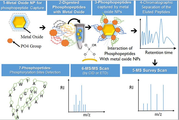
112                                                                                                              KOBEISSY ET AL.

and structure in a given cell type at a particular time point        Grant, 2004; Zhao and Jensen, 2009). For example, on the level
(Zhang et al., 2013). Protein profiles at both physiological and     of brain proteome, it is estimated that there exist around
pathophysiological processes characterize the information            20,000 brain proteins that are differentially expressed in the
flow in a cell, tissue, or organism (Petricoin et al., 2002). Pro-   various regions of the brain (Wang et al., 2005a). Furthermore,
teomics studies utilize several available techniques for the         it is difficult to associate mRNA expression to the protein
identification, validation, quantification, and expression of        expression levels (Denslow et al., 2003; Freeman et al., 2005;
certain protein(s). Such techniques are highly sensitive             Morrison et al., 2002; Wang et al., 2004). This is due to several
achieving targeted proteins analysis; among these tools are:         factors, including ‘‘alternative splicing,’’ which is highly
Western blotting, ELISA, and protein arrays, which are used          common in brain tissue, generating several copies of highly
for identification and quantification of proteins. On the other      related splices from a single gene (Hunnerkopf et al., 2007;
hand, proteomics can be of high throughput nature where a            Missler and Sudhof, 1998; Morrison et al., 2002; Wu and
set of proteins are globally evaluated (expression and quan-         Maniatis, 1999). It is estimated that a single gene can generate
tification) by methods including mass spectrometry, protein          up to 10 protein isoforms (Kim et al., 2004; Williams et al.,
arrays and 1D and 2-D gel electrophoresis (Kobeissy et al.,          2004). Thus, knowing the gene sequence is not sufficient to
2008b; Lamond et al., 2012; Smith and Figeys, 2006).                 predict the possible translation pattern of that specific protein.
    It is generally accepted that the human genome consists of       Currently, there are approximately 430 possible protein post-
around 40,000 genes (Lander et al., 2001; Yates, 2013), yet a        translational modifications (PTMs) that can contribute to this
single gene does not necessarily translate into one protein,         complexity (Khoury et al., 2011; Woodsmith et al., 2013).
and once proteins are synthesized, many undergo post-                   Post-translational modifications are defined as integral
translational modification (PTM) by phosphates, carbohydrates,       ‘‘chemical modifications of proteins that have implications on
lipids, or other groups, which tremendously complicates the          new protein functions in response to a specific cellular con-
global proteome profiling (Mann and Jensen, 2003).                   dition such as activation, turnover, downregulation, confor-
    Similar to the Human Genome Project, a Human Proteome            mation, and localization (Berretta and Moscato, 2010; Husi
Project (Cottingham, 2008) was initiated by a group of scien-        and Grant, 2001; Khoury et al., 2011; Morrison et al., 2002;
tists from the Human Proteome Organization (HUPO) and                Witze et al., 2007). Furthermore, the different expression lev-
was launched at the 2011 World Congress of Proteomics in             els of certain proteins leading to huge dynamic range differ-
Geneva, Switzerland (Omenn, 2012). In this project, scientists       ence hamper the analysis of low expression proteins (Hortin
have to deal with approximately more than 1,000,000 pro-             and Sviridov, 2010; Zubarev, 2013). For example, there is
teins, which can then be further complicated by several pro-         0.5 pg/mL of IL-6 compared to 35 mg/mL of albumin present
tein modifications. The time, effort, and money it takes for a       in the serum that exemplifies the dynamic range difference of
protein to be fully identified, sequenced, validated, and            some protein expression levels analyzed using traditional
structurally characterized impose a true challenge for re-           proteomics techniques including mass spectrometry, ELISA,
searchers (Lemoine et al., 2012; Yau, 2013). Consequently, the       Western blotting, and protein arrays (Anderson and Ander-
very early hope of characterizing the whole human proteome           son, 2002). Therefore, many of the potential biomarkers have
shifted focus on trying to find molecular differences between        concentrations in the femtomolar range while being im-
one functional state of a biological proteome system to an-          mersed in a complexity of other biological components with
other aided by systems biology analysis, which certainly             concentrations that span 12–15 orders of magnitude (Mitchell,
provided more precise comprehensive data of the proteome             2010; Rifai et al., 2006).
profile (Cox and Mann, 2007).                                           Furthermore, some protein classes are notoriously very
                                                                     difficult to analyze due to their intrinsic characteristics. For
                                                                     instance, membrane proteins constitute almost 30% of the
Challenges in Proteomics
                                                                     open reading frames in the sequenced genome (Bagos et al.,
   The rapidly growing field of proteomics has excelled in           2004; Lai, 2013; Vuckovic et al., 2013); however, they are very
several disciplines in biology, including injury, cancer, aging,     hydrophobic and buried in the lipid bilayer and tend to pre-
and different neurological conditions, as well as psychiatric        cipitate in aqueous buffers; thus, are harder to isolate. In ad-
conditions including drug/substance abuse, schizophrenia,            dition, the field of proteomics lacks a DNA–PCR-like
and depression (Abul-Husn and Devi, 2006; Becker et al.,             technique, which brings about sensitivity problems, associ-
2006; Becker, 2006; Choudhary and Grant, 2004; Cochran               ated with low abundant proteins.
et al., 2003; Dean and Overall, 2007; Dumont et al., 2004).
Proteomics is one of the fastest growing fields of biochemical
                                                                     Role of Nanotechnologies in Proteomics
sciences; a PubMed search reveals 287,021 articles published
in the past 2 years containing the word ‘‘protein,’’ compared           Nanotechnology is defined as the systematic study of a
to 156,200 articles using the term ‘‘gene’’ ( January, 2011–         particular system at the nanometer scale (1–100 nm) (Nie
April, 2013), which may also reflect a shift in genomics studies     et al., 2007; Vo-Dinh, 2005). Considering that average bond
towards proteomics investigations. Interestingly, the field of       lengths range in the picometer range (74 picometer for H–H
proteomics is in a continual growth with the introduction of         bond, 200 picometer for C–I bond); this is basically the limit of
some more specialized disciplines and subdisciplines such as         the metrics at which molecules cannot be further manipulated
neuroproteomics, psychoproteomics and nanoproteomics                 at the molecular level.
(Kobeissy et al., 2008a; 2008c).                                        Nanotechnology opens up unique opportunities, not only
   Proteomics analysis is more complicated than genomics             for material science research, but also for biology, medi-
due to a number of challenges occurring at different levels of       cine, and many other disciplines by manipulating individual
protein post-translational modifications (Choudhary and              atoms and molecules in a specific way that can fit into a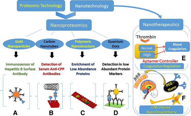
POST-GENOMICS NANOTECHNOLOGY                                                                                                      113

certain application (Petros and DeSimone, 2010). So, what is        surface area and, hence, increasing the available binding sites,
the relationship between nanotechnology and proteomics and          and (2) enhancing the accessibility of the target to the surface-
why will nanotechnology be beneficial for proteomics appli-         immobilized probe. This, in return, allows the detection of
cations?                                                            specific target(s) with higher sensitivity and faster kinetics.
   As mentioned previously, proteomics technology is chal-          Kang et al. (2005) reported that protein arrays prepared by
lenged by several limitations (PTMs, dynamic range, biolog-         silica nanotube membranes significantly improved the signal-
ical complexities, etc.), which in turn makes it incapable of       to-noise ratio compared to plain analogs. Similarly, Kim et al.
achieving some of its goals in elucidating protein changes          (2010) also showed that three-dimensional surfaces provide
unless it is coupled with other methods.                            higher aspect ratios, improving the immobilization capacity
   The attractive point of nanotechnology is that it can reduce     of the capturing probe 5-fold and enhancing its detection
these difficulties and therefore help to draw new information       ability to almost 15-fold. Another example of nanostructured
out of biological systems that otherwise would not be possible      material is porous silicon that was first discovered by Bell
by using conventional techniques. The field of nanotechnol-         Laboratories in 1960s; it has been used for many protein
ogy has been associated with several proteomics applications        sensing applications ( Jane et al., 2009). Unique fluorescence
such as phosphoproteomics/metal oxide nanoparticles, na-            properties and tremendous surface areas of porous silicon
nostructure surfaces for protein separation, and analytical         (500–800 m2/g) made it an ideal candidate for protein sensors
detection of biomarker proteins using arrays techniques             (Sailor, 2007). Ressine et al. (2007) used porous silicon as a
(Leitner, 2010; Nelson et al., 2009; Northen et al., 2007; Rissin   scaffold for antibody arrays and obtained detection limits
et al., 2010). This merging between nanotechnology and              down to 1 picomolar for IgG and 20 picomolar for prostate-
proteomics has generated nanoproteomics, which is defined as        specific antigen (PSA) in clinical samples.
a discipline of science involving the application of proteomics        Hill et al. (2009) explored the curvature effect of spherical
techniques aided by nanotechnology to enhance probing and           gold nanoparticles on the loading density of probes. They
evaluating protein systems (Archakov, 2007). As discussed by        showed that the binding to the nanoparticle surface is similar
Vo-Dinh and colleagues (2005), several basic cellular struc-        to that of a planar structure when the diameter of the particle
tures (proteins, polymers, carbohydrates, and lipids) are           is greater than 60 nm. Kelley et al. further explored this con-
molecules with similar sizes to various nanostructures. The         cept by controlling the topography of gold microelectrodes at
similarity between these biological nanosystems and nano-           the nanostructure level. Neither the increase of surface area
structures have important implications in designing and             nor the probe density were found to be the dominant factor in
manufacturing of the next generation nano-assemblies                improving on the sensitivity but the nanotopography intro-
(nanotechnology tool kits, lipid vesicles, and dendritic poly-      duced onto the microelectrodes (Bin et al., 2010; Das and
mers) that may have important medical and biotechnological          Kelley, 2011). Using the optimized topography, ovarian can-
applications (Chen et al., 2013; Zhang et al., 2009).               cer biomarker CA-125 was detected to the limit of 0.1 U/mL
   In this report, we will summarize the recent nano-               without the need to covalent label the target or the probe nor
technology applications that have been applied in different         to resort to sandwich complexes techniques (Das and Kelley,
proteomics-related applications. We will discuss nanostructured     2011).
surfaces, nanoporous particles, magnetic nanomaterials, gold           Another application of nanoparticles is femtoliter arrays.
nanoparticles, carbon-based nanomaterials, polymeric nano-          This technology has been developed in studying single en-
structures, quantum dots technology, and finally clinical           zyme molecules, detection of low abundance protein bio-
utility of nanoproteomics, along with their technology              markers in biological fluids, and single cell analysis (Malhotra
commercialization.                                                  et al., 2012; Rusling et al., 2013). Since the properties of single
                                                                    molecules significantly differ from the bulk solution, this
                                                                    method enabled detailed studies of single proteins and their
Nanostructured Surfaces
                                                                    kinetic properties, as illustrated by Gorris and Walt (2010).
   Conventionally, the size of typical structures is on the order   Also, detection of low femtomolar biomarker proteins was
of micrometers; however, technologies in the area of nano-          achieved utilizing such techniques (for reviews, see Gorris
technology have enabled us to achieve surface structure in the      and Walt, 2010; LaFratta and Walt, 2008; Rissin and Walt,
range of nanometers. Nanostructured surfaces have attracted         2006). The principle of detection enhancement basically relies
big attention owing to their unique physical, chemical, and         on Poisson Statistics, dictating that at very low concentration
structural properties, including enormously high surface area,      values, femtoliter size reaction chambers may contain either
quantum confinement, and interaction with light (Hoheisel           one or no molecules (Rissin et al., 2010). Recently, this tech-
et al., 2010; Parker and Townley, 2007; Tawfick et al., 2012; Vo-   nology has been coupled with classical enzyme-linked im-
Dinh et al., 2005). Not surprisingly, nanoparticles have found      munosorbent assay (ELISA). ‘‘Femtoliter arrays’’ platform,
widespread use in proteomics applications that can be sum-          used for protein detection, has been called ‘‘single molecule
marized into three basic areas: (a) scaffold for protein bio-       arrays,’’ which can enhance ELISA assay sensitivity up to
sensing, (b) sample purification and enrichment tool, and (c)       68,000 times, reaching zeptomolar (10 - 21) detection limits that
substrate for mass spectrometry analysis (Luong-Van et al.,         enable identification of markers specific to prostate cancer
2013). These diverse uses enhance the efficiencies of sev-          (Rissin et al., 2010).
eral proteomics applications, including ELISA and mass                 Matrix-assisted laser desorption ionization mass spec-
spectrometry-related techniques, as will be discussed.              trometry (MALDI-MS) has been a workhorse in proteomics
   Protein biosensing changes the morphology of a surface           studies; since the early days of its discovery (Karas et al.,
from plain to a nanostructured form and alters the sensing          2000). However, the presence of the matrix often causes het-
properties of that particular material by (1) increasing the        erogeneous co-crystallization of the matrix and analytes,
114                                                                                                          KOBEISSY ET AL.

resulting in significant background ion intensity in the           compared to plain analogs. It has shown that real biological
low-mass range (
POST-GENOMICS NANOTECHNOLOGY                                                                                                    115

interact with these surfaces, unique MS profiles were ob-           and affibodies) can be introduced on their surface with rela-
tained. This technique was a good demonstration of enrich-          tively easy chemistries. A third and probably most important
ment of labile and carrier-protein-bound molecules in               feature is that the separation can be easily performed with a
biological samples. Similar to this work, Hu et al. (2009) used     simple magnet. Owing to these properties, paramagnetic
mesoporous silica chips for proteome fractionation based on         particles have been widely applied for protein isolation
enhanced sieving properties of these nanostructure surfaces.        shown in Figure 2.
Hu et al showed that by surfactant-functionalized mesopor-             A widely used method for such purification is nickel ni-
ous silica, low molecular weight peptides can be specifically       trilotriacetic acid (NTA/Ni2 + )-based magnetic separation for
isolated and fractionated from complex biological samples.          histidine-tagged proteins (Gu et al., 2006). Pioneering work by
After fractionation, the response of mass spectrometry greatly      Xu et al. has been applied in developing a general strategy
improved by achieving better sensitivity in detecting low           using NTA-modified iron platinum (FePt) nanoparticles for
molecular weight peptides (Hu et al., 2009). In another study,      separation of histidine-tagged proteins at concentrations as
Finnskog et al. (2006) showed that highly improved sequence         low as 0.5 pM (Xu et al., 2004a; 2004b). Lee et al. (2004) in-
coverage for prostate-specific antigen (PSA) and human              troduced bimetallic nanorods comprising of gold and nickel
glandular kallikrein-2 protein can be achieved by trapping          to fish out proteins magnetically using the similar Ni-NTA
trypsin protein in a porous silicon nanovials with protein          chemistry. In this work Lee et al. used Ni-Au nanorods to
amounts as low as 8 fmol. The technique was not only sensi-         separate IgG antibodies. In another report, Shukoor et al.
tive but also very fast. The digestion was achieved within          (2008) employed multifunctional copolymer functionalized
30 sec, as opposed to conventional trypsin digestion protocols      superparamagnetic Fe2O3 nanoparticles for immobilizing and
that usually take up to 16–24 h with low sample amounts             subsequently isolating His-tagged recombinant protein sili-
(fmol levels). In a very recent work, Fan et al. utilized a na-     cate from marine sponge Suberites domuncula. This highly
noporous silica-based method to isolate low molecular weight        versatile biomagnetic separation methodology also allows the
peptides from high molecular weight proteins in serum bio-          re-use of the magnetic nanocrystals. Antibody-conjugated
fluids of metastatic melanoma patients as molecular signa-          magnetic particles were also used to isolate biomarker pro-
tures. This was followed by proteomics identification (Fan          teins from plasma samples of cancer patients. Ranzoni et al.
et al., 2012b). MALDI mass spectrometry analysis and rigor-         (2012) has synthesized PSA antibody conjugated nano-
ous bioistatictical analysis led to the identification of 27 pep-   particles and isolated PSA from plasma samples in conjunc-
tides that might be used as potential biomarkers in metastatic      tion with pulsed magnetic fields.
melanoma. Yin et al. (2012) used C8-modified graphene@                 Chou et al. (2005) synthesized antibody-conjugated mag-
mSiO2 nanoconjugates for enriching endogenous peptides              netic nanoparticles as a tool for isolating cancer proteins be-
prior to mass spectrometry. These nanoconjugates provide a          fore MALDI mass spectrometric detection. C-reactive protein
very huge surface area (632 m2/g) and excellent capturing           (CRP) and amyloid P component were efficiently isolated
properties that can fish out peptides from standard protein         from unfractionated human plasma and sub-nanomolar level
digests, as well as from real biological mixtures such as mouse     detection was achieved in MALDI analyses. In a follow-up
brain tissue. The Ferrari group has reported a similar strategy     study from the same group, serum amyloid A (SAA), C-
for finding low molecular weight biomarkers for breast cancer       reactive protein (CRP), and serum amyloid P (SAP) antigens
samples (Fan et al., 2012a). Peptides from the serum of nude        were captured, isolated from human plasma, and detected by
mice with MDA-MB-231 human breast cancer were isolated              MALDI-MS in a multiplexed immunoassay format. In a recent
by nanoporous silica particles and analyzed by matrix-              study, Bamrungsap et al. (2011) used aptamer-conjugated
assisted laser desorption/ionization time-of-flight mass spec-      superparamagnetic particles to detect lysozyme by using
trometry. Protein signatures unique to different stages of          magnetic relaxation upon target capture.
cancer development were identified. Their approach and re-
sults reported in this study possess a significant potential for
                                                                    Gold Nanoparticles
the discovery of proteomic biomarkers that may significantly
enhance personalized medicine targeted at metastatic breast            Another form of nanomaterials are gold (Au) nanoparticles
cancer.                                                             that have been utilized for protein immobilization owing to
   In a conceptually very similar work, Tan et al. (2012) has       their high affinity to thiol (-SH) and disulfide (S–S) groups
used nanoporous silicon particles. They tested the efficacy of      present in various molecules. Their exceptional optical, scat-
these nanoparticles for enriching the low molecular weight          tering, and agglomeration-dispersion properties render them
proteome of serum from colorectal cancer patients and found         optimal candidates for various types of biolabeling applica-
that patient samples can be can be clearly distinguished from       tions (Kneipp et al., 1999; Rosi et al., 2004). Protein-mediated
control patients by statistical analysis.                           agglomeration of Au nanoparticles generates large local
                                                                    electromagnetic fields between immediate neighbors when
                                                                    illuminated known as ‘‘hot spots‘‘ (Lee et al., 2006). This
Magnetic Nanomaterials
                                                                    technology enables researchers to analyze proteins within
   Protein expression, purification, and modification has been      interparticle spaces using surface-enhanced Raman spectro-
well established with the existing biotechnologies; however,        scopy (SERS) reaching detection levels down to single mole-
methods for low abundant protein enrichment and separation          cule level (Gunnarsson et al., 2005; Kneipp et al., 1998; Kneipp
are still challenging. Magnetic materials hold great promise.       et al., 1997; Nie and Emory, 1997; Podstawka et al., 2004;
First of all, they have high surface/volume ratio that provides     Wang et al., 2005b). Various colorimetric sensors for detecting
high surface area for coating/binding of different substrates.      metabolites, proteins, small molecules, and whole cells in
Second, several affinity tags (antibodies, aptamers, lectins,       solution as well as in real samples were developed by
116 KOBEISSY ET AL.
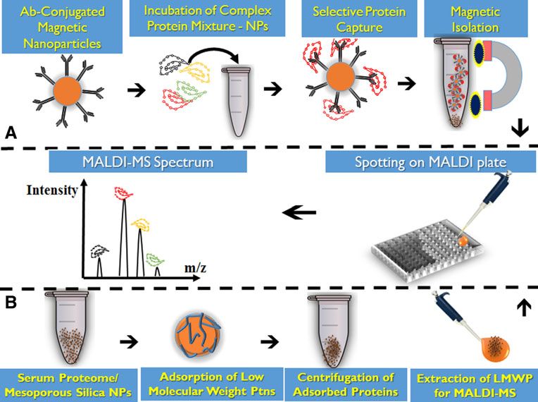
FIG. 2. (A) Antibody-conjugated nanoparticles preparation for immunoassays: Antibodies
are first tethered onto magnetic nanoparticles with a biocompatible conjugation chemistry
(e.g., avidin-biotin, amine-carboxyl coupling, and click chemistry). A complex proteome (e.g.,
serum, plasma, cell lysate, saliva) is then incubated with the antibody-conjugated nano-
particles, and a magnet is utilized for magnetic separation. Finally, particles are washed and
spotted onto sample plate and analyzed by MALDI-MS. (B) Enrichment of low molecular
weight serum proteome: In the first step, serum proteome sample is incubated with silica
nanoparticles and adsorbed or size excluded by mesopores on the silica NP, and the sample
is centrifuged and NPs are separated. The nanoparticles are spotted onto sample plate and
analyzed by MALDI-MS.
combining highly specific recognition properties of aptamers (Ocsoy et al., 2013). They used Au@MnO nanoflower-shaped
(single stranded target specific DNA molecules) and optical nanoparticle matrix for LDI-MS that was found to be an effi-
properties of Au nanoparticles (Pagba et al. 2010; Wang et al., cient nanoparticle substrate to LDI-MS as compared to other
2010). These sensors are used to develop smart strip-type nanoparticles. The same nanoflowers were also used to target
biosensors for point-of-care devices (Yoon, 2013). cancer cells and for the selective metabolite extraction and
In addition, gold nanoparticles play an important role in detection from cancer cell lysates.
the field of quantitative nanoproteomics based on the bio-
barcode assay developed by Nam and colleagues (Georga-
Carbon-Based Materials
nopoulou et al., 2005; Nam et al., 2003; Thaxton et al., 2009).
This methodology is used for enzyme-free ultrasensitive de- Elemental carbon in the sp2 hybridization can form a va-
tection of proteins and nucleic acid targets with sensitivity riety of different structures. Among these, graphite, nano-
much higher than that of conventional ELISA-based assays. diamond, carbon nanotubes, fullerene, grapheme, and
The same group also reported that PSA protein detection graphene oxide, which are different forms of carbon nano-
approached 330 fg/mL in the sera of patients who have un- structures, have been well studied in different applications.
dergone radical prostatectomy (Thaxton et al., 2009). Larsen et al. (2002) used graphite nanopowders in gel loading
Gold nanoparticles can also be applied for the enhancement tips for desalting and preconcentration of peptides prior to
of biomarker discovery strategies through enriching low mass spectrometric analysis, which is commonly used in
abundance proteins that were usually missed by conventional MALDI sample preparation or other protein clean up settings.
detection techniques. Yasun et al. (2012) used aptamers con- The same group showed that hydrophilic phosphopeptides
jugated to gold nanorods for the detection of rare proteins, can be significantly enriched using graphite powder (Larsen
and demonstrated that these gold nanorods can be used to et al., 2004). Similar to that, Li et al. (2005b) used silicon-
enrich proteins present at low abundance and increase the graphite hybrid coating for on target desalting. Among other
efficiency of their detection by 47%. The group showed that forms of carbon, carbon nanotubes (CNTs) have also attracted
the used probes were able to detect thrombin in a buffer up to great attention, since they have numerous novel and useful
10 ppb. Later, the same group was the first to test the use of properties investigated since 1991. CNTs’ length (up to sev-
hybrid Au-metal oxide nanomaterial as substrates for LDI-MS eral microns) with small diameter (a few nanometers) resultsPOST-GENOMICS NANOTECHNOLOGY 117
in a large aspect ratio. This makes CNTs very attractive ma- of the polymers used (e.g., these can be thermo-responsive
terial for proteomics applications via their wide surface area materials). For instance, N-isopropyl acrylamide is hydrophilic
that enables better immobilization and capturing of proteins below a certain temperature and hydrophobic above that par-
through functionalizing its surface with a capturing protein ticular temperature. As a result, selective capture of hydrophilic
probe. For instance, Okuno et al. (2007) exploited the property or hydrophobic proteins can be achieved. Li et al. (2007a)
of CNTs and designed a CNT-based electrochemical system showed that this thermo-responsive polymer coating in com-
to detect prostate-specific antigen from clinical samples. bination with temperature control can be used to clean up
Furthermore, Chen et al. (2008) showed that a very sensitive protein samples prior to mass spectrometry analysis.
Raman sensor that was able to detect low fM levels of protein Another interesting class of nanostructured polymeric
biomarkers can be constructed by using CNTs. material is the polymer brushes composed of a layer of
In another study, Drouvalakis et al. (2008) designed a CNT polymers attached with one end to a surface ( Jain et al., 2009).
sensor to detect autoantibodies in rheumatoid arthritis pa- Polymeric nanostructures are also used as biomarker har-
tients. In an elegant study, Guo et al. (2009) used carbon na- vester; Luchini et al. (2008) used N-isopropylacrylamide na-
notubes to improve the resolution of native PAGE detecting nogels in conjunction with affinity baits that functioned as
serum proteins. Other proteomics application include: carbon molecular size sieve for capturing abundant proteins such as
based-nanostructures, which has also been applied as sub- albumin. Longo et al. (2009) used the same nanogels to con-
strates for ionization in laser-based ionization techniques. Xu centrate and preserve platelet-derived growth factor (PDGF)
et al. (2003) used carbon nanotubes as a substrate for ioniza- from serum in order to magnify the detectable level of the
tion. Sunner et al. (1995) used graphite for the same purpose. marker. This was performed in one single step and in solution
Hsu et al. (2010) used carbon nanotubes for screening long- phase (Longo et al., 2009) and has been commercialized under
chain fatty acids in patient samples. In another study, Najam-ul- the trade name Nanotrap (Shaffer, 2011).
Haq sputter coated nanostructured diamond like carbon on
commercially available DVD disks and used this substrate for
Metal Oxide Nanoparticles
laser desorption ionization of peptides with a detection sensi-
and Phosphonanoproteomics
tivity of femtomolar levels (Najam-ul-Haq et al., 2008).
Graphene (G) and graphene oxide (GO) have been recently Another area that has been addressed by nanotechnology
used in protein science. Tang et al. (2010) employed G & GO as a and proteomics is the identification of PTMs and their struc-
scaffold for enriching DNA and proteins in solution, yielding tural characterization. More specifically, nanoparticles have
high recoveries which were four times higher than nanodia- been applied in characterization of protein phosphorylation,
mond particles. Finally, Dong et al. (2010) used graphene as a involving phosphopeptide isolation and subsequently iden-
substrate for ionization for the analysis of small peptides and tification by mass spectrometry as illustrated in Figure 3
small metabolites. Compared to conventional MALDI matrices, (Olsen et al., 2010; Oppermann et al., 2009)
graphene exhibited higher desorption ionization efficiencies Protein phosphorylation is a ubiquitous and central process
with very low background ions. Furthermore, it eliminated that regulates several different cellular functions such as sig-
fragmentation and provided better tolerance to salts. nal transduction, cell growth and differentiation. Protein ki-
Carbon-based materials are also used in field effect tran- nase activities are significantly elevated in different types of
sistors that allows for the real-time and fast detection of label- cancer and many new pharmaceuticals target phosphorylated
free proteins and biomolecules (Cui et al., 2001; Hahm Jong-in, proteins to help curb the disease progression. The state of the
2004; Patolsky et al., 2004). A change in charge or electric po- art method for identifying protein phosphorylation sites is
tential at the nanomaterial surface is induced by the binding of mass spectrometry. However analysis and characterization of
a biomacromolecule. This in turn leads to a change in the phosphoproteins from complex samples using MS is a real
charge carrier concentration in the semiconductor material that challenge. The ionization efficiency of cellular phosphopro-
can be detected by conductometry measurements. Carbon na- teins is significantly hampered in mass spectrometry owing to
notubes are relatively easy to prepare when compared to sili- their very low abundance relative to unphosphorylated pro-
cone nanowires and have a long shelf life (Tans et al., 1998). teins and to their negatively charged phosphate groups.
Chen et al. (2004) reported the detection of protein adsorption Therefore, phosphoproteins are largely missed in high through-
on single-walled carbon nanotube using field effect transistors put proteomics experiments and phosphoproteomics has not
(FETs). Furthermore, Chen et al. enhanced their work by been previously possible, due to the challenge of capturing
modifying carbon nanotubes with DNA aptamers to recognize and enrichment of low abundant phosphopeptides (Tao
specific macrobiomolecules selectively (Chen and Chen, 2005). et al., 2005). The best way to address this problem is to use a
capturing tool to pre-concentrate phosphopeptides prior to
analysis by mass spectrometry (Engholm-Keller and Larsen,
Polymeric Nanostructures
2013; Johnson and Hunter, 2004).
Polymeric materials, including polylysines, polyethyl- The chemistry behind phosphopeptide enrichment re-
enimine, and cationic dendrimers, have been used in proteomics mained immature until immobilized metal ion affinity chro-
studies (Tao et al., 2005). However, the nano-architectured na- matography (IMAC) was introduced (Feuerstein et al., 2005;
nomaterials turned out to be better alternatives to bulk poly- Jin et al., 2004; Mann et al., 2002). The underlying principle of
mers. Li et al. (2005a) used radiofrequency plasma polymers phosphopeptide enrichment is to form reversible and strong
coated on a traditional MALDI plate for selective capture and interaction between the metal and phosphorylation site of the
detection of proteins that they termed as ‘‘on probe affinity protein. The task has been originally achieved by the use of
capturing.’’ The advantage of using this technique is that it al- IMAC resins that are mostly replaced with metal oxide-based
lows the capture of different molecules by tuning the properties affinity chromatography (MOAC) (Lin et al., 2008; Lin et al.,118 KOBEISSY ET AL.
FIG. 3. Phosphopeptide capture by metal oxide nanoparticles technology: (Step 1) Com-
plex proteome sample (e.g., serum, plasma, cell lysate) is first digested with trypsin. (Step 2)
Peptide mixture is incubated metal oxide nanoparticles. (Step 3) After incubation, metal
nanoparticles are isolated from non-phosphopeptides and thoroughly washed. (Step 4)
Phosphopeptides are eluted from the metal oxide nanoparticles and fractionated by SCX
and RP-HPLC and infused into high resolution mass spectrometry. (Step 5) A survey scan is
performed to identify most abundant peptides. (Step 6) Selected peptides are fragmented by
collision induced dissociation (CID) and electron transfer dissociation (ETD). (Step 7)
Phosphopeptides are identified by proper software ( Johnson and Hunter, 2004).
2009; Thingholm et al., 2006; Zhou et al., 2007). Metal ox- veloped an on-plate enrichment protocol. This device, known
ides behave as Lewis acids and show different acidity which as T-plate, unifies the sample enrichment and mass spectro-
forms the basis of targeted-specific selection observed in metric analysis. He et al. (2011) used TiO2-coated ZnO na-
phosphoproteomics. norod arrays in a capillary microchannel to bind and enrich
Phosphoproteomics was further supported by the intro- phosphopeptides selectively.
duction of enhanced mass spectrometry instruments and Several different nanocomposites of TiO2 were additionally
dissociation chemistry that facilitated the identification of tested for phosphopeptide enrichment. As notable examples,
different PTMs. For example, the use of high resolution mass Fang et al. (2012) used titanium dioxide-multiwalled carbon
spectrometry instruments and advanced electron transfer dis- nanotube (TiO2-MWNT) nanocomposites, Zeng et al. (2012)
sociation (ETD) analysis enabled the successful identification of used TiO2-coated carbon-encapsulated iron nanoparticles, and
these phosphopeptides (Coon et al., 2005). This was widely Lu et al. (2012) has used TiO(2)/graphene composites. Com-
applied in identification of phosphorylation sites in embryonic mercially, another form of functionalized TiO2 is available
stem cells (Swaney et al., 2009). Ultimately, profiling of a large composed of ‘‘TiO2 coated magnetic nanoparticles.’’ They rep-
number of different classes of phosphoproteins became possi- resent a new hybrid class of phosphopeptide enrichment tool.
ble and so emerged a (sub)-discipline termed as ‘phosphona- Another metal oxide used for enrichment is zirconium
noproteomics’ (Najam-ul-Haq et al., 2012). dioxide (ZrO2), which was first introduced by Kweon and
Oxides of group 4 and 5 elements such as Ti, Hf, Zr, V, Nb, Hakansson (2008). Similarly, other functionalized ZrO2 na-
and Ta are the commonly used metal oxides (Najam-ul-Haq nomaterials were used for phosphoprotein enrichment (Li
et al., 2012), among which titanium dioxide (TiO2) is the fa- et al., 2007a; Li et al., 2007b; Zhou et al., 2007).
vored metal oxide for phosphoprotein enrichment (Chen and Aluminum hydroxide (AlOH)3 (Chen and Chen, 2008) and
Chen, 2005; Liang et al., 2006). Several different titanium di- iron oxide coated-Niobium oxide (Nb2O5) (Lin et al., 2009) are
oxide-based nanostructures with various shapes and geome- few other metal oxides that have been also used in selectively
tries have been used for phosphopeptide enrichment. For trapping phosphopeptides from peptide mixtures such as
example Lu et al. (2010) have used TiO2 nanocrystal clusters tryptic digest of caseins, serum, and cell lysate. Magnetic
for enriching phosphopeptides. A mixture of b-casein, materials do show some degree of affinity to phosphopep-
horseradish peroxidase, b-lactoglobulin, and fetuin was in- tides (Lee et al., 2008) and have been majorly used as the core
cubated with these nanoclusters for phosphpeptides enrich- material for other peptide capture metal oxide (MO) coatings
ment. Torta et al. (2009) used pulsed laser deposition (PLD) to such as Fe3O4@TiO2 (Chen and Chen, 2005; Li et al., 2008) or
create nanostructured thin films on a MALDI plate and de- Fe3O4@ZrO2 (Li et al., 2007a). This coating improvesPOST-GENOMICS NANOTECHNOLOGY 119
purification and separation of target peptides upon selective Nanoproteomics in Clinical Applications
binding from a complex peptide mixture, using external
magnetic field. Nanoproteomics technology has been applied to various
When the performance of metal oxide nanoparticles were clinical settings, mainly for enhancing biomarker discovery
compared to that of conventional IMAC resins, it was found capabilities. In this regard, nanoproteomics are seen to be
that metal oxide nanoparticles out-perform IMAC resins. functioning as protein amplification techniques similar to
Larsen et al. (2005) compared the performance of titanium PCR. Potential clinical applications include infectious, endo-
oxide micro-columns with IMAC resins and found that sig- crine, autoimmune, and neurodegenerative diseases, as well
nificantly higher number of non-phosphorylated peptides as brain injury and several types of tumors (Ray et al., 2011).
were observed in the IMAC experiments. They reported that Some of these applications are illustrated in Table 1 and
performance of the TiO2 based-techniques significantly sur- Figure 4.
passed the IMAC method with respect to the number of de- As applied to infectious diseases, Ansari et al. (2010),
tected phosphorylated peptides and reduction of the number Kaushik et al. (2009a), and others used nano-structured sur-
of nonphosphorylated peptides. This is attributed to the more faces for the detection of ochratoxin-A mycotoxin pro-
selective binding of phosphorylated peptides to TiO2 micro- duced by Aspergillus. Ansari et al. utilized Nano-ZnO film
columns than IMAC resins. deposited on an indium-tin-oxide (ITO) with immobilized
rabbit-immunoglobulin and bovine serum albumin to reach a
detection limit of 0.006 nM/dm of the toxin. Similarly,
Quantum Dots
Kaushik et al. used nanostructured cerium oxide to achieve a
Fluorescence-based techniques for protein sensing appli- detection limit of 0.25ng/dL of ochratoxin (Kaushik et al.,
cations have gained an ever increasing popularity due to their 2009a). Tang et al. (2004; 2005) proposed different im-
simplicity and exquisite sensitivity (De et al., 2009; Ibraheem munosensors based on gold nanoparticles for the detection of
and Campbell, 2010; You et al., 2007). In particular, quantum hepatitis B surface antibody in blood. Reported detection limit
dots (QDs), also called semiconductor nanoparticles, are of the applied techniques ranged between 5 and 15 ng/mL,
emerging as a new class of fluorescent probes (Boeneman which demonstrates a much higher sensitivity than the stan-
et al., 2009; Larson et al., 2003; Pinaud et al., 2006). When dard diagnostic serology techniques, allowing for better dis-
compared to organic dyes and fluorescent proteins, QDs have ease detection and monitoring. The same group also applied
unique optical and electronic properties that make them the immunosensors approach for the detection of serum levels
brighter and highly resistive to photobleaching and chemical of Bacillus anthracis protective antigen as well as HIV-capsid
degradation (Leutwyler et al., 1996; Murray et al., 1993). The p24 antigen (Tang and Hewlett, 2010; Tang et al., 2009). These
maximum emission depends on the size of the electron gap immunosensors approaches demonstrated rapid detection
that is tuned by the particle core diameter. Smaller nano- and much higher sensitivity compared to the traditional
particles have a maximum emission in the blue region and ELISA techniques that can reach up to 100 folds. Moreover, as
bigger particles tend to emit in the red or near-IR. part of a disease preventative measure, Yang et al. (2008) used
These unique optical properties make QDs an excellent carbon nanotubes for the detection of Staphylococcal En-
fluorescent probes for applications in the field of diagnosis terotoxin B in food products that were traditionally tested by
(Bruchez et al., 1998; Chan and Nie, 1998), in vivo and in vitro ELISA. The new technique enhanced toxin detection limit and
imaging (Kim et al., 2003; Levene et al., 2004; Rosenthal et al., raised screening sensitivity by more than six-fold.
2002), multicolor cell imaging (Hanaki et al., 2003; Jaiswal et al., In the field of autoimmune diseases, many of the current
2002; Sukhanova et al., 2004; Wu et al., 2002), cell and protein diagnostic serology techniques do not demonstrate optimal
tracking (Dahan et al., 2003; Voura et al., 2004), and DNA and sensitivity for diseases such as rheumatoid arthritis, celiac
protein sensing (Medintz et al., 2003; Zhang et al., 2005). disease, Wegener’s granulomatosis, and others. This could be
More recently, Liu et al. (2010) reported the detection and in part due to the limited ability of these techniques to detect
characterization of four low-abundant protein biomarkers low levels of autoantibodies in the sera of suspected patients.
(CD15, CD30, CD45, and Pax5) in Hodgkin’s lymphoma Here, the application of nanoproteomics techniques could be
using the multiplexing capabilities of QDs. QDs were also promising. In fact, Drouvalakis et al. (2009) utilized peptide-
used to detect apolipoprotein E, the most important known coated nanotubes for the detection of rheumatoid arthritis-
genetic risk factor for Alzheimer disease, by designing a specific cyclic citrulline-containing peptide. The proposed
sandwich immunocomplex microarray assay based on technique was capable of detecting 12 out of 32 RA patients
cadmium-selenide/zinc-sulfide (CdSe@ZnS) quantum dots. missed by ELISA and microarray (Drouvalakis et al., 2008).
The assay provided a low detection limit of 62 pg mL–1, seven Several other studies have also reported the use of nanopro-
times more than that of the ELISA (470 pg mL–1) when tested teomics for the detection of RA-related immunoglobulins (de
under the same conditions (Morales-Narváez et al., 2012). Gracia Villa et al., 2011; Jiménez et al., 2012). Carbon nano-
A major drawback, however, to the utilization of QDs in the tubes were also suggested as a screening tool for Wagner’s
field of proteomics is the nonspecific binding onto their sur- granulomatosis (Chen et al., 2008). The authors used macro-
face (Pathak et al., 2007). To minimize such nonspecific molecular single-walled carbon nanotubes (SWNTs) as mul-
binding, QDs are often modified with polyethyleneglycol ticolor Raman labels for multiplexed protein arrays for the
(PEG)-based polymers (discussed later) (Geho et al., 2005; Liu detection of anti-proteinase 3 autoantibodies that are markers
et al., 2008). More recently, Breus et al. (2009) capped QDs of Wagner’s granulomatosis. Results have shown a three-fold
with small zwiterinonic molecules. This step rendered the increase in the detection level of these proteins compared to
nanoparticles water soluble and dramatically diminished the recent fluorescent detection techniques. In addition, na-
nonspecific binding. noproteomics techniques were also assessed for utilization inTable 1. Major Studies in Nanotechnology and Clinical Applications Including Infectious,
Endocrine, Autoimmune, and Neurodegenerative Disorders
Study Tool used Disease/condition Aim Description
Detection of bacteria/viruses/toxins
Nanostructured zinc oxide Nano-ZnO film deposited on Mycotoxin Detection of ochratoxin-A A detection limit of 0.006 nM/dm of the
platform for mycotoxin an indium-tin-oxide (ITO) toxin was achieved
detection (Ansari et al., 2010)
A novel immunosensor of hepatitis Tris(2,2¢-bipyridyl)cobalt(III) Hepatitis B Detection of hepatitis B Comparable results to ELISA detection
B surface antibody and gold nanoparticles surface antibody with a limit of 0.005–0.015 lg/mL
(Tang et al., 2004) assembled on the PPF-
modified Pt electrode
Detection of anthrax toxin by an Europium nanoparticle- Bacillus anthracis Serum levels Bacillus The assay had a detection range of 0.01
ultrasensitive immunoassay based immunoassay anthracis protective to 100 ng/mL and was approximately
using europium nanoparticles antigen 100-fold more sensitive
(Tang et al., 2009)
Carbon nanotubes based optical Carbon nano-tubes Staphylococcal food Staphylococcal The assay raised the screening sensitivity
immunodetection of poisoning Enterotoxin B in by more than 6-folds
Staphylococcal Enterotoxin B food products
(SEB) in food (Yang et al., 2008)
Detection of biomarkers for autoimmune diseases
Peptide-coated nanotube-based Peptide-coated carbon Rheumatoid arthritis Detection of rheumatoid The test was capable of detecting 12 out
biosensor for the detection nanotubes arthritis-specific cyclic of 32 RA patients missed by ELISA
120
of disease-specific citrulline-containing and microarray.
autoantibodies in human peptide
serum (Drouvalakis
et al., 2008a)
Protein microarrays with Macromolecular single- Wagner’s granulomatosis Detection of anti- Three-fold increase in the detection level
carbon nanotubes as walled carbon nanotubes proteinase 3 of these proteins compared to the
multicolor Raman labels (SWNTs) as multicolor autoantibodies recent fluorescent detection techniques
(Chen et al., 2008a) Raman labels for multiplexed
protein arrays
Celiac disease detection Screen-printed carbon electrodes Celiac disease Detection of IgA and IgG The proposed technique was comparable
using a transglutaminase (SPCE) nanostructurized with type anti-tTG to ELISA
electrochemical immunosensor carbon nanotubes and gold autoantibodies
fabricated on nanohybrid screen- nanoparticles
printed carbon electrodes
(Neves et al., 2012)
Detection of cancer biomarkers
A voltammetric immunosensor Nanobiocomposite materials based Hepatocellular carcinoma, Serum alpha-fetoprotein The assay had a detection limit
based on nanobiocomposite on gold nanoparticles with germ cell tumors (AFP) detection of 3.7 ng/ml.
materials for the determination immobilized antibodies and others
of alpha-fetoprotein in serum
(Giannetto et al., 2011)
(continued)Table 1. (Continued)
Study Tool used Disease/condition Aim Description
Integrated microfluidic systems with Glassy carbon electrode modified Prostate cancer Prostate specific antigen This technique was a quick detection
an immunosensor modified with with multiwall carbon nanotubes (PSA) technique with higher sensitivity then
carbon nanotubes for detection of traditional ELISA.
prostate specific antigen (PSA) in
human serum samples
(Panini et al., 2008)
Nanogold-enwrapped graphene Nanogold-enwrapped graphene Colorectal cancer Carcinoembryonic A low detection limit of 0.01 ng/mL was
nanocomposites as trace labels nanocomposites antigen (CEA) demonstrated
for sensitivity enhancement of
electrochemical immunosensors
in clinical immunoassays:
Carcinoembryonic antigen as a
model (Zhong et al., 2010)
In situ amplified electrochemical Horseradish peroxidase- Colorectal cancer Carcinoembryonic The assay lowered the detection limit of
immunoassay for encapsulated nano-gold antigen (CEA) CEA down t0 1.5 pg/mL.
carcinoembryonic antigen using hollow microspheres
horseradish peroxidase-
encapsulated nanogold hollow
121
microspheres as labels
(Tang and Ren, 2008)
Drug delivery and disease monitoring
A logical molecular circuit for Logical circuit based on DNA Different applications Autonomous, self-sustained An example of application involves
programmable and autonomous aptamer–protein interactions and programmable monitoring the levels and activity of
regulation of protein activity manipulation of protein thrombin in plasma and delivers an
using DNA aptamer–protein activity in vitro inhibitory anti-coagulant accordingly
interactions (Han et al., 2012)
Quantum-dot-conjugated graphene Quantum-dot-conjugated Cancer chemotherapy Targeted therapy with The system can deliver the antineoplastic
as a probe for simultaneous graphene doxorubicin drug doxorubicin to cancer cells and
cancer-targeted fluorescent monitor its release out of the cell
imaging, tracking and allowing for a safer and more targeted
monitoring drug delivery therapy
(Chen et al., 2013)
Mesoporous silicon nanotechnology Multistage delivery systems using Targeted drug therapy The protein platform helps detect protein
for cancer application mesopotrous silicon and protein changes that can allow disease
(Bouamrani et al., 2009) platform chips with MS/MALDI monitoring in association with
MS/MALDI and the multistage
delivery system allows for targeted
drug delivery.122 KOBEISSY ET AL.
FIG. 4. Current clinical applications of nanoparticles/proteomics. (A) Use of gold nano-
particles for high sensitivity detection of hepatitis B surface antigen (HBsAg). (B) Use of
carbon nanotubes for the detection of anti-citrullinated peptide antibodies in rheumatoid
arthritis. (C) Use of polymeric nanostructures for enrichment of low molecular weight
proteins. (D) Use of quantum dots for detection of Apolipoprotein E detection in Alzhei-
mer’s disease. (E) Aptamer based circuit that monitors the levels and activity of thrombin in
plasma and delivers an inhibitory anti-coagulant accordingly. (F) Use of PEGylated lipo-
somes for drug delivery into the CNS across the blood brain barrier (BBB) for treatment of
brain injury, including stroke and traumatic brain injury.
celiac disease through the detection of anti-gliadin auto- the detection limit of this tumor marker down to 1.5 pg/mL
antibodies using different techniques where high sensitivity (Tang and Ren, 2008). Several studies have reported the use of
detection potentials were demonstrated (Neves et al., 2012; nanoproteomics as means for biomarker detection as in breast
Ortiz et al., 2011). cancer, prostate cancer as well as others; for a detailed review,
An important application of nanoproteomics tools resides see Liu et al., (2013). Of interest, these nanoproteomic appli-
in cancer screening ( Ji et al., 2010). Several plasma protein cations have been reported with different sensitivities ranging
biomarkers have been associated with different types of can- from 30 attoM to 2000 fMA even for the same antigen (PSA),
cer, yet their detection by current diagnostic and screening however, using different assays as shown in Table 2.
techniques is not satisfactorily sensitive. Therefore, nanopro- A further detailed comparison among different biomarker
teomics tools have been investigated for their possible detection sensitivities utilizing different nanomaterial-based
screening potentials in these cases. For example, Giannetto approaches is presented in Table 3. In this table, HIV proteins,
et al. (2011) utilized nanobiocomposite materials based on PSA, and Ochratoxin-A protein levels are compared and
gold nanoparticles with immobilized antibodies for the de- evaluated for their sensitivity detection limits using magnetic
velopment of serum alpha-fetoprotein (AFP) immunosensors nanoparticles, metal oxides, composite nanosensor assay,
(Giannetto et al., 2011). Serum levels of alpha-feto protein are Europium-based nanoparticles, carbon nanoparticles, gold
associated with various types of tumors including germ cell nanoparticles, and porous silica coupled with mass spec-
tumors, liver tumors, and others. Two other areas that show trometry-based approaches.
major potential for cancer nanoproteomics application are the Added to the value of nanoproteomics application in bio-
detection of prostate-specific antigen (PSA) and carcinoem- marker discovery, drug delivery, and personalized medicine
bryonic antigen (CEA). Panini et al. (2008) used carbon na- are other fields where nanotechnology also holds future
notubes for the detection of PSA in serum. The utilization of promise (Ferrari, 2005b; Kim et al., 2013). Nanotechnology-
glassy carbon electrode modified with multiwall carbon na- based ‘‘theranostics’’ applications were recently reviewed by
notubes provided a quick detection technique and a higher Kim et al. (2013). Several applications for nanoparticle-based
sensitivity then traditional ELISA. On the other hand, Zhong drug delivery have been proposed for therapeutics, including
et al. (2010) employed highly sensitive electrochemical im- blood coagulation monitoring, cancer therapy, and stroke
munosensor with a sandwich-type immunoassay format to treatment; an eminent example of which is the use of liposo-
detect CEA that is associated with multiple tumors including mal nano-carriers (Alaouie and Sofou 2008). Han et al. (2012)
colorectal cancer. This technique lowered the detection limit described a logical circuit that enables autonomous, self-
of CEA down to 0.01 ng/mL. Furthermore, Tang et al. (2008) sustained, and programmable manipulation of protein activ-
utilized horseradish peroxidase-encapsulated nano-gold hol- ity in vitro. An example of such application is the use of a
low microspheres as labels for the detection of CEA, lowering circuit that monitors the levels and activity of thrombin inYou can also read