Grad-seq identifies KhpB as a global RNA-binding protein in Clostridioides difficile that regulates toxin production
←
→
Page content transcription
If your browser does not render page correctly, please read the page content below
microLife, 2, 2021, uqab004

                                                                                          doi: 10.1093/femsml/uqab004
                                                                                          Advance Access Publication Date: 22 April 2021
                                                                                          Research Article

RESEARCH ARTICLE

                                                                                                                                                       Downloaded from https://academic.oup.com/microlife/article/doi/10.1093/femsml/uqab004/6245135 by guest on 27 July 2021
Grad-seq identifies KhpB as a global RNA-binding
protein in Clostridioides difficile that regulates toxin
production
Vanessa Lamm-Schmidt1,¶ , Manuela Fuchs1,@ , Johannes Sulzer1,# ,
Milan Gerovac1,♀ , Jens Hör1,$ , Petra Dersch3,! , Jörg Vogel1,2,† and
Franziska Faber1, *,‡
1
 Faculty of Medicine, Institute of Molecular Infection Biology (IMIB), University of Würzburg,
Josef-Schneider-Straße 2/D15, 97080 Würzburg, Germany, 2 Helmholtz Institute for RNA-based Infection
Research (HIRI), Helmholtz Centre for Infection Research (HZI), Josef-Schneider-Straße 2/D15, 97080
Würzburg, Germany and 3 Centre for Molecular Biology of Inflammation, Institute for Infectiology, University
of Münster, Von-Esmarch-Straße 56, 48149 Münster, Germany
∗
  Corresponding author: Institute of Molecular Infection Biology (IMIB), University of Würzburg, Josef-Schneider-Straße 2/D15, 97080 Würzburg,
Germany. Tel: +49 931 3186 268; E-mail: franziska.faber@uni-wuerzburg.de
One sentence summary: Gradient centrifugation of native cell lysates coupled to sequencing and mass-spectrometry that reveals complexes of RNA
and proteins and guides identification of a globally acting RNA-binding protein in C. difficile.
†
  Jörg Vogel, http://orcid.org/0000-0003-2220-1404
‡
  Franziska Faber, http://orcid.org/0000-0002-8700-6522
¶
  Vanessa Lamm-Schmidt, http://orcid.org/0000-0003-4281-8006
@
   Manuela Fuchs, http://orcid.org/0000-0002-7804-1959
#
  Johannes Sulzer, http://orcid.org/0000-0002-2800-9328
$
  Jens Hör, http://orcid.org/0000-0003-3052-4430
!
  Petra Dersch, http://orcid.org/0000-0001-8177-3280
♀
   Milan Gerovac, http://orcid.org/0000-0002-6929-7178

ABSTRACT
Much of our current knowledge about cellular RNA–protein complexes in bacteria is derived from analyses in
gram-negative model organisms, with the discovery of RNA-binding proteins (RBPs) generally lagging behind in
Gram-positive species. Here, we have applied Grad-seq analysis of native RNA–protein complexes to a major Gram-positive
human pathogen, Clostridioides difficile, whose RNA biology remains largely unexplored. Our analysis resolves in-gradient
distributions for ∼88% of all annotated transcripts and ∼50% of all proteins, thereby providing a comprehensive resource
for the discovery of RNA–protein and protein–protein complexes in C. difficile and related microbes. The sedimentation
profiles together with pulldown approaches identify KhpB, previously identified in Streptococcus pneumoniae, as an
uncharacterized, pervasive RBP in C. difficile. Global RIP-seq analysis establishes a large suite of mRNA and small RNA

Received: 2 April 2021; Accepted: 14 April 2021

C The Author(s) 2021. Published by Oxford University Press on behalf of FEMS. This is an Open Access article distributed under the terms of the

Creative Commons Attribution-NonCommercial License (http://creativecommons.org/licenses/by-nc/4.0/), which permits non-commercial re-use,
distribution, and reproduction in any medium, provided the original work is properly cited. For commercial re-use, please contact
journals.permissions@oup.com

                                                                                                                                                   1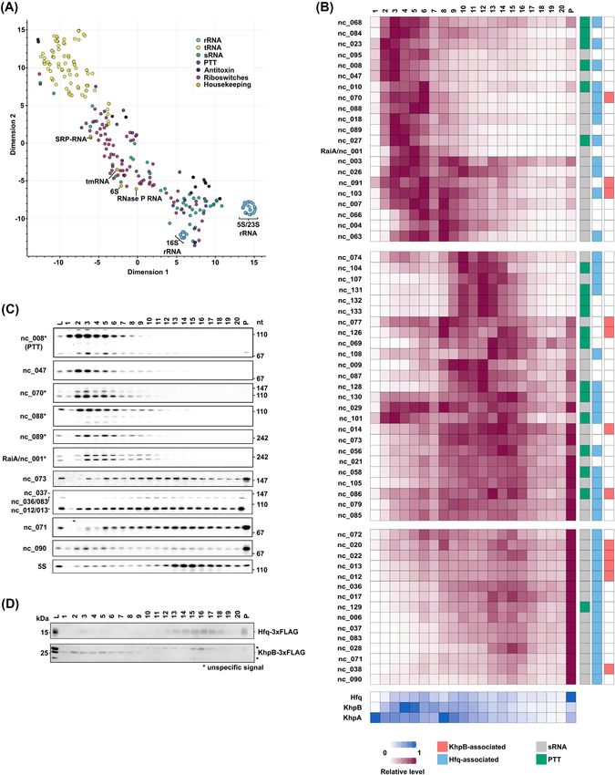
2      microLife, 2021, Vol. 2

targets of KhpB, similar to the scope of the Hfq targetome in C. difficile. The KhpB-bound transcripts include several
functionally related mRNAs encoding virulence-associated metabolic pathways and toxin A whose transcript levels are
observed to be increased in a khpB deletion strain. Moreover, the production of toxin protein is also increased upon khpB
deletion. In summary, this study expands our knowledge of cellular RNA protein interactions in C. difficile and supports the
emerging view that KhpB homologues constitute a new class of globally acting RBPs in Gram-positive bacteria.

Keywords: Clostridioides difficile; Grad-seq; RNA-binding protein; Jag; KhpB; KhpA; small RNA; toxin

INTRODUCTION                                                            regulators that control toxin expression in response to environ-
                                                                        mental signals (summarized in Bouillaut et al. (2015)).

                                                                                                                                                     Downloaded from https://academic.oup.com/microlife/article/doi/10.1093/femsml/uqab004/6245135 by guest on 27 July 2021
RNA–protein complexes serve important functions in all cellu-
                                                                             Genome-wide profiling studies have provided general tran-
lar processes associated with gene expression, from the reg-
                                                                        scriptome maps (Scaria et al. 2011, 2013; Antunes et al. 2012;
ulation of transcription to protein synthesis. Unsurprisingly,
                                                                        Janoir et al. 2013; Kansau et al. 2016; Jenior et al. 2017; Berges et al.
there have been long-standing efforts to understand the number
                                                                        2018; Giordano, Hastie and Carlson 2018; Neumann-Schaal et al.
and nature of RNA-binding proteins (RBPs) in model systems of
                                                                        2018; Fuchs et al. 2020; Fletcher et al. 2021) and protein invento-
molecular biology. Thanks to several recently developed global
                                                                        ries (Otto et al. 2016; Neumann-Schaal et al. 2018) under several
techniques, nearly saturated RPB catalogues are now available
                                                                        infection-relevant growth conditions. In contrast, experimental
for eukaryotic yeast and human cells (Huang et al. 2018; Queiroz
                                                                        evidence for protein complexes, alone or with RNA, is still scarce
et al. 2019; Shchepachev et al. 2019; Trendel et al. 2019). Ironi-
                                                                        in C. difficile (Jackson et al. 2006; Ciftci et al. 2019; Touchette et al.
cally, even though bacteria are often considered much simpler
                                                                        2019; Unal et al. 2019), and given the bacterium’s limited genetic
organisms, their RBP repertoire seems to be much harder to
                                                                        tractability, often based on heterologous protein expression in
catalogue (Holmqvist and Vogel 2018). The main reason being
                                                                        E. coli or B. subtilis (Aboulnaga et al. 2013; Demmer et al. 2017;
that as bacterial transcripts lack functional poly(A) tails, purifi-
                                                                        Valenčı́ková et al. 2018).
cation is less straightforward and global RBP co-purification after
                                                                             The limitations of working with C. difficile notwithstanding,
oligo(T)-based capture cannot be transferred from eukaryotes.
                                                                        this species offers promising leads towards a better understand-
    While Escherichia coli currently has ∼180 annotated RBPs,
                                                                        ing of functional ribonucleoprotein particles (RNPs) in bacteria.
many of which have a ribosomal function (Holmqvist and Vogel
                                                                        It is the only Gram-positive species in which the RNA chaperone
2018), far fewer RBPs are known in other bacterial species. These
                                                                        Hfq significantly impacts gene expression and bacterial physi-
under-researched species broadly include Gram-positive bacte-
                                                                        ology, leading to increased sporulation rates upon hfq deletion
ria, including many human pathogens of high medical inter-
                                                                        (Boudry et al. 2014; Maikova et al. 2019). Moreover, recent exper-
est. Important well-characterized RBPs of the Gram-negative
                                                                        imental annotation efforts have identified a large number of
model organisms E. coli and Salmonella enterica, such as Hfq,
                                                                        cis-regulatory RNA elements and sRNAs in this organism (Sou-
either are absent from Gram-positive bacteria (e.g. S. pneumo-
                                                                        tourina et al. 2013; Fuchs et al. 2020). In addition, Hfq exhibits
niae) or have different functions. For example, the RNA chaper-
                                                                        a surprisingly broad RNA-binding activity, interacting with at
one Hfq globally promotes small RNA (sRNA)-mediated regula-
                                                                        least 10% of all C. difficile transcripts including dozens of sRNAs
tion of mRNAs in Gram-negative enteric bacteria, and its dele-
                                                                        (Boudry et al. 2021; Fuchs et al. 2020).
tion usually results in pronounced phenotypes (Chao and Vogel
                                                                             Grad-seq is a recently introduced approach to discover RBPs
2010). By contrast, many of the known sRNAs in Gram-positive
                                                                        and their complexes in a poly(A)-independent manner (Smirnov
bacteria seem to be Hfq-independent. In addition, hfq disrup-
                                                                        et al. 2016). The method is based on the separation of soluble
tion in Listeria monocytogenes (Christiansen et al. 2004), Staphylo-
                                                                        cellular complexes by a classical glycerol gradient, followed by
coccus aureus (Bohn, Rigoulay and Bouloc 2007) or Bacillus subtilis
                                                                        high-throughput RNA-seq and mass spectrometry (MS) analy-
(Rochat et al. 2015) produces no obvious growth defects or influ-
                                                                        ses of the individual gradient fractions. Potential RBPs are then
ences the intracellular stability of sRNAs (Geissmann et al. 2009;
                                                                        predicted by a ‘guilt-by-association’ logic, searching for correla-
Toledo-Arana et al. 2009; Hammerle et al. 2014). The cellular RNA
                                                                        tion between in-gradient behavior of cellular proteins and tran-
degradation enzymes and machinery also differ significantly in
                                                                        scripts. Originally developed for S. enterica, Grad-seq guided the
Gram-positive species (Durand et al. 2015), limiting extrapola-
                                                                        discovery of a hitherto overlooked global RBP, the FinO-domain
tion of established knowledge from E. coli or S. enterica. There-
                                                                        containing protein ProQ (Smirnov et al. 2016). A more recent pio-
fore, systematic searches are needed to understand the general
                                                                        neering application of Grad-seq in a Gram-positive bacterium
landscape of RNA–protein interactions in Gram-positive species,
                                                                        helped to identify a new mechanism of exonucleolytic sRNA
which includes the human pathogen Clostridioides difficile.
                                                                        activation in competence regulation of Streptococcus pneumonia
    C. difficile is the leading cause of nosocomial diarrhoea fol-
                                                                        (Hör et al. 2020b).
lowing antibiotic treatment. Infections are inherently difficult
                                                                             In the present study, we applied Grad-seq to systematically
to treat using conventional antibiotic therapy (Peng et al. 2017;
                                                                        identify RNA–protein and protein-protein complexes in C. diffi-
Guery, Galperine and Barbut 2019), mainly due to C. difficile’s abil-
                                                                        cile. Using this new approach, we identify the broadly conserved
ity to form resistant spores. This has led to heightened inter-
                                                                        KhpB homologue Jag (to which we will refer as KhpB), originally
est in the molecular biology of the species itself with a grow-
                                                                        identified in S. pneumoniae (Ulrych et al. 2016; Stamsås et al. 2017;
ing number of mechanistic studies on how C. difficile physiology
                                                                        Zheng et al. 2017), as a new globally acting sRNA binding RBP in
and virulence are regulated (Paredes-Sabja, Shen and Sorg 2014;
                                                                        C. difficile. We show that the pervasive activity of KhpB resem-
Smits et al. 2016; McKee, Harvest and Tamayo 2018). For example,
                                                                        bles the scope of Hfq activity and includes the regulation of
numerous studies addressing the regulation of the clostridial
                                                                        toxin expression. As such, our findings support a view that KhpB
toxins TcdA and TcdB, the central virulence factors of C. difficile
                                                                        homologues constitute a new class of conserved bacterial RBPs
responsible for the symptoms of C. difficile infections (CDI; Smits
                                                                        with global RNA-binding activity.
et al. 2016) have revealed a complex network of transcriptional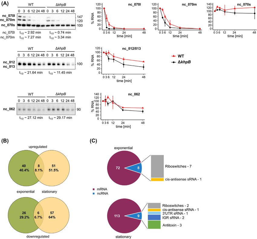
Lamm-Schmidt et al.          3

RESULTS                                                              RNAs (sRNAs, riboswitches, transcriptional attenuators resid-
                                                                     ing in 5 UTRs and type-I antitoxins) showed a very broad gradi-
Grad-seq analysis captures RNA-protein complexes in                  ent distribution indicating their existence in different molecular
C. difficile                                                         weight complexes.
To capture the landscape of cellular complexes in C. difficile we        Comparison of the sedimentation profiles of well-known
applied Grad-seq to a native cell lysate of the 630 wild type (WT)   RNA–protein complexes showed congruent profiles for the
strain grown to late-exponential phase in rich BHI broth. We         respective RNA and protein components, reflecting the preser-
chose the widely used reference strain 630, because it offers        vation of complexes in the gradient. For example, profiles of SRP,
the most comprehensive genome annotation to date. We use             the signal recognition particle formed by 4.5S RNA and protein
the original, most familiar genome annotation (e.g. CD0001)          Ffh, and of various tRNAs and their associated aminoacyl-tRNA
throughout the manuscript but datasets can be searched using         synthetases were highly similar (Fig. 1E). Further, proteins of the

                                                                                                                                             Downloaded from https://academic.oup.com/microlife/article/doi/10.1093/femsml/uqab004/6245135 by guest on 27 July 2021
all three available gene identifiers (e.g. CD0001, CD630 00010 and   small and large ribosomal subunits exhibited a strong correla-
CDIF630 00001; see Materials and Methods). Proteins, RNAs and        tion with their associated 16S and 5S/23S rRNAs, respectively
complexes were biochemically separated on a linear glycerol          (Fig. 1E).
gradient based on their size and shape yielding 20 individual
fractions and a pellet (Fig. 1A).                                    Evidence for a functional 6S-RNAP complex in C. difficile
    Conventional A260 nm analysis of the resulting gradient frac-    First discovered in E. coli (Wassarman and Storz 2000), functional
tions showed a bulk peak at low-molecular weight fractions and       associations of the abundant 6S RNA with RNA polymerase
two peaks for the small and large ribosomal subunits (Fig. 1B)       (RNAP) have been reported in multiple species, including the
which recapitulated Grad-seq profiles previously obtained for        Gram-positive bacteria B. subtilis (Trotochaud and Wassarman
E. coli, Pseudomonas aeruginosa, S. enterica and S. pneumoniae       2005; Beckmann, Burenina and Hoch 2011) and S. pneumoniae
(Gerovac et al. 2020a,b; Hör et al. 2020a,b; Smirnov et al.         (Hör et al. 2020b). Here, northern blot probing of 6S RNA in the
2016). Separation of RNA-samples on a conventional ethidium          gradient fractions revealed two different RNA species of approx.
bromide-stained polyacrylamide gel visualized the most abun-         200 nt and 175 nt (Figure S1B, Supporting Information), similar
dant cytosolic RNAs that are associated with the 30S (16S rRNA)      to previous signals in C. difficile (Soutourina et al. 2013), B. sub-
and 50S (5S/23S rRNAs) ribosomes as well as tRNAs (Fig. 1C). In      tilis (Trotochaud and Wassarman 2005) and S. pneumoniae (Acebo
addition, an abundant transcript likely corresponding to tmRNA       et al. 2012). Importantly, these 6S RNA species clearly co-migrate
is visible, suggesting its presence in a stable complex with its     with the subunits of C. difficile RNAP in the gradient (Fig. 1E),
cognate protein partner SmpB. Complementing this picture are         which strongly indicate that they are present in a stable com-
protein profiles characteristic of 30S and 50S ribosomal subunits    plex. In addition, functional 6S RNA is known to be used by RNAP
that were obtained by SDS-PAGE analysis of the gradient frac-        as a template, leading to the synthesis of 14–20 nt RNA products
tions (Fig. 1D).                                                     (pRNAs; Wassarman 2018). Importantly, our transcriptome map-
    We noticed two additional highly abundant RNA species            ping of C. difficile detects such pRNAs and shows that they do
peaking in fractions 3–5, which could not be assigned to             initiate within the central bulge, as expected (Figure S1C, Sup-
any known or predicted housekeeping RNA species (Fig. 1C,            porting Information; arrow; Fuchs et al. 2020). Combined, these
orange boxes). We propose that they correspond to RaiA               data provide evidence for this to be a functional 6S RNA that
(CDIF630nc 001), an exceptionally abundant sRNA recently iden-       associates with RNAP in vivo.
tified by RNA-seq based annotation of the C. difficile strain 630         To facilitate a straightforward analysis of this complex RNA
(Fuchs et al. 2020). The level of RaiA in the cell lysate is com-    and protein data, all profiles can be viewed in an online browser,
parable to that of rRNA species (Table S1, Supporting Informa-       which is available at https://helmholtz-hiri.de/en/datasets/grad
tion), which would explain its potential detection by ethidium       seqcd. The browser allows easy access and comparison with pre-
bromide-staining. Northern blot probing of RaiA in the gradient      viously published Grad-seq datasets for other Gram-positive and
fractions revealed two strong signals at approx. 260 nt and 220 nt   Gram-negative species (Gerovac et al. 2020a,b; Hör et al. 2020a,b;
that matched the staining pattern in the RNA gel (Fig. 3B). Our      Smirnov et al. 2016).
RNA-seq based annotation of the raiA gene identified one TSS
and two associated termination sites, which would account for
                                                                     Functional implications of in-gradient protein profiles
the different lengths of the two detected transcripts. RaiA rep-
resents the first experimentally validated member of the ncRNA       Having established proof-of-concept for Grad-seq in C. difficile,
family RaiA (Weinberg et al. 2017) whose members can be found        we examined our data for potential new RBPs outside of con-
in Firmicutes and Actinobacteria. So far, their potential func-      served housekeeping RNPs. We focused on small proteins of
tion(s) remains unknown. That said, our detection of RaiA in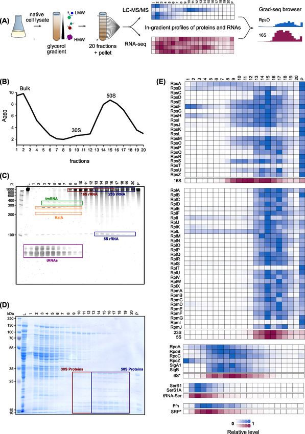
4      microLife, 2021, Vol. 2

                                                                                                                                                                           Downloaded from https://academic.oup.com/microlife/article/doi/10.1093/femsml/uqab004/6245135 by guest on 27 July 2021

Figure 1. Grad-seq visualizes the RNA/protein complexome of C. difficile. (A) Grad-seq workflow. (B) A260 absorbance profile of gradient fractions. Low-molecular-weight
complexes (bulk peak) and ribosomal subunits (30S, 50S) are highlighted. (C) Ethidium bromide stained PAA gel showing gradient distribution of housekeeping RNAs
(tmRNA, tRNAs, 5S rRNA, 16S rRNA and 23S rRNA) and RaiA. (D) Coomassie stained SDS gel showing gradient distribution of proteins. Ribosomal subunit proteins (30S
and 50S) are highlighted. (E) Heatmaps showing normalized sedimentation profiles of selected housekeeping RNA-protein complexes. Data were obtained by RNA-seq
and LC-MS/MS analysis of gradient fractions. Sedimentation profiles of proteins and transcripts are scaled in the range from 0 to 1. Fractions 1–20 and the pellet (P)
are shown. ∗ not available in previous genome annotations.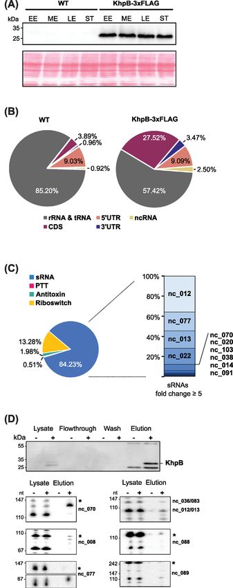
Lamm-Schmidt et al. 5
Downloaded from https://academic.oup.com/microlife/article/doi/10.1093/femsml/uqab004/6245135 by guest on 27 July 2021
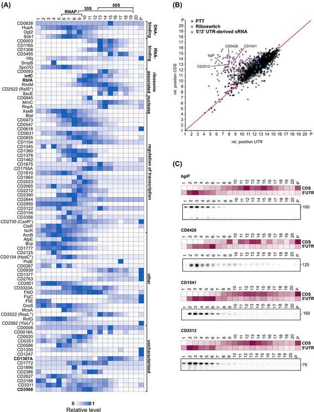
Figure 2. Grad-seq sedimentation profiles facilitate functional predictions for proteins and RNA transcripts. (A) Heatmaps showing normalized sedimentation profiles
of small proteins ( fraction 3). A fast sedimentation of small proteins can indicate that the proteins are part
of a larger complex. Predicted protein functions (Uniprot) and the positions of RNA polymerase (RNAP) and ribosomal subunits (50S and 30S) are indicated. Proteins
discussed in the main text are indicated in bold letters. (B) Correlation of sedimentation profiles for UTR–CDS pairs reveals 5 /3 -UTRs with divergent sedimentation
behavior. The relative position for each UTR was plotted against the relative position of its corresponding CDS. For calculation of relative positions see Materials
and Methods. Data points are colored according to UTR association with a riboregulatory element. UTRs that were validated by Northern blot in (C) are labeled. (C)
Heatmaps of normalized UTR/CDS sedimentation profiles and northern blot validation of gradient fractions using radioactively labeled DNA probes specific for the
5 -UTR regions of bglF (CDIF630nc 008, PTT), CD0426 (CDIF630nc 010, PTT), CD1541 and CD2512.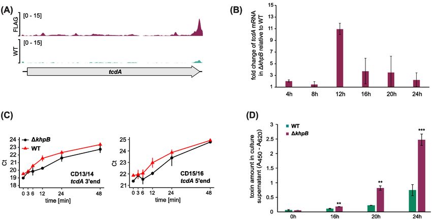
6      microLife, 2021, Vol. 2

                                                                                                                                                                        Downloaded from https://academic.oup.com/microlife/article/doi/10.1093/femsml/uqab004/6245135 by guest on 27 July 2021

Figure 3. Analysis of ncRNA sedimentation profiles. (A) Scatterplot showing global RNA-seq data of all known ncRNA in C. difficile after dimension reduction with
tSNE. ncRNAs with similar gradient sedimentation profiles cluster together, t-SNE perplexity = 40. Housekeeping genes are highlighted. (B) top: Heatmaps showing
normalized sedimentation profiles of all predicted sRNAs and PTTs in C. difficile. sRNAs were clustered into three big clusters of similarity with k-means algorithm.
Known Hfq-binding sRNAs are marked in light blue and transcripts enriched in KhpB-3×FLAG RIP-seq are marked in light red. Bottom: Heatmaps showing normalized
sedimentation profiles of Hfq and the two putative RNA-binding proteins KhpB and KhpA are shown below. (C) Northern blots of gradient fractions. Blots were probed
for different sRNAs with radioactively labelled DNA oligos. sRNAs marked with an asterisk were chosen for RNA-bait dependent pulldown. (D) Western blots of gradient
fractions of C. difficile 630 Hfq-3×FLAG and C. difficile 630 KhpB-3×FLAG. Western blots were incubated with anti-FLAG antibody.
Lamm-Schmidt et al.          7

non-spore-forming bacteria (e.g. Lactobacillus; Figure S2A, Sup-      (t-Distributed Stochastic Neighbor Embedding) dimension
porting Information). In B. subtilis (Fukushima et al. 2003; Lei      reduction (van der Maaten and Hinton 2008). Using this anal-
et al. 2013) the Veg protein is expressed from a predicted σ A -      ysis, high dimensional datasets can be visualized by giving
dependent promoter both during vegetative growth and sporu-           each datapoint a location within a two- or three-dimensional
lation, indicating a potential housekeeping function (Fukushima       plot, with similarly behaving datapoints clustering together.
et al. 2003). Here, our Grad-seq profiles show a distinct peak        This analysis revealed distinct clusters of functionally related
of CD3568 in fractions 10 and 11 (Fig. 2A) containing the free        housekeeping RNAs, such as the 5S/23S rRNAs, as well as tRNAs
30S subunit along with many mRNAs which could point to a              (Fig. 3A). As described above, their sedimentation behavior
potential ribosome- or mRNA-associated regulatory function of         mirrored that of their corresponding RNPs (Fig. 1E). By contrast,
CD3568 during vegetative growth and sporulation.                      sRNAs and PTTs displayed heterogeneous behavior. The normal-
    Another interesting example is CD1307A (Q18BH1), a 10.4 kDa       ized sedimentation profiles revealed one cluster of sRNAs/PTTs

                                                                                                                                                 Downloaded from https://academic.oup.com/microlife/article/doi/10.1093/femsml/uqab004/6245135 by guest on 27 July 2021
protein of the COG2740 family which is characterized by a             that peaked strongly in the pellet fraction, which contains
conserved motif GRGA(Y/W) (Figure S2B, Supporting Informa-            not only 70S ribosomes but also Hfq (Fig. 3B). Importantly, all
tion). The S. pneumoniae homologue (PDB entry: 1G2R) is struc-        sRNAs/PTTs in this cluster were recently shown to associate
turally dissimilar to any known protein but possesses a posi-         with Hfq (Fuchs et al. 2020; Boudry et al. 2021), reaffirming the
tively charged patch that suggests nucleic acid binding (Osip-        role of Hfq as a general sRNA RBP in C. difficile. A second large
iuk et al. 2001). The B. subtilis homologue YlxR has recently been    sRNA cluster associated partially with the pellet fractions but
implicated in post-transcriptional regulation of the fructosely-      also with 30S and contained several Hfq-dependent sRNAs. The
sine utilization operon (Ogura and Kanesaki 2018; Ogura, Sato         co-sedimentation of Hfq-dependent sRNAs with 30S ribosomes
and Abe 2019; Ogura, Shindo and Kanesaki 2020). Our Grad-seq          suggests a classical mode-of-action, i.e. as modulators of mRNA
analysis shows CD1307A to peak sharply in fraction 15 along           translation initiation through their Hfq-facilitated binding to
with the 50S subunit (Fig. 2A), and this ribosome association         trans-encoded mRNA targets. A third cluster of sRNAs and
might be considered additional support for a potential function       PTTs was distinct from the ribosomal fractions (Fig. 3B). In silico
in post-transcriptional control.                                      profiles were readily confirmed by northern analysis of several
                                                                      selected sRNAs from all three clusters and 5S rRNA (Fig. 3C).
                                                                      In their sum, these results suggested the existence of another
In-gradient RNA profiles reveal potential UTR-derived                 sRNA/PTT-binding RBP in C. difficile.
ncRNAs                                                                    To identify this candidate RBP, we sought to co-purify pro-
                                                                      teins from C. difficile lysate with in vitro-transcribed sRNAs (Hör
Similarly to proteins, sedimentation profiles of transcripts can
                                                                      et al. 2020b). As bait RNAs we selected several sRNAs with
point at molecular functions. To illustrate this, we focused on
                                                                      peaks outside the ribosomal fractions (marked with an asterisk
the mRNA 5 UTRs and 3 UTRs, many of which harbor cis- and
                                                                      in Fig. 3C). This set included three Hfq-binding sRNAs (whose
trans-acting regulatory elements with functions independent of
                                                                      names are highlighted with blue boxes in Fig. 4A and Figure
their parental mRNAs (Serganov and Nudler 2013; Dar et al.
                                                                      S3, Supporting Information) and two Hfq-independent sRNAs.
2016; Adams et al. 2021). Calculating the Spearman’s rank cor-
                                                                      Each of these bait sRNAs enriched several proteins (Fig. 4A, B
relation of sedimentation profiles for 5 /3 UTRs and their cor-
                                                                      and Figure S3, Supporting Information) and, reassuringly, all
responding mRNAs revealed strong correlation for the major-
                                                                      three Hfq-binding sRNAs enriched Hfq. While Spearman’s corre-
ity of transcripts, i.e. a UTR and its associated CDS exhibited
                                                                      lation analysis showed good agreement (R > 0.8) for several pro-
the same sedimentation profile (Fig. 2B). Interestingly, however,
                                                                      teins, the most convincing candidate from all pull-downs was
there were numerous UTRs showing a low correlation with their
                                                                      the KhpB protein (Fig. 3B).
parental mRNA (R < 0.2); these often harbored a conserved
                                                                          KhpB contains two RNA binding domains, a well character-
riboswitch, a premature transcription termination (PTT) ele-
                                                                      ized KH-domain and a putative R3H (Grishin 1998; Valverde,
ment, or a 5 /3 UTR-derived sRNA (Fig. 2B and Table S2, Sup-
                                                                      Edwards and Regan 2008; Nicastro, Taylor and Ramos 2015),
porting Information). A case in point are two recently identi-
                                                                      as well as a Jag domain mediating protein-protein interactions
fied premature termination events in the 5 UTRs of the bglF
                                                                      (Winther et al. 2021; Fig. 3F). This domain composition together
and CD0426 genes (Fuchs et al. 2020; Fig. 2C). Additional inter-
                                                                      with our pull-down findings strongly suggests that C. difficile
esting UTR candidates include the 5 UTRs of CD1541 (putative
                                                                      KhpB function as an RNA-binding protein. KhpB is broadly con-
drug/sodium antiporter of the MATE family) and CD2512 (PTS
                                                                      served having homologues, for example, in S. pneumoniae (a.k.a.
system, maltose-specific IIA component). Northern blotting of
                                                                      EloR/KhpB) and Lactobacillus plantarum (EloR; Figure S4, Support-
gradient fractions detected stable transcripts for all 5 UTRs that
                                                                      ing Information). In S. pneumoniae it undergoes heterodimeriza-
formed LMW complexes (Fig. 2C). While most of them likely
                                                                      tion with another KH-domain containing protein, called KhpA,
represent stable products of transcription attenuation or mRNA
                                                                      and binds a variety of transcript classes in vivo (Zheng et al. 2017;
degradation processes, this suggests that they could have addi-
                                                                      Winther et al. 2019). Interestingly, we also identified the C. difficile
tional functions in trans, independent of the translation of their
                                                                      KhpA homologue CD1254 in our pulldowns (Fig. 4A, B and Fig-
parental mRNA.
                                                                      ure S3, Supporting Information), but it was not always enriched
                                                                      above threshold (log2 = 4). Similar to KhpB homologues, gene
Identification of KhpB as a non-coding RNA-binding                    synteny and sequence conservation is very high for KhpA homo-
protein in C. difficile                                               logues (Figure S5, Supporting Information); therefore, we will
                                                                      refer to this protein as KhpA hereafter. Since KhpA peaked pre-
Next, we specifically searched for C. difficile non-coding RNAs       dominantly in fraction 1 and in higher molecular weight (HMW)
with an unknown RBP partner. To find similarly behaving               fractions, we decided to focus our further analysis on KhpB
ncRNAs in our global RNA-seq data, we performed t-SNE                 itself.
8 microLife, 2021, Vol. 2
Downloaded from https://academic.oup.com/microlife/article/doi/10.1093/femsml/uqab004/6245135 by guest on 27 July 2021
Figure 4. Identification of the RNA-binding protein KhpB. (A) LC-MS/MS analysis of RNA-bait dependent pulldowns with Hfq-dependent sRNA CDIF630 070. The Log10
LFQ intensities of the sample plus control were plotted against the log2 ratio for the sample versus control. Thresholds were set to log10 = 5 for LFQ intensity and
to log2 = 4 for the ratio. All proteins significantly enriched are highlighted in yellow or red. RNA-binding proteins Hfq, KhpB and KhpA are highlighted in red. (B)
Spearman’s correlation coefficient matrix of sedimentation profiles of sRNA baits and proteins that were enriched in the RNA-bait dependent pulldown. Proteins that
were specifically enriched with each bait sRNA are marked with an asterisk. (C) Genomic location and domain structure of the RNA-binding protein KhpB.
C. difficile KhpB is a global RBP KhpB homologues in cell division in S. pneumoniae and L. plan-
tarum (Ulrych et al. 2016; Stamsås et al. 2017; Zheng et al. 2017;
To determine potential in vivo target transcripts of KhpB, we Myrbråten et al. 2019; Winther et al. 2019). To test this further,
took a RIP-seq (RNA immunoprecipitation followed by deep we constructed a khpB null mutant (khpB). To acquire insight
sequencing) approach after genetically tagging the protein with into potentially overlapping functions of KhpB and KhpA, we
3×FLAG epitope at the C-terminus as previously published for S. also included a khpA knockout strain. Both mutant strains phe-
pneumoniae KhpB (Zheng et al. 2017). Western blot analysis nocopied each other during growth, exhibiting a reduced growth
showed KhpB-3×FLAG to be abundant throughout different rate during exponential phase in comparison to the WT (Figure
phases of growth (Fig. 5A), and the KhpB-3×FLAG strain showed S8A, Supporting Information). By contrast, cell morphology of
identical growth to the wild type in three different media (Fig- the khpA strain essentially resembled WT, whereas the khpB
ure S6A, Supporting Information). Western blot analysis of gra- mutant exhibited slightly increased cell lengths and widths (Fig-
dient fractions generated with the KhpB-3×FLAG strain revealed ure S8B, Supporting Information).
a sedimentation profile comparable to that of the WT protein Among enriched non-coding transcripts, reads mapping to
(compare Fig. 3B and D) and confirmed its co-sedimentation sRNAs (10/42) and riboswitches (9/80) were overrepresented
with the bait RNAs. Overall, these data suggested that the (compared to 4/15 type I antitoxins and 2/19 PTTs; Fig. 5C).
recombinant protein behaved like WT KhpB and was suitable Northern blot analysis of KhpB WT and KhpB-3×FLAG coIP
for RIP-seq analysis. fractions independently confirmed binding of KhpB to selected
RIP-seq revealed extensive RNA-binding by KhpB-3×FLAG enriched sRNAs from the RIP-seq analysis (Fig. 5D). These
with ∼1400 transcripts being significantly enriched (FC ≥ 5). included a group of sRNAs with highly similar primary sequence
Of the different transcript classes, CDS were clearly overrep- (nc012, nc013, nc036 and nc083). For the nc070 and nc077 sRNAs,
resented, whereas enrichment of rRNAs or tRNAs was not we observed significant enrichment of only the larger or small
observed (Fig. 5B). The large number of enriched CDS might be transcript variants, respectively. Further, we confirmed KhpB
partially attributable to the fact that we often observed enrich- binding to three of the bait sRNAs used to co-purify KhpB from
ment for all the genes of long operons, such as the flgB flagellar C. difficile cytosol. Interestingly, these three sRNAs (nc008, nc070
operon, the atpZ operon or the cooS operon, the latter of which and nc088) had also enriched KhpA in their pulldowns suggest-
functions in the Wood–Ljungdahl pathway (Figure S7C, Support- ing that KhpB and KhpA cooperate in RNA binding similarly to
ing Information). their homologues in S. pneumoniae (Zheng et al. 2017).
Enriched transcripts encoded for various physiological func-
tions including the related pathways for flagellar assembly and Impact of KhpB on cellular transcripts
chemotaxis (Figure S7A and Table S3, Supporting Information).
Further, we found several enriched mRNAs of cell division pro- Next, we investigated if and how KhpB might influence the
teins, including ftsZ (Figure S7B, Supporting Information), minE, fate of those transcripts found to be enriched in the RIP-seq
minC, sepF and ftsK (Table S3, Supporting Information), which experiment. We used rifampicin to determine RNA stability
was reminiscent of the previously described regulatory role of changes of KhpB-bound sRNAs. Northern blot analysis showedLamm-Schmidt et al. 9
different stabilities between the WT and the khpB deletion
strain (Fig. 6A), with KhpB appearing to exert both positive and
negative effects on steady-state levels (see CDIF630nc 062 and
CDIF630nc 070, respectively). In the case of the CDIF630nc 070
sRNA, we observed decreased stability only for the two larger
transcripts of three detectable transcripts. Interestingly, only
the larger transcript was enriched in the KhpB-3×FLAG pull-
down (Fig. 5D), suggesting the short form of this sRNA represents
a KhpB-independent stable degradation product. Together, the
data pointed to a role of KhpB as an sRNA stability factor in C.
difficile.
Downloaded from https://academic.oup.com/microlife/article/doi/10.1093/femsml/uqab004/6245135 by guest on 27 July 2021
To obtain a global picture of KhpB-dependent transcript
changes, we performed RNA-seq on khpB, comparing it to the
WT strain in the late-exponential and stationary growth phases.
Deletion of khpB impacted the steady-state levels of 80 tran-
scripts in late-exponential phase (gradient condition) and 122
transcripts in stationary phase (log2 FC > 1 or log2 FC < −1;
Fig. 6B and Table S4, Supporting Information). The large major-
ity of differentially expressed genes were protein coding genes,
but many riboswitches, type-I antitoxins and sRNAs were also
affected; by contrast, housekeeping rRNAs or tRNAs showed no
changes (Fig. 6C and Table S4, Supporting Information). Interest-
ingly, the set of up- and down-regulated genes differed signif-
icantly between the two growth phases, suggesting that KhpB
regulates distinct physiological pathways during growth.
Pathway enrichment analysis of regulated genes indicated
increased transcript levels for metabolic pathways during sta-
tionary phase that cooperate in the fixation of atmospheric
or glycolysis-derived CO2 into acetyl-CoA (Figure S9, Sup-
porting Information): the Wood–Ljungdahl pathway and the
glycine cleavage system (cooS-cooC-fhs-fchA-folD-metV-metF-lpdA,
CD0724-acsD-acsC-acsE-cdhC-gcvH and gcvPB; Fonknechten et al.
2010; Kopke, Straub and Durre 2013; Song et al. 2020). In addi-
tion, the acetyl-CoA carboxylase (accBCDA) operon whose pro-
teins catalyse the first step in fatty acid biosynthesis from acetyl-
CoA showed reduced transcript levels during stationary phase
along with the transcriptional repressor of fatty acid biosyn-
thesis fapR. All these individual transcripts or operons, respec-
tively, were also enriched in the RIP-seq dataset suggesting
they might be directly regulated by KhpB. Further, the succinate
to butyrate pathway (CD2344-cat1-sucD-abfD-CD2340-cat2) that
promotes intestinal colonization of C. difficile in a mouse model
of C. difficile infection (Ferreyra et al. 2014), was up-regulated dur-
ing late-exponential growth, in addition to being enriched in the
RIP-seq dataset (Figure S9, Supporting Information).
Figure 5. RIP-seq identifies KhpB as a global RNA-binding protein. (A) West- KhpB is a regulator of toxin A
ern blot analysis of KhpB protein levels across different growth phases in BHI
medium. Equal OD units of total cell lysates of C. difficile 630 WT and KhpB- Induction of the identified KhpB-dependent metabolic pathways
3×FLAG were loaded. Western blot membranes were incubated with anti-FLAG were known to highly correlate with maximum production of
antibody. As a loading control, the blotting membrane was stained with Pon-
the central virulence factors of C. difficile, the clostridial toxins
ceau S. EE-early exponential, ME—mid exponential, LE—late exponential, ST—
TcdA and TcdB (Karlsson, Burman and Akerlund 2008). The tcdA
stationary (B) Distribution of mapped reads across RNA classes in KhpB WT and
KhpB-3×FLAG RIP-seq libraries. (C) Pie-chart with distribution of mapped reads mRNA (note that toxin A is annotated as toxA in CP010905.2)
in the KhpB-3×FLAG library across ncRNAs classes (sRNA, PTT, riboswitches and was also enriched in the RIP-seq dataset (log2 FC = 2.21; Fig. 7A)
antitoxins). Distribution of mapped reads across significantly enriched sRNAs although slightly below the set cutoff (log2 FC = 2.5). How-
in KhpB-3×FLAG RIP-seq are shown as a stacked bar graph. Read counts are ever, considering that toxin genes are generally repressed during
normalized to transcript per million. (D) Western blot (top) and Northern blot
exponential growth in rich medium, it is possible that tcdA tran-
(bottom) analysis of KhpB-3xFLAG co-immunoprecipitation (co-IP). Lysates of C.
script levels were just too low for significant enrichment. Toxin
difficile 630 WT (-) and KhpB-3×FLAG (+) grown to late-exponential phase were
subjected to immunoprecipitation with anti-FLAG antibodies. Western blot anal- synthesis is subject to control by multiple transcriptional regu-
ysis shows lysate, flowthrough, wash and eluates of co-IP. KhpB protein is high- lators of the tcdA and tcdB toxin genes, which act in response to a
lighted. Additional bands in the eluates correspond to the anti-FLAG antibody variety of environmental signals (summarized in (Bouillaut et al.
used for co-IP. Northern blots show total RNA extracted from lysate and eluates 2015)). By contrast, post-transcriptional mechanisms of these
of WT (-) and KhpB-3×FLAG (+) co-IPs. Blots were probed for indicated sRNAs
genes were unknown. Therefore, we sought to further explore
with radioactively labelled DNA oligos.
the role of KhpB in tcdA regulation.10 microLife, 2021, Vol. 2
Downloaded from https://academic.oup.com/microlife/article/doi/10.1093/femsml/uqab004/6245135 by guest on 27 July 2021
Figure 6. Impact of KhpB on cellular transcript levels and stabilities. (A) Rifampicin assay to determine half-lives of KhpB-bound sRNAs in vivo. Samples from C. difficile
630 WT and khpB were grown to late exponential phase. Samples were taken at the indicated timepoints after addition of rifampicin. Extracted RNA was analyzed by
Northern blotting, a representative of three biological replicates is shown. Error bars show the standard deviation of the mean. Approximate half-lives were calculated
with GraphPad Prism and are shown next to each corresponding Northern blot. (B) Venn diagrams for genes differentially expressed in C. difficile khpB compared to
WT. Cultures were grown to late-exponential or stationary phase. Upper diagram shows upregulated genes, lower diagram shows downregulated genes. (C) Fractions
of transcript classes that were significantly differentially regulated (up and down) between WT and khpB.
The tcdA mRNA was both enriched in the RIP-seq analysis protein level, we quantified toxin levels by ELISA. In agreement
and increased in the khpB deletion strain (Tables S3 and S4, with the RIP-seq, transcriptome and rifampicin data, toxin lev-
Supporting Information). Quantification by RT-qPCR confirmed els in culture supernatants were increased ∼3.3-fold in the khpB
increased tcdA mRNA levels in the absence of KhpB predomi- mutant (Fig. 7D). Taken together, these data point to a role of
nantly in the early stationary growth phase (12 h; Fig. 6B). Next, KhpB in toxin regulation, possibly through post-transcriptional
we quantified the tcdA mRNA by qPCR in rifampicin-treated regulation of toxin production, adding a new layer of complexity
samples of the WT and khpB strains. Because of the large size to virulence regulation in C. difficile.
of this transcript (∼ 8kb), we performed RT-qPCR with two dif-
ferent primer pairs in either the tcdA 5 or 3 region. Differences
DISCUSSION
in Ct values over time confirmed general increases in tcdA lev-
els in khpB and indicated slight changes in mRNA stabilities The Grad-seq analysis of C. difficile presented in this paper pro-
(Fig. 7C), although they did not clearly reveal a stabilizing activity vides the first global map of cellular RNA and protein complexes
of KhpB as observed for KhpB-bound sRNAs (Fig. 6A). Finally, to in this important human pathogen. In complementing pub-
determine whether KhpB also impacts toxin production on the lished Grad-seq data sets for the three Gram-negative speciesLamm-Schmidt et al. 11
Downloaded from https://academic.oup.com/microlife/article/doi/10.1093/femsml/uqab004/6245135 by guest on 27 July 2021
Figure 7. Functional characterization of KhpB identifies a role in toxin regulation. (A) Reads from KhpB WT (turquois) and KhpB-3xFLAG (magenta) RIP-seq libraries
mapping to the tcdA region. (B) qPCR-based quantification of tcdA mRNA levels in C. difficile 630 WT and khpB across different growth phases. Error bars show standard
deviation of the mean of three biological replicates. (C) Total RNA was extracted from C. difficile 630 WT and khpB samples that were taken at the indicated timepoints
after inhibition of transcription by the addition of rifampicin in late-exponential growth phase. Transcript levels of tcdA were quantified by qPCR. Error bars show
standard deviation of the mean Ct values of three biological replicates. (D) ELISA-based toxin quantification in culture supernatants of WT and khpB grown in BHI.
Samples were taken at the indicated timepoints. Error bars show standard deviation of the mean of three biological replicates. P values were obtained by students
t-test. ∗ P < 0.05, ∗∗ P < 0.01 and ∗∗∗ P < 0.001.
E. coli, P. aeruginosa and S. enterica, and Gram-positive S. pneumo- domains (Figure S4B, Supporting Information; Ulrych et al. 2016;
niae (Smirnov et al. 2016; Gerovac et al. 2020a; Hör et al. 2020a,b), Zheng et al. 2017), in addition to a conserved genomic loca-
it will be worthwhile to identify both commonalities and dif- tion adjacent to rnpA and yidC (CD3678; Figure S4A, Support-
ferences for orthologous proteins in these phylogenetically dis- ing Information). Similar to previous observations with KhpB
tant bacteria. Specifically, gradient sedimentation profiles add homologues in S. pneumoniae and L. plantarum (Stamsås et al.
confidence to protein function annotations that are commonly 2017; Myrbråten et al. 2019; Winther et al. 2019), we also find evi-
achieved by homology-based inference. One such example is the dence that these RBPs impact cell division. In other words, KhpB
50S-ribosome associating heat shock protein Hsp15 in E. coli and homologues seem to be involved in the regulation of cell divi-
its orthologues in S. pneumoniae and C. difficile. All of them were sion in both ovococcoid and rod-shaped bacteria, which is quite
found to co-sediment with 50S ribosomes by Grad-seq which astonishing given the great differences in the underlying cell
strongly suggests a conserved function of Hsp15 orthologues in division mechanisms (Massidda, Nováková and Vollmer 2013;
these distantly related bacteria. In addition, the example of 6S Eswara and Ramamurthi 2017).
RNA and its consistent co-detection with RNAP highlights that Our data reveal binding of KhpB and Hfq to an overlapping set
conserved functions of non-coding regulators can be inferred in of sRNAs which is reminiscent of the overlapping and compet-
the absence of existing primary sequence conservation (Figure ing roles of the global RNA chaperones Hfq and ProQ in E. coli and
S1, Supporting Information). Further, similarly to a recent com- S. enterica (Holmqvist et al. 2018; Melamed et al. 2020). Currently,
parative Grad-seq analysis of RNase-sensitive gradient profiles we still know too little about either Hfq- or KhpB-mediated regu-
(GradR) in S. enterica (Gerovac et al. 2020a), comparative Grad- lation in C. difficile to draw precise conclusions on potential sim-
seq analysis could also be applied to bacterial cultures subjected ilarities or differences in their modes of action. However, both
to specific stress conditions. We are confident that this type of RBPs have the ability to stabilize their bound sRNA ligands. On
analysis will not only provide a comprehensive resource for the the other hand, enrichment patterns in the RIP-seq data for Hfq
investigation of RNA-protein complexes in C. difficile but that it (Fuchs et al. 2020; Boudry et al. 2021) and KhpB point towards
will shed light on conserved complexes among Gram-positive some major mechanistic differences. While Hfq enriched for
and Gram-negative bacteria. many sRNAs as well as predominantly 5 - and 3 -UTR regions
Our study suggests that the widely conserved KhpB homo- of mRNAs, the vast majority of KhpB-bound transcripts com-
logue in C. difficile acts as a second global RNA-binding protein, prises the CDS of mRNAs and even entire operons (Fig. 5B). Thus,
in addition to Hfq, and that one of its functions is to regulate unlike Hfq, KhpB might not function primarily in matchmaking
toxin production. KhpB is part of an RBP family that is widely of sRNA-target interactions. Also, the conserved regulatory func-
conserved among gram-positive bacteria, especially Firmicutes. tion of KhpB in cell division sets it apart from Hfq. Therefore, at
This family is characterized by their shared domain structure this early stage of our understanding of these two global RBPs
(Fig. 4C) and conserved catalytic residues in 3 domains: the Jag it seems that they likely engage in different aspects of C. difficile
domain and the well-established RNA binding KH and the R3H RNA biology.12 microLife, 2021, Vol. 2
Further, the modular domain structure of KhpB with its two of KhpB in regulating physiological adaptation to the intestinal
RNA-binding domains and the Jag domain that was recently environment and virulence. In S. pneumoniae KhpB also seems
shown to mediate protein–protein interactions in S. pneumoniae to be involved in the regulation of virulence. More precisely, a
(Winther et al. 2021) suggests engagement of KhpB in various Tn-Seq screen of the TIGR4 strain showed that KhpB is required
protein complexes. For example, proteins containing a single to cause disease in a murine model of pneumonia (van Opijnen
KH domain usually cooperate with other RNA-binding domains and Camilli 2012). However, in both S. pneumoniae and C. difficile
or proteins to achieve high-affinity and specific RNA binding our understanding of the underlying mechanisms is still in its
(Valverde, Edwards and Regan 2008; Nicastro, Taylor and Ramos infancy.
2015). In line with that, streptococcal KhpB forms a heterodimer We note that the majority of KhpB-bound transcripts showed
with another KH domain protein, KhpA, and both proteins bind no changes in steady-state levels, at least with the stringent
to an overlapping set of sRNAs and mRNAs in vivo (Zheng et al. threshold (log2 FC > 1 or log2 FC < −1) applied. While this does
Downloaded from https://academic.oup.com/microlife/article/doi/10.1093/femsml/uqab004/6245135 by guest on 27 July 2021
2017). Since S. pneumoniae lacks homologues of the known global not exclude potential KhpB-dependent effects on the half-lives
RBPs CsrA, Hfq and ProQ, the KhpA/B proteins have been sug- of those transcripts, it indicates that binding by KhpB might
gested to serve a function analogous to Hfq. Interestingly, both have different consequences, potentially depending on the type
proteins co-sedimented in fractions 2–4 in a Grad-seq analysis of of complex that KhpB is involved in. A discrepancy between
S. pneumoniae (Hör et al. 2020b). Our RNA pulldown results sug- bound and differentially expressed transcripts could also be
gest the existence of a potential KhpB/KhpA complex in C. dif- explained by indirect effects of KhpB activity, analogous to what
ficile as well, since both proteins were enriched with 3 out of 5 was observed for KhpA in S. pneumoniae, where deletion of khpA
bait RNAs. Their gradient profiles, however, are more complex, resulted in cell wall stress that in turn induced expression of the
with KhpB showing a broader distribution between fractions 2– two-component system WalRK (Zheng et al. 2017). At least under
8, whereas the smaller KhpA protein peaked predominantly in the conditions analyzed in our study, the transcriptome profiles
HMW fractions 8–9 in addition to being present in fraction 1, of the khpB mutant did not reveal a clear stress signature.
possibly as unbound monomer or as a homodimer. Of course, However, we did observe increased expression of the sin locus
this does not exclude a possible heterodimer formation between (CD2214–2215, log2 FC ∼ 3) in both growth conditions, which
the two proteins, but it does suggest that these two proteins might provide an alternative explanation for the increased toxin
independently engage in additional complexes. Follow-up stud- production we observed in the khpB mutant. The sin locus
ies will be necessary to investigate the possibility that certain encodes the transcriptional regulator SinR and its antagonis-
cellular pathways are regulated by their joint activity, while oth- tic partner SinR’ which blocks SinR activity through protein–
ers might be regulated in a KhpB- or KhpA-specific manner. Our protein interaction (Girinathan et al. 2018; Ciftci et al. 2019). How-
initial characterization of KhpA or KhpB deletion strains already ever, deletion of the entire sin locus resulted in an asporogenes
suggests only partially overlapping functions in some cases. strain, which also produced less toxin in the hypervirulent iso-
Regarding the scope of RNA-binding and gene regulation by late R20291 (Girinathan et al. 2018). Various transcriptome stud-
KhpB, our RIP-seq analysis suggests truly pervasive binding as ies of regulator mutants including sigH (Saujet et al. 2011), tcdR
inferred from the enrichment of transcripts from ∼37% of all (Girinathan et al. 2017), codY (Nawrocki et al. 2016), spo0A (Pet-
genomic loci. In stark contrast with that, RNA-seq analysis of tit et al. 2014) and fur (Berges et al. 2018), as well as two con-
the khpB strain revealed only 188 genes to be differentially served oligopeptide permeases (Edwards, Nawrocki and McBride
expressed in the absence of KhpB. This discrepancy could imply 2014), found the sin locus to be regulated, but changes in expres-
a large number of non-specifically bound targets in our RIP- sion did often not correlate with sporulation phenotypes. The
seq analysis, as seen previously with cold shock domain pro- SinRR’ regulon has been determined in C. difficile R20291. Dele-
teins in S. enterica (Michaux et al. 2017). Alternatively, it could tion of the sin locus, directly or indirectly, affects the expression
point to a complex situation where KhpB facilitates RNA-binding of ∼1 000 genes including that of global transcriptional regu-
in the context of different complexes, with differential conse- lator CodY (Girinathan et al. 2018). If the observed increases of
quences on the respective target RNAs. In this scenario, KhpB sinRR’ expression in our transcriptome analysis had functional
itself might have high but unspecific RNA-binding activity which consequences, one might expect broader changes in the overall
is changed through its interaction with other protein partners transcriptome profile. Therefore, further experiments, such as a
such as KhpA. combined deletion of both khpB and the sin locus, are needed to
Altered steady-state levels were observed with some of the clarify a potential involvement of the sin locus in KhpB-mediated
KhpB-bound mRNAs, and some of the KhpB-associated sRNAs toxin regulation.
exhibited reduced half-lives in the knockout strain. Integrating Another interesting toxin-related observation from our study
the RIP-seq and whole-transcriptome RNA-seq data points to is that tcdA and tcdB are not equally regulated by KhpB. First, only
several converging metabolic pathways as well as toxin A mRNA tcdA was enriched in the RIP-seq analysis, and second, only tcdA
as targets of KhpB activity (Figure S9, Supporting Information). transcript levels were increased in a khpB deletion strain. At least
These included the Wood–Ljungdahl pathway along with the on the transcriptional level, both genes are generally consid-
glycine cleavage system and succinate to butyrate pathway. The ered to be coordinately regulated, although each is transcribed
co-existence of these pathways is a unique metabolic feature from its own promoter (Martin-Verstraete, Peltier and Dupuy
of a limited number of bacteria such as C. sticklandii and C. 2016). Transcript levels of tcdA tend to be higher than those for
drakei (Fonknechten et al. 2010; Song et al. 2020). These pathways tcdB (Dupuy and Sonenshein 1998), which might explain why we
enable C. difficile to grow autotrophically on CO2 and H2 (Kopke, only observed enrichment of tcdA in the RIP-seq dataset. At this
Straub and Durre 2013), albeit only poorly, and likely provide an point, we cannot explain the seemingly differential regulation
advantage during glycolytic growth as produced CO2 can be fixed of tcdA and tcdB by KhpB. The data argue against an indirect
and re-incorporated into carbon metabolites (Neumann-Schaal, effect through known global transcriptional regulators, because
Jahn and Schmidt-Hohagen 2019; Krautkramer, Fan and Bäckhed this would likely affect both toxin genes equally. However, exam-
2020). The regulation of these pathways along with succinate ples for specific transcriptional regulation of tcdA over tcdB exist.
utilization and toxin production points toward a potential role For example, the global repressor of the SOS response network,Lamm-Schmidt et al. 13
LexA, binds only to the tcdA promoter region containing a LexA its individual members have evolved to also regulate species-
binding motif in the hypervirulent isolate R20291 (Walter et al. specific functions such as the regulation of toxin production in
2014; Walter et al. 2015). This interaction results in increased C. difficile.
toxin A production upon lexA deletion and under specific stress
conditions, such as the presence of sub-inhibitory levofloxacin
concentrations. MATERIALS AND METHODS
Interestingly, khpB is co-transcribed in an operon along
with yidC (CD3678) encoding the membrane protein insertase. Bacterial strains and growth conditions
Because this gene synteny is conserved, KhpB and YidC might
A list of C. difficile and E. coli strains that were used in this
be functionally related. Homologues of YidC play a central
study is provided in Table S5 (Supporting Information). The ref-
role in the insertion and/or folding of membrane proteins in
erence strain C. difficile 630 deposited at the German Collec-
Downloaded from https://academic.oup.com/microlife/article/doi/10.1093/femsml/uqab004/6245135 by guest on 27 July 2021
bacterial membranes and eukaryotic organelles (Hennon et al.
tion of Microorganisms and Cell Cultures (DSM 27543) was used
2015) by facilitating co-translational insertion of membrane pro-
for all experiments. We chose this strain because it offers the
teins together with the Sec machinery. Therefore, it is tempt-
most updated genome annotation (CP010905.2). However, we
ing to speculate that KhpB might interact with YidC to facil-
have used the familiar gene identifiers of the original genome
itate membrane targeting of transcripts whose protein prod-
annotation throughout the manuscript (e.g. CD0001) to facil-
ucts are the substrate of YidC. In such case binding by KhpB
itate accessibility of the data. Whenever a gene ID was not
would not necessarily impact transcript stability. Indeed, we
available for the original annotation, we have listed the new
find several KEGG pathways with membrane-associated func-
ID instead and marked it with an asterisk. Whenever the new
tions enriched in our RIP-seq dataset, including the KEGG path-
annotation had a gene name that was not present in the orig-
ways for flagellar assembly (e.g. flgB—flagellar basal-body rod
inal annotation, we added the gene name in brackets behind
protein), chemotaxis (e.g. CD0538—putative methyl-accepting
the original gene ID (marked with an asterisk; e.g. CD2522
chemotaxis receptor) and protein export (e.g. secY—pre-protein
(RsfS∗)).
translocase, secA1–protein translocase subunit and ftsY—signal
C. difficile was routinely grown under anaerobic conditions
recognition particle receptor). Furthermore, we also observed
inside a Coy chamber (85% N2 , 10% H2 and 5% CO2 ). Unless
KhpB binding to the yidC transcript itself, as well as to numerous
indicated otherwise Brain Heart Infusion (BHI) broth or BHI
other transcripts encoding (putative) membrane proteins, trans-
agar plates (1.5% agar) were used for C. difficile culture. If
porters, two-component sensor histidine kinases and ATPases
necessary, antibiotics were added at the following concentra-
(Table S3, Supporting Information).
tions: thiamphenicol 15 μg/mL, cefoxitin 8 μg/mL, cycloser-
Several open questions remain regarding the mechanisms of
ine 250 μg/mL. E. coli was grown in Luria-Bertani (LB) broth
gene regulation by KhpB. What is the specific contribution of the
(10 g/L tryptone, 5 g/L yeast extract and 10 g/L NaCl) or on
KH and R3H domain, respectively, to the RNA-binding activity of
LB agar plates (1.5% agar) supplemented with chloramphenicol
KhpB? Whereas, almost nothing is known about R3H domains
(20 μg/mL).
and their functions (Grishin 1998), there is a growing body of lit-
erature on the mechanism of RNA recognition by KH-domains
revealing a broad landscape of sequences that can be bound.
Plasmid and strain construction
This is partially achieved by cooperative binding with other KH-
domains (reviewed in (Nicastro, Taylor and Ramos 2015)) and is Plasmids and oligos used in this study are listed in Table S5 (Sup-
in line with the demonstrated dimerization between KhpB and porting Information). All PCRs carried out for plasmid construc-
KhpA, an interaction that is also suggested by our RNA pull- tion were done with Phusion High-Fidelity PCR Master Mix with
down results and initial functional characterizations of both cor- GC Buffer (New England BioLabs, Ipswich, Massachusetts, USA).
responding deletion strains in C. difficile. However, how much of Plasmid propagation was done in E. coli TOP10 according to stan-
KhpB’s RNA-binding activity relies on its interaction with KhpA dard procedures (Sambrook, Fritsch and Maniatis 1989). In brief,
remains unanswered. Another question is also whether it inter- 32 μL of competent cells were mixed with appropriate concen-
acts with RNA-binding proteins other than KhpA? Finally, the tration of plasmid or ligation product and incubated on ice for
function of the Jag domain is little understood so far, but recent 30 min. This was followed by a heat-shock for 1 min at 42◦ C with
work in S. pneumoniae suggests it acts as a protein–protein inter- subsequent incubation on ice for 1 min. Recovery of transformed
action domain, enabling incorporation of KhpB into different plasmids was done in LB for 1 h at 37◦ C before streaking. Plas-
protein complexes. Specifically, the results suggest that the Jag mid DNA was isolated with Plasmid purification Kit (Macherey-
domain is crucial for KhpB recruitment to the cell membrane by Nagel, Düren, Germany) according to the manufacturer’s proto-
the transglycosylase MltG (Winther et al. 2021). MltG belongs to col.
the YceG-like family (Pfam02618) of proteins which has a mem- All in vitro transcription sRNA templates were cloned into
ber also in C. difficile (CD1226). In addition, they show interaction Strataclone TA-cloning vector according to manufacturer’s
of the Jag domain with the conserved membrane insertase YidC, instructions (Strataclone PCR Cloning Kit, Agilent, Santa Clara,
which we discussed above, and whose gene is co-transcribed California, USA) using the oligos listed in Table S5 (Supporting
together with khpB in one operon in both S. pneumoniae and C. Information; RNA-bait dependent pull down).
difficile. However, sequence identities between homologues are C. difficile mutant strains FFS-271 (630khpB), FFS-275
low (< 40%) for both proteins and therefore a conservation of (630khpA) and FFS-273 (630::khpB-3×FLAG were constructed
these interactions in C. difficile will require careful experimental via homologous recombination as previously published (Cart-
validation. man et al. 2012). In brief, allelic exchange cassettes were
In conclusion, our analysis of the KhpB protein in C. diffi- designed with approx. 1.2 kB of homology to the chromoso-
cile substantiates that homologues of the KhpB family are global mal sequence flanking the up- and down-stream regions of the
RBPs that regulate conserved physiological functions in cell wall knockout/insertion sites. Homology regions were amplified via
synthesis of Gram-positive bacteria. In addition, it shows that high fidelity PCR with 5% DMSO and purified from 1% agaroseYou can also read